INTRODUCTION
Gallbladder perforation requiring an emergent treatment is usually a complication of cholecystitis1). The incidence of gallbladder perforation in acute cholecystitis has been reported to range from 2 to 18%1) Between calculous and acalculou cholecystitis, the overall incidence of gallbladder perforation due to acalculous cholecystitis is higher, reaching approximately 10 to 20%2, 8). Although there are differences in the etiology, progression, treatment and prognosis between the two forms of cholecystitis, the clinical presentations of gallbladder perforation are similar irrespective of the underlying cause3, 7, 9).
However, cases reproted as idiopathic or as spontaneous gallbladder perforation are not only rare but also have features that are different from those occurring as a complication of cholecystitis. Their different features can be described as peritonitis caused by gallbladder perforation lacking the clinical manifestations, radiological and histopathological characteristics of cholecystitis or gallbladder perforation. As a result, diagnosis is often delayed or even missed. However, the clinical outcome of the reported cases has been good despite the severity of the peritonitis because of the prompt recognition of the peritonitis and the emergency surgery within a short time interval12, 13).
We report this rare case of peritonitis caused by spontaneous gallbladder perforation without any indication of it, with a review of the related literature.
CASE
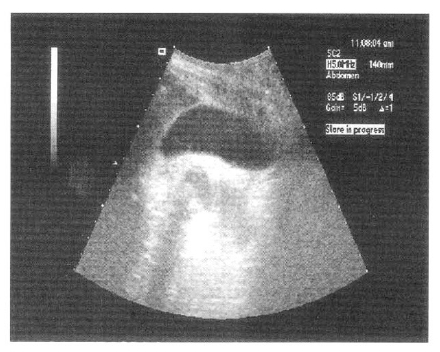
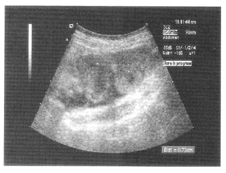
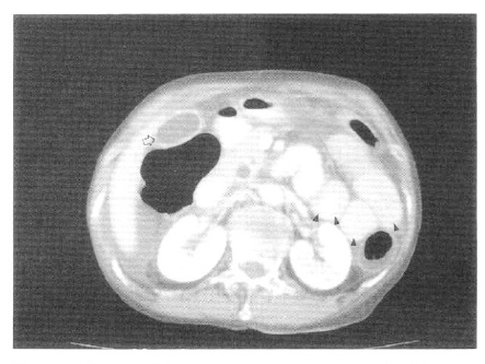
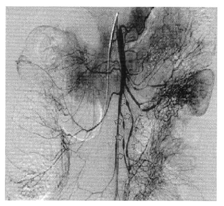
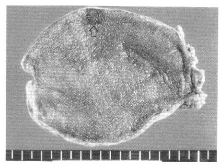
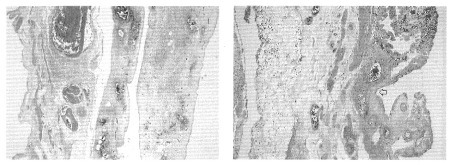
A 70-year-old woman was admitted with severe abdominal pain, which began a few hours before admission. She had neither a specific past medical history nor any underlying illness. The abdominal pain was continuous and diffuse without localization. The initial blood pressure was 120/80 mmHg, the pulse rate was 84 beats per minute, the temperature was 36.4┬░C, and the respiratory rate was 32 breaths per minute. On physical examination, the abdomen was soft but moderately tender throughout without rebound tenderness. The laboratory values were as follows: white blood cell count, 11,900/mm3; AST, 26 IU/L; ALT, 11 IU/L; amylase, 210 IU/L; lipase, 316 IU/L. An ultrasonographic examination showed that the gallbladder had no abnormal findings but the small intestine showed diffuse dilatation and wall thickening (Figure 1, 2). A computed tomographic examination of the abdomen and pelvis performed with the intravenous administration of contrast media showed severe thickening of the small intestinal wall and small quantity of ascites without any evidence of bowel perforation or acute appendicitis (Figure 3). During the next three hours, the patientŌĆÖs blood pressure fell to 80/50 mmHg and the temperature rose to 38┬░C. Her abdominal pain worsened with newly developed rebound tenderness and rigidity on abdominal palpation. After deliberating on the patientŌĆÖs age, clinical manifestation and radiological findings, abdominal angiography was performed to rule out the possibility of acute intestinal ischemia. Abdominal angiography showed multiple stenoses at the superior and inferior mesenteric artery and extensive spasms at the branches of the superior mesenteric artery (Figure 4). An exploratory laparotomy was performed on with the impression of small bowel necrosis due to acute intestinal ischemia. On exploration, there were neither intestinal necrosis nor intestinal perforation. Only bile-colored ascites was noted. Although the gallbladder appeared normal, bile leaked on a localized focus of the gallbladder with gentle squeezing. Therefore, the exploration was stopped after performing the cholecystectomy. The excised gallbladder appeared grossly normal except for the focal necrotic area without a frank perforation or stone (Figure 5). On a pathologic examination, the gallbladder showed a focal area of inflammation and necrosis confined to the microscopically perforated site at the body (Figure 6). The results of the bile culture were negative. After surgery she recovered and was discharged 16 days later.
DISCUSSION
Even though there are many reports concerning gallbladder perforation, some controversy still remains regarding the tools of the early diagnosis and the therapeutic modalities3). Unlike gallbladder perforation, as a complication of cholecystitis, cases of gallbladder perforation without an apparent cause are rare and are reported vaguely as being either idiopathic or spontaneous12, 13). A literature search found only 17 cases worldwide reported as idiopathic or as spontaneous gallbladder perforation. In addition, no case has been reported in Korea. While reporting a case of idiopathic gallbladder perforation, which had been misdiagnosed as peritonitis secondary to appendicitis preoperatively, Jose EC et al. insisted that almost all cases of gallbladder perforation were in fact secondary to a coexistent disease such as inflammation, trauma or obstruction13). They also proposed a classification system, which categorized the gallbladder perforation into three groups as spontaneous, traumatic and iatrogenic13) The spontaneous group is further subdivided into an idiopathic group and a secondary group, which includes lithiasis, inflammation, infection, congenital obstruction, anticoagulants, etc13). Although there is little consensus about this classification, we compared these idiopathic cases to secondary cases, particularly those caused by the complications of cholecystitis.
The most plausible mechanisms for gallbladder perforation complicating acute cholecystitis are: 1) bile stasis due to cystic duct obstruction, fasting, dehydration, total parenteral nutrition, which leads to a change in the bile content and concentration; 2) vascular impairment of the gallbladder due to distension of the viscus, underlying systemic illness such as sepsis, shock, atherosclerosis; and 3) ischemic necrosis and perforation of the gallbladder wall. Gallbladder perforation occurs most commonly at the fundus, which has the least blood supply. This fact denotes the importance of the ischemic mechanism of gallbladder perforation1, 3, 11). However, the mechanisms of spontaneous or idiopathic gallbladder perforation in an otherwise normal gallbladder have not been reported15ŌĆō17).
The diagnosis of an idiopathic case is often delayed or missed, the majority being made only at surgery by observing the bile-colored ascites13, 14). It is believed that the reasons for this are a lack of clinical manifestations pertinent to gallbladder perforation or cholecystitis and the limited usefulness of imaging studies including ultrasonography (US) and computed tomography (CT). It has been known that a US, CT, hepatobiliary scan and endoscopic retrograde cholangiopancreatography (ERCP) can be used to detect gallbladder perforation or cholecystitis3, 5, 9, 10ŌĆō12). However, diagnosis is difficult in idiopathic cases in the absence of the typical radiological findings such as a defect of the thickened gallbladder wall (hole sign), a pericholecystic fluid collection and an abscess formation10ŌĆō13).
Minoru T et al. reported that the insensitivity of these imaging studies was due to the mild inflammation of the gallbladder12). Indeed, the inflammation of the gallbladder was mild and was usually confined to the area of the perforation in the idiopathic cases12, 13). Although a hepatobiliary scan and ERCP may be useful4, 5, 13, 14), it is believed that it is almost impossible to perform such studies without any prior indication pointing to the diagnosis.
The mortality rate of gallbladder perforation with cholecystitis is high3, 4, 6). The reasons related to such a poor prognosis appear to be a delayed diagnosis and treatment due to the clinical similarities of acute cholecystitis without perforation and the doctor being unaware of the emergent situation. Therefore, it is important to suspect perforation clinically and to proceed with the proper treatment rather than to do unnecessary and ineffective studies to confirm perforation3, 4, 9, 11). However, surgery related mortality has not been reported despite the seriousness of the bile-induced peritonitis in idiopathic cases12ŌĆō14) Such a good clinical outcome can be explained by the prompt recognition of the peritonitis, the timely surgery and the negative results of the bile culture12, 13).
In this case, the exact diagnosis was elusive because of the scanty clues suggesting gallbladder perforation. The bile colored ascites, which led us to perform the cholecystectomy, was the only clue. Considering the result of the imaging studies, ischemia of the small intestine, though not severe enough to cause necrosis, was probably present as well. It is believed that this might be caused by an unknown systemic ischemic insult or secondary circulatory insufficiency due to bile-peritonitis. A gross examination of the excised gallbladder was normal without a frank perforation. In addition, a pathologic examination showed only a focal area of inflammation with ischemic necrosis and vascular congestion confined to the microscopically perforated site.
Compared to the case reports of idiopathic gallbladder perforation, this case had many similarities in regard to the clinical manifestation, the radiological findings and progression. From the pathologic findings of ischemic necrosis of the gallbladder wall and the radiological findings of small bowel ischemia, it is possible that the ischemic insult played a pivotal role in this case. The presumptive causes of the ischemia might be atherosclerosis, focal vasospasm or localized vasculitis or some other unknown factors. Therefore, it is believed that this is a case of spontaneous gallbladder perforation, probably due to an ischemic cause. Therefore, clinicians should take this disorder into consideration when encountering cases of elderly patients presenting with an acute abdomen with an unknown origin.


 PDF Links
 PubReader
 ePub Link
 Full text via DOI
 Download Citation
 Print
Print


