 |
 |
| Korean J Intern Med > Volume 1(1); 1986 > Article |
|
Abstract
Ferrokinetic studies were performed with 59Fe-citrate to evaluate erythropoietic activity in CAPD patients and to investigate the mechanism(s) by which the hematocrit increases in CAPD patients. Plasma iron disappearance rate (PID), plasma iron turnover rate (PIT), red cell iron utilization (RCIU), red cell iron turnover rate (RCIT) and marrow transit time (MTT) were all “normal” in uremic patients not yet on dialysis (Hct 23.8±3.4%), CAPD patients with persistently low hematocrit (Hct 24.9±1.8%) and CAPD patients with improved hematocrit (Hct 32.4±3.1%). Compared to these uremic patients, patients with iron deficiency anemia and normal renal function (Hct 28.0±5.1 %) had significantly faster PID and MTT and significantly higher RCIU and RCIT. Plasma volume was significantly reduced (to normal level) in CAPD patients with improved hematocrits.
The results of this study suggest that erythropoietic activity is inadequate for the degree of anemia in CAPD patients as well as uremic patients not on dialysis and further suggest that the hematocrit increases in CAPD as a result of decreased plasma volume.
The major mechanisms that have been recognized to contribute to the anemia of uremia are increased destruction1–3) and decreased production of red blood cells.2–3) Inadequate erythropoietin production3–5) and retained inhibitors of erythropoiesis6–7) are probably important in ineffective erythropoiesis. Folate8) or iron deficiency9) has also been implicated.
Radioactive iron turnover studies have been done in uremic patients to measure erythropoietic activity or to investigate the effect of hemodialysis on iron metabolism. Red cell iron utilization was significantly decreased,10–12) “efficient”13) or variable.2–3) Plasma iron turnover was normal or increased13) or decreased.2,3,11)
Despite reports of improvement in iron metabolism3,13,14) and decrease in inhibitor of erythropoiesis6) in some patients on hemodialysis, anemia continues to be a problem in most patients on hemodialysis.15) In many patients on continuous ambulatory peritoneal dialysis (CAPD) the hemotocrit rises early16–18) and remains elevated for up to 2 years on CAPD. Furthermore, many patients previously treated with hemodialysis show an increase in hematocrit while on CAPD. A rise in the hematocrit was accompanied by an increase in red cell mass, thus a “true” improvement of anemia17) or by an increase in serum erythropoietin level.18)
We performed ferrokinetic studies in patients on CAPD to measure erythropoietic activity and to investigate the effect of CAPD on iron metabolism in these patients. Erythropoiesis was less than adequate for the degree of anemia in CAPD patients and in uremic patients not yet on dialysis and a significant reduction in plasma volume (to normal value) was observed in CAPD patients with higher hematocrit values.
Studies were performed on 11 patients who had been on CAPD for at least 3 months: 7 patients with hematocrit 30% or above (CAPD I), and 4 with hematocrit below 30% (CAPD II), 6 patients with uremia not yet on dialysis (uremia), 6 patients with iron deficiency anemia (IDA) and normal renal function and 5 healthy volunteers (normal). Clinical data of study groups are shown in Table 1. IDA group and 3 uremic groups had comparable degree of anemia. Informed consent was obtained from all patients prior to the study. No patient received blood transfusion within 3 months of study.
A blood sample was obtained in the morning on the first day of ferrokinetic studies for the usual hematologic parameters. Hematocrit and reticulocyte count were obtained by conventional methods. Serum iron and total iron binding capacity were measured by colorimetric method using Fe B test kit and supplementary reagent kit (Wako). Serum was also analyzed for urea and creatinine by autoanalyzer and ferritin by radioimmunoassay.
Ferrokinetic studies used 59Fe-citrate (Amersham, U.K., 100 μCi/ml) and were done by techniques described by Huff et al.19) and Finch et al.20) Blood samples were withdrawn into syringes containing acid citrate dextrose and plasma was separated. To 8 ml of plasma, 20 μCi of 59Fe-citrate was added and allowed to stand at room temperature for 30 minutes, and exactly 5ml of this was reinjected intravenously. The remaining radiolabelled plasma was kept as a standard. Blood samples were obtained at 15, 30, 45, 60, 120 and 180 minutes after reinjection and plasma was separated. The radioactivity of plasma samples and standard were counted in a well type scintillation counter (Picker Spectroscaler 4) for 10 minutes and the plasma radioactivity data were plotted on semilogarithmic paper. Plasma volume (PV) was determined from the initial dilution of injected radioactive iron by extrapolation of the disappearance slope of the iron in plasma to zero time:
Total blood volume (TBV) and red cell volume (RCV) were derived from this PV and hematocrit (Hct) of the first day’s sample:
Plasma iron turnover (PIT) was calculated from the serum iron concentration and the initial rate of disappearance of radioiron(PID):
The initial slope of PID was determined from the individual radioactivity points drawn on semilog paper, and the half-time was expressed in minutes.
Whole blood samples were then obtained in 1, 3, 5, 7, 9 and 15 days after reinjection of the 59Fe-labelled plasma and was stored at room temperature until the last day when the radioactivity of whole blood samples and the standard was counted. Red cell iron utilization (RCIU) was calculated by comparing the whole blood activity with the radioactivity injected:
Red cell iron turnover (RCIT) was calculated from PIT and RCIU:
Marrow transit time (MTT) was derived from the RCIU(%) subtracted form 100 plotted on semilog paper as the time interval (days) required for release of 50% of the radioiron measured in circulation.
Statistical analysis: Unpaired t-test, two tailed, was used to compare the differences between groups. The level of significance was 0.05.
Serum iron and total iron binding capaity (TIBC) are shown in Table 2. Serum iron was significantly reduced and TIBC significantly increased in the iron deficiency anemia group when compared to other groups. There were no differences in serum iron and TIBC among normal control, uremia and CAPD groups I and II.
Serum ferritin was low in iron deficiency anemia and elevated in uremia and CAPD groups as shown in Table 3.
Plasma volume, as shown in Table 4, was significantly larger in uremia (53.3 ±8.3 ml/kg) and CAPD group II (57.7 ±6.4 ml/kg) than in normal control (39.7 ± 4.2 ml/kg) and CAPD group I (40.1 ± 7.6 ml/kg). There was no difference in plasma volume between normal control and CAPD group I. Red cell volume (Table 4) was significantly smaller in IDA, uremia and CAPD groups I and II than in normal control. CAPD group I had slightly larger red cell volume than in uremia and CAPD group II but the difference did not reach statistical significance.
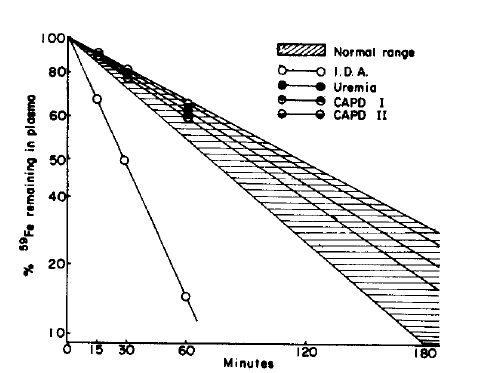
Plasma iron disappearance rate (PID) and plasma iron turnover rate (PIT) are shown in Table 5 and rate of disappearance of injected 59Fe during the first 60 minutes is shown in Fig. 1. PID was significantly faster in IDA (28.5 ± 9.3 min) than in the rest of the groups. PIT was significantly faster in IDA than in CAPD groups I and II. There were no differences in both PID and PIT among normal control, uremia and CAPD groups I and II.
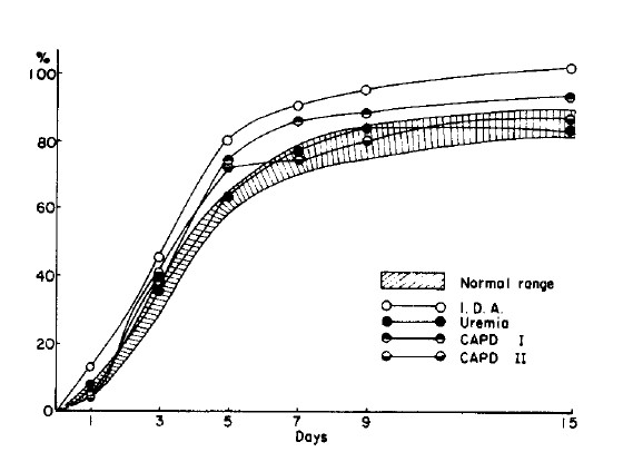
Red cell iron utilization (RCIU) and red cell iron turnover rate (RCIT) 15 days after injection of 59Fe are shown in Table 6 and percent incorporation of 59Fe into circulating erythrocytes from 1 to 15 days after injection of 59Fe is shown in Fig. 2. RCIU was significantly higher in IDA (104.1 ±14.6%) than in normal control (84.4 ±3.8%). RCIT was significantly higher in IDA (0.80 ± 0.27 mg/kg/day) than in normal control (0.35 ± 0.12), uremia (0.25 ±0.11) and CAPD group II (0.33±0.07).
Marrow transit time (MIT) is shown in Table 7 and was significantly shorter in IDA (3.1 days) than in normal control (4.0 days) and uremia (4.0 days). No differences were observed among normal control, uremia and CAPD groups I (3.9 days) and II (3.8 days).
A substantial increase in hematocrit is commonly observed in uremic patients early after they go on CAPD16–18) and, in some, the hematocrit values reach normal range.18) De Paepe et al.17) observed that the early rise of hematocrit in CAPD patients was due to a combination of an increase in red cell mass, thus a “true” improvement of anemia and a decrease in plasma volume. Zappacosta et al.18) reported that serum erythropoietin level was higher in patients with normalized hematocrit than in patients with persistently low hematocrit. Since uremic toxins were shown to have no inhibitory effect on erythropoietin production by kidneys previously exposed to hypoxia,21) CAPD probably had no role in increased production of erythropoietin by diseased kidneys. It is more likely that those patients who normalized their hematocrit values on CAPD had higher erythropoietin level to begin with since 3 of Zap-pacosta’s 4 patients had polycystic kidney disease.
Recent studies6,7) suggest that retained inhibitors or toxic metabolites in uremic serum may be the primary factor responsible for the anemia of chronic renal failure by more directly inhibiting erythroid progenitor cell formation. The increase in hematocrit associated with decrease in inhibitor levels during long-term hemodialysis suggest that inhibitory substances may be more efficiently removed by CAPD if these substances were in the “middle” molecular range since clearance of some middle molecular substances is 6 times greater with CAPD than with hemodialysis.22)
Effects of hemodialysis on erythropoietic activity and iron metabolism have been reported previously.3,13,14) Eschbach et al.3) observed doubling of red cell iron turnover in 5 patients on hemodialysis for 10–35 months. Increased red cell production was observed in 4 patients. A sharp increase in plasma iron turnover was observed by Carter et al.13) in 2 of 5 patients on hemodialysis but the effect only lasted for 2 months. Kurtides et al.14) reported improvement in ferrokinetics and effective erythropoiesis after a single hemodialysis in all 6 patients.
This study was conducted to evaluate erythropoietic activity in CAPD patients and to gain insight into the mechanisms by which the hematocrit increases in CAPD patients. Ferrokinetic studies were carried out with 59Fe-citrate. Plasma iron disappearance rate, plasma iron turnover rate, red cell iron utilization rate, red cell iron turnover rate and marrow transit time were all “normal” in uremic patients not yet on dialysis, CAPD patients with persistently low hematocrit (less than 30%) and CAPD patients with improved hematocrit (30% or above). Despite “normal” values, total and effective erythropoiesis in the 3 uremic groups are still less than adequate for the degree of anemia since the group with iron deficiency anemia and normal renal function showed significantly more active erythropoiesis under the same study condition. A significant reduction in plasma volume (to normal range) was observed in CAPD patients with hematocrit at or above 30% when compared with uremic patients not yet on dialysis and CAPD patients with hematocrit below 30%. Red cell volume was slightly larger in CAPD patients with higher hematocrit than in uremia patients not on dialysis and CAPD patients with persistently low hematocrit but the difference was not significant.
Ferrokinetic data in this study are valid since normal control data are in good agreement with previous reports in the literature2,3,10–14,20,23) and those of patients with iron deficiency anemia and normal renal function are entirely appropriate with rapid plasma iron disappearance and marrow transit time and increased red cell iron utilization and red cell iron turnover. 20) Concomittent iron deficiency may explain “normal” iron kinetics in uremia and CAPD patients. This was unlikely because serum ferritin when checked were elevated in these patients.
The results of this study suggest that erythropoiesis is inadequate for the degree of anemia in CAPD patients as well as uremic patients not on dialysis and further suggest that the hematocrit increases in CAPD not so much as a result of improved erythropoiesis but, in large part, due to decrease in plasma volume.
Table 1.
Clinical data in study groups
| N | Mean age(range) Years | Sex M/F | Hematocrit % | BUN mg/dl | Creatinine mg/dl | |
|---|---|---|---|---|---|---|
| Normal | 5 | 27.2(26–30) | 5/0 | 44.4 ± 0.9** | 14.4± 2.1 | 1.0±0.1 |
| I.D.A.* | 6 | 39.3(16–65) | 1/5 | 28.0 ± 5.1 | 16.9 ± 2.9 | 0.8±0.2 |
| Uremia | 6 | 46.3(32–61) | 4/2 | 23.8±3.4 | 111.0 ± 40.2 | 12.3 ± 5.1 |
| CAPD I | 7 | 42.1(32–59) | 6/1 | 32.4 ± 3.1 | 56.0±13.1 | 12.5±4.5 |
| CAPD II | 4 | 49.3(38–63) | 3/1 | 24.9 ± 1.8 | 59.1 ± 4.1 | 15.1 ±2.3 |
Table 2.
Serum iron and total iron binding capacity (T.I.B.C.)
| N | Serum iron μg/100ml | T.I.B.C. μg/100ml | |
|---|---|---|---|
| Normal | 5 | 103.6±26.2 | 277.3±23.9 |
| I.D.A. | 6 | 39.8±10.1* | 372.4±63.2** |
| Uremia | 6 | 68.4±29.5 | 239.3±26.8 |
| CAPD I | 7 | 92.2±38.5 | 277.9±53.8 |
| CAPD II | 4 | 80.1 ±22.6 | 230.9 ±80.8 |
Table 3.
Serum ferritin (ng/ml)
Table 4.
Plasma volume and red cell volume
| N | Plasma volume ml/kg | Red cell volume ml/kg | |
|---|---|---|---|
| Normal | 5 | 39.7 ± 4.2 | 26.4 ± 2.3 |
| I.D A. | 6 | 44.7±6.9 | 16.7±3.0** |
| Uremia | 6 | 53 3±8.3*# | 1 4.6 ± 3.1 ** |
| CAPD I | 7 | 40.1 ±7,6 | 17.1 ±5/8** |
| CAPD II | 4 | 57 7±6.4#*# | 16.8±3.4** |
Table 5.
Plasma iron disappearance rate (P.I.D.) and plasma iron turnover rate (P.I.T.)
| N | P.I.D. min | P.I.T. mg/kg/day | ||
|---|---|---|---|---|
| Normal | 5 | 101.0 | 20.7# | 0.4176 ± 0.1312 |
| I.D.A. | 6 | 28.5 | 9.3 | 0.6735 ± 0.2490 |
| Uremia | 6 | 108.7 | 45.6# | 0.4156 ± 0.3256 |
| CAPD I | 7 | 99.5 | 27.3# | 0.3475 ± 0.1585## |
| CAPD II | 4 | 117.1 | 20.9# | 0.3854 ± 0.0184## |
Table 6.
Red cell iron utilization rate (R.C.I.U.) and red cell iron turnover rate (R.C.I.T.) 15 days after injection of 59Fe
| N | R.C.I.U % | R C.I T. mg/kg/day | |
|---|---|---|---|
| Normal | 5 | 84.8 ± 3 8 | 0.35 ± 0.12## |
| I.D.A. | 4 | 104.1 ± 14.6 | 080 ± 0.27 |
| Uremia | 4 | 83.1 ±11.8 | 0 25 ± 0.11 # |
| CAPD I | 3 | 93.3 ±18 1 | 0.38 ± 0.30 |
| CAPD II | 4 | 86.6 ± 20.4 | 0.33 ± 0.07## |
REFERENCES
3. Eschbach JW, Funk D, Adamson J, Kuhn I, Scribner BH, Finch CA. Erythropoiesis in patients with renal failure undergoing chronic dialysis. N Engl J Med 276:653. 1967.


4. Gallagher NI, McCarthy JM, Lange RD. Observations on the erythropoietic stimulating factor (ESF) in the plasma of uremia and nonuremic anemia patients. Ann Intern Med 52:1201. 1960.


5. Naets JP, Heuse AF. Measurement of erythropoietic stimulating factor in anemia patients with or without renal disease. J Lab Clin Med 60:365. 1962.

6. Wallner SF, Vautrin RM. Evidence that inhibition of erythropoiesis is important in the anemia of chronic renal failure. J Lab Clin Med 97:170. 1981.

7. McGonigle RJS, Wallin JD, Shadduck RK, Fisher JW. Erythropoietin deficiency and inhibition of erythropoiesis in renal insufficiency. Kidney Int 25:437. 1984.


8. Hampers CL, Streiff R, Nathan DG, Snyder D, Merrill JP. Megaloblastic hematopoiesis in uremia and in patients on long-term dialysis. N Engl J Med 276:552. 1967.

9. Eschbach JW, Cook JD, Scribner BH, Finch CA. Iron balance in hemodialysis patients. Ann Intern Med 87:710. 1977.


10. Loge JP, Lange RD, Moore CV. Characterization of the anemia associated with chronic renal insufficiency. Am J Med 24:4. 1958.


12. Lawson DH, Boddy K, King PC, Linton AL, Will G. Iron metabolism in patients with chronic renal failure on regular dialysis treatment. Clin Sci 41:345. 1971.


13. Carter RA, Hawkins JB, Robinson BHB. Iron metabolism in the anemia of chronic renal failure. Effect of dialysis and parenteral iron. Brit Med J 3:206. 1969.


14. Kurtides ES, Rambach WA, Alt HL, Del Greco F. Effect of hemodialysis on erythrokinetics in anemia of uremia. J Lab Clin Med 63:469. 1964.

16. Lee HB. Current status and future of CAPD in Korea. Korean J Nephrol 2:161. 1983.
17. De Paepe MBJ, Schelstraete KHG, Ringoir SMG, Lamerire NH. Infuence of continuous ambulatory peritoneal dialysis on the anemia of end-stage renal disease. Kidney Int 23:744. 1983.


18. Zappacosta AR, Caro J, Erslev A. Normalization of hematocrit in patients with end-stage renal disease on continuous ambulatory peritoneal dialysis. Am J Med 72:53. 1982.


19. Huff RL, Elmlinger PJ, Garcia JF, Oda JM, Cockrell MC, Lawrence JH. Ferrokinetics in normal persons and in patients having erythropoietic disorders. J Clin Invest 30:1513. 1951.


20. Finch CA, Deubelbeiss K, Cook JD, Eschbach JW, Harker LA, Funk DD, Marsaglia G, Hillman RS, Slichter S, Adamson JW, Ganzoni A, Giblett ER. Ferrokinetics in man. Medicine 49:17. 1970.


21. Erslev AF. The effect of uremic toxins on the production and metabolism of erythropoietin. Kidney Int 7:S129. 1975.
22. Popovich RP, Moncrief JW, Nolph KD, Ghods AJ, Twardowski ZJ, Pyle WK. Continuous ambulatory peritonal dialysis. Ann Intern Med 88:449. 1978.


23. Lee MH. Clinical Nuclear Medicine. In: Lee MH, ed. 204. Ryo Moon Gak, Ltd, 1982.


 PDF Links
 PubReader
 ePub Link
 Full text via DOI
 Download Citation
 Print
Print


