


INTRODUCTION
Intercellular adhesion molecule-1 (ICAM-1), a 80–110 kD glycoprotein, has been found to be a ligand for the lymphocyte function associated antigen-1 (LFA-1)molecule1–3). It plays an important role in a variety of inflammatory and immune mediated mechanisms, including lymphocyte recruitment targeting, antigen presentation and recognition and lymphocyte cytotoxicity.4–7) ICAM-1 is expressed on thyroid follicular cells8–13) of patients with Hashimoto disease11) and cultured thyroid monolayer cells12) derived from thyroid surgical specimen. The release of various cytokines at the site of inflammation results in local augumentation of ICAM-1 expression14,15). In addition to the expression of ICAM-1 on the surface of cells, soluble variants of several adhesion molecules have been reported16–23). A soluble form of ICAM-1 of mol. wt 82000 has been described that binds LFA-1 and blocks rhinovirus infection18). This circulating form of ICAM-1, found in increased levels in patients with inflammatory, immune or malignant diseases, has also been associated with metastatic disease in adults with melanoma19–24). In the present study, high serum levels of ICAM-1 were associated with autoimmune thyroid disease. Graves’ disease and Hashimoto disease and positively correlates with levels of antithyroperoxidase antibody.
MATERIALS AND METHOD
Sera were collected from 58 patients with autoimmune thyroid disease, 28 patients with Graves’ disease and 30 patients with Hashimoto disease. Diagnosis of Graves’ disease was based on clinical assessment, elevated serum T3 and T4 levels and increased homogenous 99mTc uptake on the thyroid scan. Graves’ patients had no clinically significant inflammatory ophthalmopathy. 18 patients with Graves’ disease were clinically and biochemically hyperthyroid(TSH<0.04 mU/ml) and the others were euthyroid with antithyroid drug(propylthiouracil)treatment. The diagnosis of Hashimoto lisease was based upon the combined presence of hypothyroidism with an elevated serum TSH level(TSH>4mU/ml) and a significant autoantibody titer against thyroglobulin and thyroperoxidase(both above 3 U/ml). All blood samples, obtained from patients with Graves’ disease and Hashimoto disease, were processed immediately for centrifugation and sera were stored frozen at−20°C until used in the ICAM-1 assay.
Serum concentrations of circulating ICAM-1 were determined with a commercially available ICAM-1 sandwich enzyme immunoassay(Cell-free, T cell diagnostics, Cambridge MA). Briefly, serum samples were diluted in 1:100 and applied to 96 polystylene microwells precoated with murine monoclonal antibody to human ICAM-1. A horeseradish peroxidase-conjugated anti-mouse monoclonal antibody with neutralizing function that binds to the ICAM-1 captured by primary antibody was then added. Following incubation on a rotator(150 RPM) for 2 h at 25°C and extensive washing, the reaction product was developed in O-phenylene diamine substrate solution for 30 mins, after which the enzyme reaction was terminated with 4 N sulphuric acid. Absorbance for samples and ICAM-1 standards were determined on a spectrophotometerx using 490 nm as the primary wavelength. Concentrations for circulating ICAM-1 in serum samples were determined by comparing the mean absorbance of duplicate samples with the standard curve for each assay. A random selection of 20 normal sera were assayed. Intraassay variance of circulating ICAM-1 enzyme immunoassay was 2.1% at 215 ng/ml.
Serum total T4 and T3 were measured by radioimmunoassay using the T4 Tetrabead kit and T3 Riabead kit from Abbott(IL, USA). Serum TSH was measured by immunoradiometric assay using the TSH Riabead II kit from Abbott. Serum antithyroperoxidase antibody(APA) and antithyroglobulin antibody(AGA) were measured by direct radioimmunoassay, using recombinant thyroperoxidase and thyroglobulin respectively. Thyrotropin binding inhibiting immunoglobulin (TBII) activity was expressed as the percentage inhibition of 125I-bTSH binding to the TSH receptor. A TBII value exceeding 15%, which was greater than the two standard deviations of the mean value for 64 normal samples, was considered to be abnormal or positive. The intra-assay variance of TBII activity was 1.7–24.5%.
An analysis of variance was used to compare serum circulating ICAM-1 levels in the study groups. Data were analysed by t-test for comparing group means and correlation analysis were performed by SPSS/PC + program. All tests of significance were two-tailed, and p values of 0.05 or less were considered significant.
RESULTS
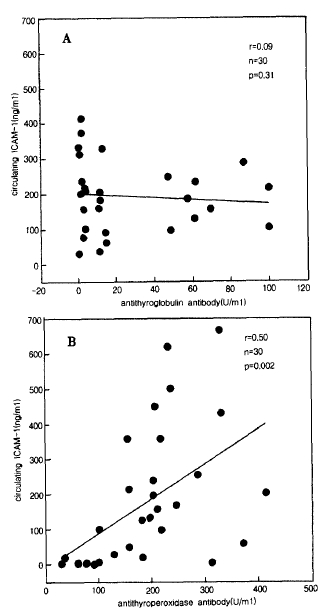
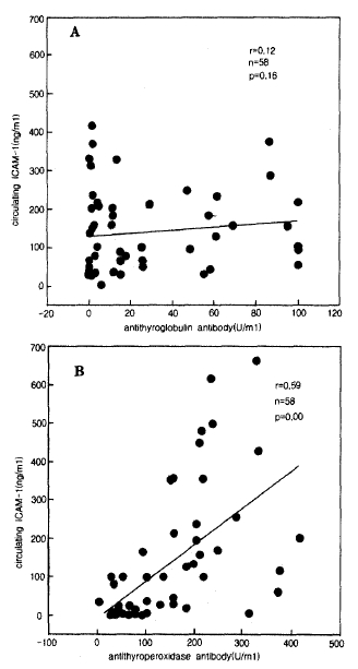
Serum concentrations of circulating ICAM-1 in samples derived from Graves’ disease and Hashimoto disease, as well as normal individuals, are demonstrated in table 1. Compared with normal individuals, mean serum concentrations of circulating ICAM-1 were significantly elevated in patients with Hashimoto disease and antithyroperoxidase-positive Graves’ disease. No significant difference in circulating ICAM-1 concentrations was observed between patients with Graves’ disease, without antithyroperoxidase antibody, and healthy individuals. Patients with antithyroperoxidase-positive Graves’ disease revealed significantly higher serum circulating ICAM -1 concentrations than antithyroperoxidase-negative Graves’ disease. The high levels of circulating ICAM-1 in the sera of patients with Graves’ disease were attributable to the positivity of antithyroglobulin antibody and antithyroperoxidase antibody in these sample, since circulating ICAM-1 showed significant positive correlation with serum titers of antithyroglobulin and antithyroperoxidase antibody(r = 0.44, n = 28, p= 0.009, and r = 0.55, n = 28, p = 0.001 repectively. Fig. 1). Serum concentrations of circulating ICAM -1 were highest in patients with Hashimoto disease(table 1)but circulating ICAM-1 levels were not significantly higher in the hypothyroid group compared with the euthyroid group of Hashimoto disease. There was a significant positive correlation between circulating ICAM-1 levels and serum antithyroperoxidase level in the group of autoimmune thyroid disease(Fig. 3), and also circulating ICAM-1 levels were significantly correlated with serum antithyroperoxidase antibody levels in antithyroperoxidase antibody-positive Graves, disease(r = 0.55, n = 28, p = 0.001)and in Hashimoto disease(r = 0.5, n = 30, p = 0.002). The titers of anti thyroglobulin autoantibody were not significantly correlated with secrum levels of ICAM-1 in Hashimoto disease and Graves’ disease(Fig. 2, 3). The thyrotropin binding inhibiting immunoglobulins(TBII) showed no significant correlation with circulating ICAM-1 levels(data not shown).
DISCUSSION
Intercellular adhesion molecule-1 (ICAM-1), a 80–110kD glycoprotein, has been found to be a ligand for the lymphocyte function-associated antigen-1 molecule2). ICAM-1 plays an important role in inflammatory disease4–6). The release of cytokines, such as interferon-gamma, interleukin-1 and tumor necrosis factor at sites of inflammation and immune responses, causes cell activation and results in locally augmented expression of adhesion molecules including ICAM-1 on the cell surface10). Furthermore, the ICAM-1 antigen has recently been reported to be a member of the immunoglobulin supergene family with five domain structures6). Expression of ICAM-1 on thyroid cells can also be stimulated by various cytokines10,12,15). Some of locally expressed ICAM-1 molecules shedded into circulations and these shedding can be induced in vitro by exposure of cells to active cytokines16–21).
In this study, we report the presence and concentration of circulating ICAM-1 in sera derived from patients with Graves’ diseae and Hashimoto disease. Patients with Hashimoto disease and antithyroperoxidase antibody-positive Graves’ disease revealed significantly elevated mean serum concentrations for circulating ICAM-1, and the level in patients with Hashimoto disease were significantly higher than those measured in patients with Graves’ disease. Heufelder et al24) reported that the circulating level of ICAM-1 in patients with graves’ disease with opthaImopathy and in patients with Riedel's invasive fibrous thyroiditis revealed markedly elevated circulating ICAM-1 concentrations compared with normal controls. In this study, patients with Graves’ disease showing negative antithyroperoxidase antibody, circulating ICAM-1 was not significantly elevated compared to that of normal controls. This discrepancy may due to the difference of characteristics of patients. In this study, the patients with Graves’ disease showed no clinically obvious thyroid-associated inflammatory opthalmopathy.
Antithyroperoxidase autoantibody could interfere with the endogenous thyroperoxidase activity25), leading to impaired thyroglobulin iodination and thyroid homone synthesis and titers of the autoantibody also reflects a more active autoimmune reaction and functional damage in the thyroid gland. But regarding antithyroglobulin antibody, it is usually not stressed that high antithyroglobulin antibody titiers may be an expression of a more active cellular infiltration in the thyroid gland.
The potential functions of soulble forms of adhesion molecules can, at present, only be the subjects of speculation. Release of adhesion molecules or shedding of their extracellular portions from the cell surface may generate competitive soluble inhibitors of the membrane-bound forms, thereby regulating the cytodine-induced increase in their expression and adhesive properties. Cell adhesion studies suggest that the soluble ICAM-1 molecule present in sera of patiernts with Graves’ opthalmopathy possess functional activity24).
The origin of ciculating ICAM-1 in sera of patients with autoimmune thyroid disease is not known. Because the adhesion molecule ICAM-1 is normally expressed in endothelial cells, thymus and other lymphoid organ, all of the above are potential candidates for the origin fo circulating ICAM-1. Shedding of ICAM-1 can be induced in vitro by exposure of Graves’ retroocular fibroblast monolayers to interferon-γ and tumor necrosis factor-α. We think that the circulating ICAM-1 is originated from thyroid follicular cells and retro-orbital fibroblast after stimulation of local cytokine produced during autoimmune inflammation. We need further study for confirmation of circulating ICAM-1 as a marker of ongoing autoimmune thyroid inflammation.

 PDF Links
 PubReader
 ePub Link
 Full text via DOI
 Download Citation
 Print
Print


