


INTRODUCTION
Acute pyelonephritis is a common acute bacterial infectious disease of the kidneys accompanying fever, tenderness on the costovertebral angle, and urinary symptoms, such as dysuria, frequency, and can cause renal or perirenal abscess and may leave some sequelae such as cortical scar, hypertension and renal insufficiency. Sometimes, it is not easy to differentiate the upper urinary tract infection from other febrile disease accompanying pyuria.
Recently, many better imaging methods including ultrasonography, intravenous pyelography, computerized tomography and magnetic resonance imaging were developed. However, such diagnostic imaging does not show active pyelonephritic lesions in the kidneys1,2).
Normally, Tc-99m DMSA used in the static renal scintigraphy accumulates on the renal cortical proximal tubule cells, 2–3 hours after intravenous injection. Tc-99m DMSA has been used in renal imaging to estimate the functional renal mass and relative renal function, especially in pediatric patients with urinary tract infection3–5). Recently, several authors reported that Tc-99m DMSA renal scan frequently showed cortical defects even in patients with acute pyelonephritis who did not show abnormal findings in the ultrasonography and intravenous pyelography(IVP)5–10). The pathophysiologic mechanisms about the renal cortical defects of Tc-99m DMSA renal scan in the patients with acute pyelonephritis were not known. But several upkae mechanisms 11) such as 1) direct tubular defect due to bacterial infection12), 2) interstitial edema and tubular defect by intratubular aggregation of white blood cells13),3) perinephronal capillary occlusion by aggregated inflammatory cells had been postulated14).
We designed a clinical study to evaluate the utilities of Tc-99m DMSA renal scan and the clinical meaning of cortical defects of the Tc-99m DMSA renal scan in the patients with acute pyelonephritis.
MATERIALS AND METHODS
Ninety two patients with acute pyelonephritis showing typical symptoms and signs such as acute fever, urinary symptoms, tenderness on costovertebral angle areas and pyuria were included in this study from March 1991 to February 1994. But the patients finally diagnosed with chronic pyelonephritis or renal tuberculosis were excluded. Patients with a vesicoureteral reflux or severe obstructive uropathy were also excluded because of the possibility of chronic pyelonephritis.
For the Tc-99m DMSA renal scan, all patients were injected 5mCi of Tc-99m DMSA intravenously and, two or three hours after injection, we took renal images with planar image gamma camera(Siemens Co.) on anterior, posterior, left posterior and right posterior oblique view. All Tc-99m DMSA renal scintigraphys were performed within 1 week after admission.
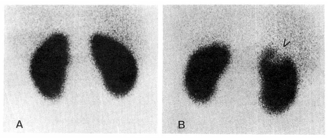
Patients were subdivided as Group A(n=42): patients showing normal Tc-99m DMSA renal scan, and Group B(n=50):patients with the definite cortical defects on the Tc-99m DMSA renal scan(Fig. 1) We compared clinical characteristics such as age and sex, recurrency, duration of fever, bacterial culture study, incidence of renal insufficiency and the results of renal ultrasonography and intravenous pyelography between the two groups.
All patients were treated with the same regimens of antibiotics for 2 weeks or more, such as cephalosporin and aminoglycosides. IVP and/or renal ultrasonographic examinations were performed within 1 week before or after Tc-99m DMSA renal scan.
RESULTS
Group A was 42 patients and Group B was 50 patients. Mean age of Group A and B patients was 44.3±16.5(range 19–81) and 46.8±17.5 (range 21–79), respectively(p>0.05). Most of the patients in the two groups were women (Table 1). On past history, the patients who experienced acute pyelonephritis more than two episodes was 11(22%) in Group A and 10(23%) in Group B(p>0.05). Duration of fever(>37.2’C oral)in the pre-admission period was not significantly different between the two groups. However, duration of fever after admission in Group A and B patients were 2.3±1.1 and 3.9±3.3 days, respectively(p<0.05, Table 2-A). White blood cell counts of the blood between the two groups was not significantly different at the time of the admission(p>0.05). Of Group B patients, 7 patients(16%) showed elevation of serum creatinine levels above 1.5mg/dl and that was more frequent than Group A patients(2%)(p<0.01). On the urine microbiologic culture study, 18 patients of 42 Group A patients(43%) showed positive culture results and 38patients of Group B(76%)disclosed positive culture results(p<0.01). Group B patients showed more frequent positive blood culture results(23%)than Group A patients, significantly(p<0.01, Table 2-B). Most of pathogen in blood and urine was E. coli.
IVP examinations were performed on 55 patients of all patients with acute pyelonephritis. Of Group A patients, 18 patients were given IVP study but there was no abnormality. Of Group B patients, 37 patients were gvien IVP study and 11 patients(30%) showed abnormalities. Among the 11 patients with abnormalities in the IVP study, delayed renal opacification was noted in 6 patients, calyceal blunting in 3 patients, double ureter and mild hydronephrosis in one patient, each respectively(Table 3-A.).
The kidney ultrasonographic examinations were performed on 69 patients. Of 34 Group A patients, only 3 patients(8%) showed abnormalities such as two cases of enlarged kidneys and one case of focal irregular increased renal echogenicity. However, 11 patients of 35 Group B patients(31%) disclosed abnormal results. six patients showed enlarged involved kidneys, 3 patients focal increased echogenicities and one patient showed an irregular renal cortical margin and the other a mild hydronephrosis(Table 3-B.).
Therefore ultrasonographic abnormalities in the Group B were significantly more frequent than Group A patients(P<0.05).
DISCUSSION
Acute pyelonephritis is a kind of bacterial infection of the upper urinary tract and sometimes it is not easy to differentiate it from the lower urinary tract infection. Although Ga-67 scan15,16) can be used in the diagnosis of acute bacterial infection in the kidneys, it takes usually two or three days or more to get the scan image and there are many conditions of abnormal renal Ga-67 accumulation such as acute interstitial nephritis, renal abscess, perirenal abscess, tumor and glomerunonephritis. Tc-99m DMSA is an chelating radiopharmaceutical accumulating in renal proximal tubule cells3). Tc-99m DMSA renal scan can be performed easily within several hours. After Davis et al5) firstly reported clinical utilities of Tc-99m DMSA or Tc-99m glucoheptonate renal scan in patients with urinary tract infections, both renal scanning radiopharmaceuticals heve been commonly used as static renal imaging agents. The uptake ratio of renal cortex vs. medulla in Tc-99m DMSA scan is known as 22:13,5). Tc-99m DMSA renal scan has been used also on peiatric patients to estimate the functional mass in kidneys with vesicoureteral reflux17,18). There are only a few reports about Tc-99m DMSA renal scan in adult patients with acute pyelonephritis5,6,7). Although exact mechanisms were not known, several pathophysiologic mechanisms11–14) about renal cortical defects on Tc-99m DMSA renal scintigraphy-1) direct tubular renal cortical defects on Tc-99m DMSA renal scintigraphy-1) direct tubular defect due to bacterial infection, 2) interstitial edema and tubular dysfunction by intratubular aggregation of white blood cells, and 3) perinephronal capillary occlusion by aggregated inflammatory cells were postulated. Because these conditions may decrease focal renal perfusion in an involved area, that may cause focal cortical defects. Majd et al19) and Wikstad20) reported that Tc-99m DMSA renal scan could detect early focal ischemic lesions of the renal cortex defore functional tubular cell defects developed. Of course, other space-occupying lesions, such as renal tumor, cyst and scar, can induce focal renal cortical defects in Tc-99m DMSA renal scan.
Some authors2) reported that less than 25% of the patients with acute pyelonephritis showed some abnormalities, such as focal decreased opacification, enlarged kidney or hydronephrosis in the IVP study. In the renal ultrasonography, most patients with acute pyelonephritis showed also nonspecific results unless there were obstructive uropathy, vesicoureteral reflux, abscess or other mass lesions1). In this study, 25% of the patients with the acute pyelonephritis showed abnormal results of IVP and most of them were delayed opacifications. Only twentyfive percent of the patients showed abnormalities in the renal ultrasonographic examination. Most of abnormalities on the renal ultrasonography were enlarged kidneys and focal increased echo-genicity in the involved kidneys. These abnormalities on the IVP or renal ultrasonographic examinations were more frequently found in Group B than Group A patients. Therefore, we thought that IVP or ultrasonography may be only useful to evaluate the complications or underlying anatomical urinary tract abnormalities.
The incidence of renal cortical defects on the Tc-99m DMSA renal scan performed on patients with acute pyelonephritis is variable. Rushton et al8) reported that the sensitivity and specificity of Tc-99m DMSA renal scan for the detection of acute pyelonephritis in experimental animal study were 91% and 98%, respectively. In this study, 47 of 92 acute pyelonephritis patients showed unilateral cortical defects and 3 patients had bilateral defects. Therefore, the total incidence of cortical defects was 54% in this study. This is somewhat lower than those of the other studies 6,7). We thought that the reason for this low incidence rate is due to low resolution power of our single head planar gamma camera and exclusion of the patients with vesicoureteral reflux, obstructive uropathy and patients with chronic pyelonephritis. One study7) using SPECT(single photon emission computerized tomograpy)in Korea showed 83% of sensitivity in acute pyelonephritis.
All patients included in this study recovered completely and even the patients showing transient renal insufficiency or bacteremia showed the recovery of the renal function also. The patients showing cortical defects(Group B)disclosed a longer febrile period after admission, higher positive rates of the urine and blood culture study, higher incidence of renal insufficiency significantly than those of Group A patients. Therefore we thought that Tc-99m DMSA renal scan in the acute pyelonephritis may predict the patients group with more severe disease course and the cortical defects may suggest the focal functional defects insted of anatomical lesions. Because we did not perform follow up Tc-99m DMSA renal scintigraphy, we could not examine whether the cortical defects could disappear or not after recovery. Mok et al6) and Ryu et al7) reported that the patients who initially showed focal cortical defects in the involved kidneys showed a tendency of resolution of cortical defects several weeks after clinical recovery.
In summary, Tc-99m DMSA renal scan was a more sensitive imaging test than ultrasonography and intravenous pyelography to detect pylonephritis lesions and may be useful to predict the patient group with severe disease course. These patients may need more careful management and further studies to evaluate the possibility of complications.
 PDF Links
 PubReader
 ePub Link
 Full text via DOI
 Download Citation
 Print
Print


