


INTRODUCTION
Aspergillus is a ubiquitous organism that is associated with a number of pulmonary syndromes, one of which is aspergilloma. Most cases of aspergilloma are thought to arise from saprophytic colonization and proliferation of the fungus in pre-existing, poorly drained intrapulmonary cavities that result from various underlying diseases1,2). Its occurrence in association with pulmonary sequestration is extrmely rare3,4). We present a case of aspergilloma developed within intralobar pulmonary sequestration.
CASE REPORT
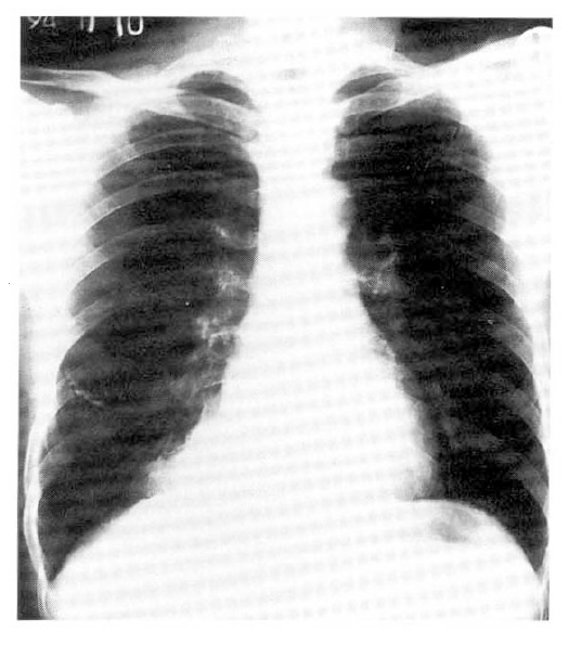
A 41-year-old male was admitted with fever, dry cough and pleuritic chest pain for 10 days. He had always considered his health to be good except that he had a subtotal gastrectomy due to peptic ulcer bleeding 20 years before admission. On examination, his blood pressure was 130/80 mmHg, pulse rate was 90/min and body temperature was 100.8 ┬░F. Breathing sounds were diminished over the lower half of the right posterior chest. Laboratory data included a hemoglobin of 13g%, hematocrit of 44%, white blood count of 12,400/mm3 with 60% neutrophil, 35% lymphocyte, 4% monocyte and 1% eosinophil. IgE was 20 IU/ml. No pathogen was found in the smear and culture of the sputum. Immediate skin test to Aspergillus antigen was nonreactive. Serum precipitins to Aspergillus fumigatus were positive. Chest roentgenogram showed a well-defined mass in the posteromedial portion of the right lower lobe (Fig. 1). The pulmonary perfusion scan using 99mTc-macroaggregate albumin showed a perfusion defect in the right lower lung field, corresponding to the lesion in the chest roentgenogram. A computed tomographic scan of the chest showed a mass with soft tissue density in the posterior segment of the right lower lobe (Fig. 2). CT-guided biopsy specimens from the lesion showed aggregations of hyphae of Aspergillus.
During the operation, one artery arose from the descending aorta just above the diaphragm and passed wihin the leaves of the pulmonary ligament to the medial border of the right lower lobe. A right lower lobe lobectomy was performed.
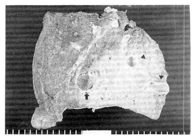
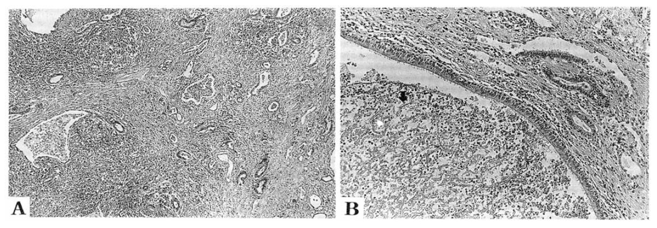
Cut section of the resected lobe revealed a large feeding artery measuring 0.8 cm in diameter that entered a poorly defined area of consolidation, measuring 8.0├Ś6.5 cm. The consolidation contained multiple small cysts with pus and a large cyst measuring 4├Ś3 cm that was filled with dark brownish mud-like fragile materials (Fig. 3). The adjacent parenchyma to the large cyst showed scattered small cysts of the same appearance. Microscopically, the parenchyma was distorted and largely replaced by dense fibrosis and chronic inflammation. Scattered bronchiol-like structures lined by cuboidal-to-columnar epithelium were present. The large cystic wall was lined with ciliated columnar epithelium. Hyphae of the Aspergillus were found within the cystic lumen (Fig. 4).
DISCUSSION
Bronchopulmonary sequestration is a region of lung parenchyma that has an incomplete or no connection with the airways and is supplied by an aberrant artery arising from the aorta or one of its branches5). It is generally divided into intralobar sequestration, which is part of a lobe, and extralobar sequestration, which has its own pleural covering. Intralobar sequestration is less often associated with other congenital anomalies than is extralobar sequestration6).
Pryce et al.5) classified the extent of blood supply from aberrant artery in intralobar sequestration: abnormal artery without sequestration (type 1), abnormal artery supplying the sequestered as well as the adjacent normal lung (type 2), and abnormal artery that supplies only the sequestered lung. Iwai et al.7) classified the bronchoalveolar structure of the sequestered area according to the extent of development of the bronchoalveolar tree: sequestered lung consists of large bronchial wall (grade 1), of poly-cystic changes (grade 2), of cystic and alveolar structure (grade 3), and of alveolar structure (grade 4). This patient is type 3 according to PryceŌĆÖs classification and grade 3 according to IwaiŌĆÖs classification.
The clinical picture of intralobar sequestration is nonspecific. Symptoms, when present, are usually secondary to a superimposed infection presenting as acute or recurrent pneumonia. Infections are usually pyogenic. The development of an aspergilloma in a sequestered cyst is extremely rare4,6). In a review of 540 published cases of lung sequestration, Savic et al.6) reported six occasions of tuberculous disease and a case of nocardial infection within the sequestration. However, there was no case of infection associated with Aspergillus species. As detailed previously, this patient is a typical case of intralobar bronchopulmonary sequestration. A fungus ball due to Aspergillus species was detected in the cystic lumen within the sequestered lung.

 PDF Links
 PubReader
 ePub Link
 Full text via DOI
 Download Citation
 Print
Print


