


INTRODUCTION
Choledochal cyst, or cystic dilatation of the hepatobiliary tree, was first described in 17231). Although there is a predominance of females with the condition (75%), though not in Asia, only 24 cases of choledochal cysts during pregnancy have been reported2). Choledochal cyst is frequently encountered in infancy and childhood, but in 20% of patients with choledochal cyst, the diagnosis is not established until adulthood3). Surgical treatment of choledochal cysts in adults is much more difficult than in the pediatric population. Adult choledochal cyst in pregnancy is difficult to diagnose radiologically and surgically, and is an obstetrical challenge. However, with sonography, the diagnosis of choledochal cyst is no longer a diagnostic problem, even in a pregnant patient. If left untreated, choledochal cyst almost uniformly causes death.
The following is a report of our recent experience with acute pancreatitis complicating pregnancy with choledochal cyst. We offer guidelines, based on review of literature, for the management of choledochal cysts that is found during pregnancy.
CASE REPORT
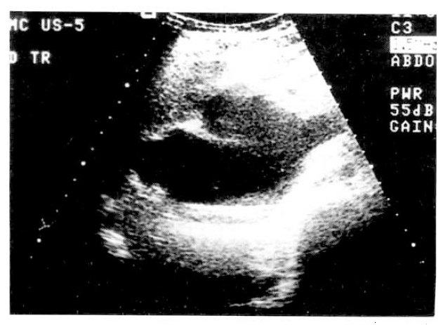
A 29-year-old nulliparous woman at 32nd week gestation began experiencing epigastric burning and back pain associated with nausea and vomiting. She had no significant gynecological, past medical history. She denied any drug ingestion, foreign travel or recent contact with infectious disease and there was no history of fatty food intolerance or long standing indigestion. On examination, the patient was afebrile with normal vital signs. She was not anemic or jaundiced. Abdominal palpation confirmed tenderness in the epigastrium. Fetal movements were felt and the uterus was of an appropriate size for gestational age. The full blood count showed leukocytosis of 12000/mm3 and serum amylase 174U/L (normal 30ŌĆō110U/L) and lipase 1297U/L (normal 23ŌĆō300 U/L) were elevated. Laboratory values revealed total bilirubin of 1.2mg/dl, direct bilirubin 0.7mg/dl, alkaline phosphatase 189IU/L, AST 71U/L, ALT 73U/L. The hepatitis screens were negative. All other laboratory values were within normal limits. A diagnosis of acute pancreatitis was made. Ultrasonography of the abdomen identified the gall bladder to be normal and the intrahepatic ducts were not dilated. A large choledochal cyst was noted high in the portahepatis (Fig. 1). The pancreas appeared swollen and echolucent consistent with acute pancreatitis. An ongoing intrauterine pregnancy was confirmed, fetal ultrasonography was consistent with that of a 34th week fetus and showed no evidence of growth retardation. On the 15th hospital day, the patient began experiencing abdominal pain. Because the fetus was known to be mature, it was elected to proceed with cesarean section. With the patient under general endotracheal anesthesia, the infant was delivered without difficulty. On the 27th hospital day, endoscopic retrograde cholangiopancreatogram was performed. The injected contrast material showed a markedly dilated choledocal cyst in the common bile duct (Fig. 2). On the 33rd hospital day, arrangement was made for laparotomy to operate on the choledochal cyst. During the operation, she was found to have a large type I choledochal cyst. Resection of the choledochal cyst was performed and a Roux-en-Y hepatico-jejeunostomy was carried out to provide biliary drainage. The procedure was well tolerated and she made an uncomplicated recovery.
DISCUSSION
Acute pancreatitis is an uncommon cause of abdominal pain in pregnancy. The incidence is variable, quoted at among 1 in 3800 pregnancies4)and 1 in 11000 live births5). The morbidity and mortality from severe pancreatitis in both the pregnant and the nonpregnant population remain high. However, mortality is higher in pregnant than in nonpregnant women of similar age and is quoted at about 20%6). The disease occurs most commonly in the third trimester in the puerperium. In most instances, the disease is idiopathic, but the most commonly known cause is undoubtedly primary disease of the gall bladder. During the pregnancy, the gall bladder is larger than in the nonpregnant state, empties more sluggishly and is hypomotile and, consequently, there is a greater likelihood of precipitation of solids in the bile and the formation of gallstones. The role of pregnancy as an etiological factor remains controversial7). The treatment of pancreatitis in pregnancy is similar to that in the nonpregnant state and is principally supportive; the objectives of therapy are prevention and treatment of shock, suppression of pancreatic excretion, pain relief, the prevention of infection and the treatment of any complications that may arise. Our patient demonstrated no complication of pancreatitis in pregnancy. Therefore, there is no reason to terminate pregnancy in patients who develop acute pancreatitis unless severe fetal compromise ensues during, the course of the illness itself.
A choledochal cyst is localized dilatation of the biliary tree and is generally considered to be congenital. Most conditions are diagnosed during childhood, although persons so affected may have no symptoms into adulthood3). More than 60% of all reported cases have occurred in Asians8). The scarcity of cases involving pregnancy can perhaps be best explained by the fact that most choledochal cysts are diagnosed before adolescence. There have been less than 25 reported cases of choledochal cysts during pregnancy. Our patient, presenting at 34th week of gestation, delivered safely with good fetal outcome 2 weeks later. The mode of delivery remains controversial.
Asymptomatic cysts discovered during ultrasonography for evaluation of jaundice during pregnancy can be observed, with expansion documented by serial ultrasound examinations. Cholangitis, complicating a choledochal cyst, may be treated initially with antibiotics, but probably will require cyst decompression to prevent recurrent bouts of sepsis. If possible, nonoperative management should be carried out until the fetus is sufficiently mature to tolerate cesarean section9). This will allow definitive surgery at the time of section. That is how we treated our patient. Accordingly, any symptomatic or rapidly enlarging choledochal cyst during pregnancy should be treated, and labor-with its further increase in intraabdominal pressure and its known association with cyst rupture-should be avoided. In the past, however, definitive treatment of a choledochal cyst during pregnancy has been associated with a prohibitive fetal and maternal morbidity and mortality10). Several cases described in literature showed that antepartum cyst drainage, either by cholecystostomy2) or by internal drainage11), has permitted subsequent normal vaginal delivery without the morbidity of definitive therapy. Cyst decompression, however, does not eliminate the possibility of subsequent episodes of cholangitis, nor does it obviate the need for a subsequent postpartum operation for definitive treatment of the cyst, which is now considered to be cholecystectomy, complete excision of the cyst and Roux-en-Y hepaticojejunostomy12).
The mortality of this procedure is 7%, which is comparable to that of internal drainage13). Complications, including recurrent pain, jaundice, stricture formation and cholangitis occur in 50% of patients after internal drainage, and in 8% of patients after excision. Malignant changes in cysts occur in 0.7% before the age of 10, 6.8% in the second decade of life, and 14.3% in the older age group with internal drainage13). Review of literature reveals only one reported case of cancer after excision13).
In this pregnant adult with choledochal cyst, our goal was to excise the cyst, if feasible, without compromising the life of the mother or the fetus. We hope that this approach will decrease future complications and subsequent development of cancer. It also highlights the importance of fully investigating epigastric pain in pregnancy, a symptom frequently encountered in the antenatal period.

 PDF Links
 PubReader
 ePub Link
 Full text via DOI
 Download Citation
 Print
Print


