INTRODUCTION
Since the initial description of eosinophilic leukemia, there has been much controversy over the existence of such entity. Hardy and Anderson1) proposed the term ŌĆ£hypereosinophilic syndromeŌĆØ, which is a heterogeneous syndrome in its presentation. Chusid et al.2) concluded that there was a continum of hypereosinophilic diseases with eosiniphilic leukemia existing at one pole. A chromosome analysis has served to differentiate ŌĆ£eosinophilic leukemiaŌĆØ from many others constituting the hypereosinophilic syndrome.
The occurrence of malignancy of non-neural origin in patients with neurofibromatosis has received much attention in recent years3).
In this paper, the authors describe a case of eosinophilic leukemia, which was associated with neurofibromatosis.
REPORT OF A CASE
A 17-year-old boy was admitted to the hospital in March, 1986, due to high fever. He was well until seven months prior to admission, when intermittent pustules developed in the left thumb, accompanied by fever, night sweats, and dry cough. Three weeks before hospitalization, an ache in his left upper quadrant, high fever, chill, dry cough, and general weakness developed. On admission, the temperature was 38.2┬░C, the pulse 94, the respiration 32, and the blood pressure 120/70 mmHg. He was pale and slightly dyspneic. He had multiple petechiaes, especially over the abdominal wall and many large and small c─üfe-au-lait spots over the trunk and extremities of his body. There also was a family history of neurofibromatosis (father, 2 brothers, and a sister).
Nontender firm enlarged lymph nodes, up to 3 cm in diameter, were palpated in bilateral periauricular, submandibular, cervical, axillary, and inguinal regions. The neck was supple. The lungs were clear except for a few inspiratory crackles at the right base, and the heart was normal. The edge of the liver descended 8 cm below the right costal margin with vertical span of 18 cm, the spleen was felt 10 cm below the left costal margin with tenderness. Examination of the nervous system was not remarkable. Laboratory studies included a hemoglobin 7.4 gm/dl, white cell count of 25,500/mm3 with 9% blasts, 5% neutrophils, 8% lymphocytes, 19% monocytes, 54% eosinophils, and 23 normoblasts/100 WBC. Most of the eosinophils showed abnormal featrues: hypersegmentation of nuclei and hypogranulation and vacuoles in the cytoplasm (Fig. 1). The reticulocyte was 1.2%, the leukocyte alkaline phosphatase (LAP) score 52, the platelet count 35,000/mm3 and the erythrocyte sedimentation rate 73 mm/hour. The bone marrow was hypercellular and had marked diminished megakaryocytes with a myeloid/erythroid ratio of 3:1; and the differential counts disclosed 20% myeloblasts and 35% eosinophils (bone marrow aspiration and differential counts from the peripheral blood are listed in detail in Table 1).
Serum iron was 96 ╬╝g/ml, transferrin 161 ╬╝g/dl, ferritin 708 ng/ml and serum vit. B12 1,475 pg/ml (normal range: 200ŌĆō950 pg/ml). Alkaline phosphatase was 531 IU/L, SGOT 58 IU/L, SGPT 42 U/L, total bilirubin 1.7 mg/dl, HBsAgŌłÆ, Anti-HBs+, and the serum LDH 1,311 U/L. IgG was 2,430 mg/dl (normal range: 700ŌĆō1, 500 mg/dl), IgA 365 mg/dl (normal: 0ŌĆō450 mg/dl), IgM 288 mg/dl (normal: 40ŌĆō200 mg/dl), IgE 4,635 IU/ml (normal: 0ŌĆō450 IU/ml), and serum protein electrophoresis disclosed polyclonal gammopathy (gammaglobulin fraction was 34.8%). Skin test, sputum, and stool examination for parasites were negative. X-ray films and a computerized tomographic scan of the chest showed a diffuse enlargement of hilar and mediastinal lymph nodes, and extensive infiltrations of both lung bases.
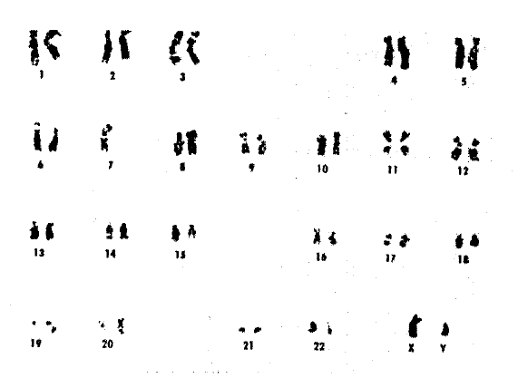
The cytogenetic analysis of bone marrow cells with G-banding showed a karyotype of 45, XY, ŌłÆ7, however, the peripheral blood leukocytes showed a consistent number of 46 with a normal male karyotype (Fig. 2).
After admission, the patientŌĆÖs condition rapidly deteriorated and he died 10 days later. The right cervical lymph node biopsied during the fourth hospitalization day, and liver, spleen, and lung tissues taken postmortemly revealed multiple single-budding encapsulated organisms, cryptococci, with evidence of splenic infarction. However, there were no recognizable eosinophilic infiltrations.
DISCUSSION
Eosinophilia is found in a variety of benign and malignant diseases. The differentiation of reactive eosinophilia from the rare neoplastic eosinophilic leukemia is considerably difficult, as both conditions may present with increased peripheral blood and bone marrow eosinophilic leukocytes, and there are no available morphologic criteria.
In 1961, Bently et al.4) defined eosinophilic leukemia, as a marked eosinophilia in the peripheral blood with blasts above normal in the bone marrow and/or peripheral blood during the course of the illness. Benvenisti and Ultmann5) defined more rigid criteria: hepatosplenomegaly, lymphadenopathy, and marked persistent eosinophilia, usually accompanied by anemia and thrombocytopenia, and accepted 43 previously reported cases as eosinophilic leukemia. On the other hand, Hardy and Anderson1) applied the term ŌĆ£hypereosinophilic syndromeŌĆØ rather than eosinophilic leukemia, and considered that the disease may represent a hypersensitivity reaction. In 1975, Chusid et al.5) reviewed the English literature extensively in search of eosinophilic leukemia cases, and also the hypereosinphilic syndrome: he accepted the point of view that there is a continum in hypereosinophilic disease with eosinophilic leukemia existing at one pole as a myeloproliferative disorder.
Chromosome analysis is a useful strategy in differentiating benign and malignant diseases. However, it has been performed only in a limited number of patiens with eosinophilic leukemia. Recent cytogenetic studies report that eosinophilic leukemia is not a mere variant of chronic myelocytic leukemia with eosinophilic proliferation6).
This patientŌĆÖs chromosomal analysis of bone marrow cell with G-Banding techniques, showed a karyotype of 45, XY, ŌłÆ7. C-monosomy myeloproliferative disorders have distinctive features: hepatosplenomegaly, lymphadenopathy, leukocytosis, thrombocytopenia, leukoerythroblastic anemia, hypercellularity of bone marrow, decreased chemotaxis of neutrophils, and frequent transition to AML7,8): and also several recent studies report on an increased susceptibility to infection in monosomy-77,9). Although the authors favor the opinion that the patient exhibited eosinophilic leukemia with monosomy-7, it must be stated that it was not possible to exclude the possibility that the present case was a preleukemic phase of monosomy-7 myeloproliferative disorder from the onset with a prominent eosinophilic reaction.
Neurofibromatosis is a frequent autosomal dominant disorder of neuroectodermal abnormality. In recent years, it was established that the link between non-neural tumors and neurofibromatosis, especially childhood leukemia, is highly possible and could be interpreted as illustrating the general effect of this mutation upon cancer risk. Hapex et al.10) documented the evidence of chromosomal instability in neurofibromatosis and held the opinion that this lead to the susceptibility of malignancy and the tendency to malignancies can also be provoked or enhanced by any of the carcinogens.
In summary, the authors presented a boy with blastic eosinophilic leukemia associated with neurofibromatosis. Chromosomal analysis of the bone marrow cells showed monosomy-7. Several biopsies of multiple organs showed cryptococci. The authors believe the disseminated crytococcosis to be a secondary infection in this compromised host, and not a primary porcess.


 PDF Links
 PubReader
 ePub Link
 Full text via DOI
 Download Citation
 Print
Print


