


CASE REPORT
A 49-year-old Korean woman presented intermittent swallowing difficulty, especially when eating solid foods, for the duration of 3 years. The symptom had been aggravated for the last 5 months. Anorexia, nausea and weight loss developed in the 3 months before admission. Other complaints were palpitation and chest discomfort for a year before admission.
Physical examination revealed that the vital signs were stable except the body temperature which was 38.8┬░C. She appeared slightly pale. Neck veins and superficial veins of the upper anterior chest wall were slightly engorged. The patient appeared unremarkable otherwise.
Laboratory data revealed microcytic and hypochromic anemia with hemoglobin of 86 g/L. The white blood cell count was 12.3├Ś10/L. Serum protein was 55 g/L and albumin was 24 g/L. Other chemistry results, including thyroid function test, were within normal range. The urinalysis, serum antimicrosomal antibody and antithyroglobulin antibody were within normal limits.
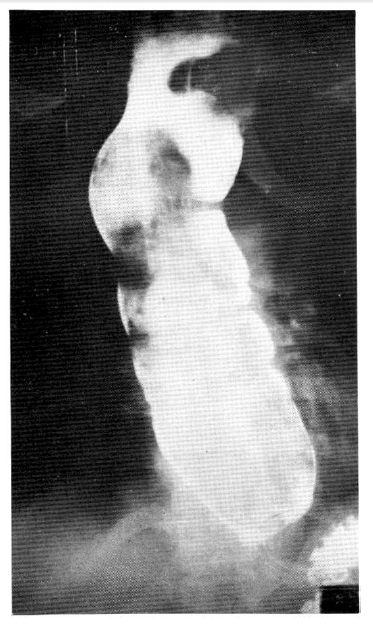
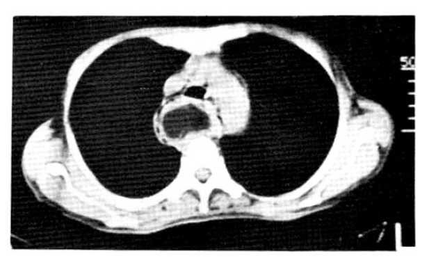
The chest x-ray showed mediastinal widening with an air shadow within it. Esophagography revealed massive dilatation of the esophagus with a huge sausage-shaped filling defect along almost the entire length of the esophagus (Fig. 1). On esophagoscopy, a huge smooth elongated mass, filling almost the entire lumen of the esophagus, was found. The tumor appeared to be covered by smooth mucosa resembling the normal esophageal mucosa. The proximal end of the mass was lobulated and there was bluish discoloration at its distal end. The tumor was too large to be removed by an endoscopic procedure. The computerized tomogram of the chest demonstrated a huge esophageal intraluminal mass. Fatty component was suggested to be the main constituent of its upper portion (Fig. 2).
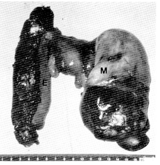
A total esophagectomy and esophago-cologastrostomy were performed. The mass was 20 cm in length, 7 cm in diameter and had a stalk measuring 3 cm in length and 1 cm in diameter just below the pharyngoesophageal junction (Fig. 3). The proximal end of the mass was lobulated. It was mostly covered by grossly unremarkable smooth mucosa with ulceration and necrosis at the distal end. The pathological examination disclosed lipoma with sarcomatous change at the distal end with interstitial infiltration of the well differentiated liposarcoma cells into the adjacent lipomatous area (Fig. 4).
There was no evidence of invasion at the stalk or metastasis to other sites. Except for a leakage from the proximal anastomosis site, which required another operation on the 8th postoperative day, the patient recovered and was discharged from the hospital. She remained well and was on a regular diet for the following 7 months until now.
DISCUSSION
Benign tumors of the esophagus usually arise from the lower portion of the esophagus. But the pedunculated tumors commonly arise from the upper fourth of the esophagus.3ŌĆō5) The wall of the upper portion of the esophagus is thinner and is habitually approximated by tonic muscular contractions. The peristaltic action of the esophagus tends to elongate and mold the tumor and gives rise to a pedicle.3,4) These intraluminal pedunculated tumors are largely mesenchymal in origin. The reported cases were lipomas, pedunculated lipomas, fibrolipomas, fibrovascular polyps, and combinations of these terms.11)
Squamous cell carcinoma, adenocarcinoma and leiomyosarcoma were also reported in the pedunculated tumors of the esophagus. But it has not been proved whether they are malignant tranformations from benign tumors or malignacy de novo.3,6,13)
Primary liposarcomas may arise wherever adipose tissue is present. The esophageal wall does have a small amount of adipose tissue. As expected, there are several reports of esophageal lipomas1,2,4,6ŌĆō8) and only one report of liposarcoma of the esophagus.14) However, there has been no report of liposarcoma arising in the lipomatous polyp. This is the first case to be reported.
The symptoms of the pedunculated tumors are sometimes so minimal that leads to a delay in the diagnosis until they became large enough to produce serious symptoms. The symptoms of all neoplasms of the esophagus are practically the same and one cannot make a diagnosis from the symptoms. Neoplasms, when large enough, may produce stenosis with severe dysphagia, regurgitation of food and marked dilatation of the esophagus. A pedunculated tumor itself may be regurgitated into the oral cavity or into the larynx causing cough, hoarseness, dyspnea or even death.1ŌĆō4, 7ŌĆō12)
In the esophagogram, the dilatation of the esophagus can incorrectly suggest achalasia if the tumor itself is overlooked. Even endoscopy can miss the tumor, and a biopsy may miss the exact nature of the tumor as it is covered with normal epithelium.12)
Once the pedunculated tumor of the esophagus is diagnosed, resection is indicated because of progressive dysphagia, possible fatal regurgitation, possible bleeding and possible chance of malignancy. Surgical removal through cervical esophagotomy for the larger tumors or endoscopic removal for the smaller ones is usually known to be sufficient. But because of the frequent presence of large vessels in the stalk, the direct surgical approach is more prudent and is the treatment of choice for most pedunculated esophageal polyps except for smaller ones.9ŌĆō12) There is frequent local recurrence of liposarcomas of other organs14) and meticulous follow-up should be done.

 PDF Links
 PubReader
 ePub Link
 Full text via DOI
 Download Citation
 Print
Print


