


INTRODUCTION
Seoul virus is one of the newly identified etiologic agents of hemorrhagic fever with renal syndro (HFRS) and was isolated by Lee et al. in 1982.1) Urban cases of HFRS in Korea, China, Japan and Southeast Asia and laboratory infections in Korea and Japan are caused by the Seoul virus. Urban commensal rats (Rattus norvegicus and Rattus rattus) and laboratory rats are the main reservoir hosts and transmit the disease to man.
Previously some investigators reported on urban type Korean hemorrhagic fever (KHF)2), but there are no reports on the clinical features of Seoul virus infection based on serological diagnosis. So we retrospectively evaluated the clinical findings of 29 patients with HFRS caused by Seoul virus infection, who were diagnosed by hemagglutination inhibition test. We also compared the clinical features of Seoul virus infection with those previously reported of classic KHF.
MATERIALS AND METHODS
The subjects were 29 HFRS patients who had admitted to general hospitals in the Seoul metropolitan area and were diagnosed as Seoul virus infection by hemagglutination inhibition test3) during the period of 1 year from January to December 1984.
All the hospital records were reviewed and the first day of febrile episode was regarded as the first day of illness. The symptoms and signs were all reviewed from the first day of illness and laboratory examinations including complete blood count and blood chemistry were analysed.
The clinical findings and laboratory results were compared with those of classic KHF previously published.
RESULTS
1. Demographic Characteristics
Demographic distributions of the study population and KHF patients of a previous report are summarized in Table 1. About half (48.3%) of the patients were in their third and fourth decade of life and males outnumbered females by 4 to 1.
2. Seasonal Incidence
Monthly incidences of Seoul virus infection and KHF are shown in Table 2. About 75% of Seoul virus infections occurred from Oct. to Dec. There was another small peak in the early summer season.
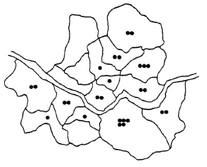
3. Geographic Distribution
Table 3 shows the geographic distribution of patients with a map of metropolitan Seoul. The occurrence of the disease was evenly distributed throughout all districts of the Seoul area.
4. Clinical Symptoms and Signs
General symptoms on admission were fever (93.1%), myalgia (41.4%) headache (27.6%) and asthenia (20.7%) in order of frequency. Gastrointestinal symptoms were noted in the order of vomiting (51.7%) nausea (34.5%) and diarrhea (17.2%). Respiratory symptoms were cough (10.3%), sore throat (10.3%) and dyspnea (3.4%). Neurologic symptoms such as mental change (6.9%), dizziness (3.4%) and convulsion (3.4%) were also noted.
These clinical symptoms are summerized in Table 4 with those of classic KHF from a previous publication.
Physical findings on admission were noted in order of petechia (41.4%), CVA tenderness (41.4%), pharyngeal injection (34.5%) and conjunctival injection (17.2%). Bleeding diasthesis, such as subconjunctival hemorrhage (10.3%), and nasal or vaginal bleeding (10.3%), was also noted. Major abdominal signs were abdominal tenderness (10.3%), hepatomegaly (6.9%) and ascites (6.9%). These physical findings are shown with those of classic KHF in Table 5.
The clinical stages of classic KHF (febrile, hypotensive, oliguric, diuretic and convalescent phases) were also applicable to the study subjects (Table 6). Febrile phase was noted in 21 cases and the mean duration was 6.2 days. Hypotensive phase was noted in only 3 cases and lasted for 2 days. Oliguric phase was noted in 19 cases and the mean duration was 6.1 days (range; 2ŌĆō15 days). Diuretic phase was also noted in 19 cases and the mean duration was 11.1 days (range; 4ŌĆō26 days). In 10 cases (35%) of the study subjects, oliguric phase was not noted.
5. Laboratory Findings
Thrombocytopenia was observed transiently in 28 patients and in 9 patients (32.1%), the platelet count was under 40,000/mm3 (Table 7ŌĆō1). Leukocytosis was observed in 22 cases (75.9%) and the leukocyte count was over 30,000/mm3 in 3 cases (10.3%) (Table 8).
Peak BUN levels were over 80mg% in 8 patients (36.4%) and peak creatinine levels were over 5mg% in 24 patients (82.8%) (Table 9-1, 9-2).
Maximum SGOT and SGPT levels were under 100 U in 77% of the cases, but significant elevations of transaminase were noted in 6 patients (23%). (Table 10).
6. Management and Prognosis
Twenty-two cases out of 29 study subjects recovered successfully with conservative treatments exclusively. Only in 7 cases (24.1%) were dialysis therapies needed. (Table 11).
There was no fatal case in 29 study subjects.
DISCUSSION
Since the isolation of Hantaan virus, an etiologic agent of HFRS, by Lee et al. in 19766), the diagnosis of HFRS became more reliable by an indirect immunofluorescent technique.7) The natural reservoir of Hantaan virus is a wild rat (Apodemus agrarius coreae) and the disease has long been believed to occur in rural areas only, with farmers and soldiers being the most likely victims.
However, several investigators reported an urban type of HFRS in Japan8) and also in Korea1) and house rats were presumed to be a natural reservoir of HFRS. In 1982 Lee et al. isolated another strain of Hantavirus from Rattus norvegicus, urban commensal rats, and proposed to name it Seoul virus.1) The antigenecity and RNA sequence of Seoul virus are somewhat different from Hantaan virus, but these have enough cross-reactivity to be reactive in the indirect immunofluorescent test.9) For the serologic identification of Seoul virus infection, plaque reduction neutralization test (PRNT) and hemagglutination inhibition (HI) test are useful.10) Our study subjects were identified by HI test. This paper is based on a retrospective analysis of hospital records of several general hospitals in the Seoul area, so there were some difficulties in analysing clinical findings and in uniformity of laboratory examinations.
However, the demographic characteristics of patients with Seoul virus infection were very similar to those with classic KHF of previous reports (Table 1). Most patients were in their third to fifth decades of life in both groups and the sex ratio was also comparable. Peak incidences of both diseases were also in November, but with regard to Seoul virus infection, there was also a small peak in June.
Clinical symptoms at presentation were high fever, abdominal pain or back pain, vomiting, myalgia and nausea. Except for high fever, the other major symptoms were somewhat lower in frequency than in previously reported KHF. (Table 4). General symptoms such as myalgia, headache and asthenia were noted in 20ŌĆō40% of subjects, which is much lower than that of 60ŌĆō90% noted in previous studies.4) Gastrointestinal symptoms such as nausea and vomiting were also less frequently noted in Seoul virus infection than in classic KHF. Respiratory symptoms such as cough, sore throat, and dyspnea were much lower in frequency than in KHF. Convulsive episode was noted only in one case of 29 study subjects.
The physical findings of Seoul virus infection were also less conspicuous than those of KHF. The main physical findings of KHF, which were considered diagnostically important, such as petechia, CVA tenderness, and conjunctival injection were noted in less than 50% of cases with Seoul virus infection. For example, conjunctival injection, which was noted in almost all patients with KHF,4) was noted in only 17% of these urban cases with Seoul virus infection (Table 5).
In laboratory examinations, the degrees of thrombocytopenia and leukocytosis were also much milder in Seoul virus infection. But about a quarter of the patients with Seoul virus infection showed significant elevations of serum transaminase levels, in contrast to the cases with KHF (Table 10). Viral hepatitis is prevalent in Korea, so it may be difficult to differentiate Seoul virus infection from non-A, non-B hepatitis, especially in cases with symptomless Seoul virus infection.
Most patients (75.9%) recovered with conservative treatment only and all of the other patients also successfully recovered with dialytic supports. In classic KHF patients, the average mortality rates are over 5% (Table 12), but in our study no fatal cases were noted.
These above findings suggest that the clinical course of Seoul virus infection is much more benign than that of classic KHF.
 PDF Links
 PubReader
 ePub Link
 Full text via DOI
 Download Citation
 Print
Print


