


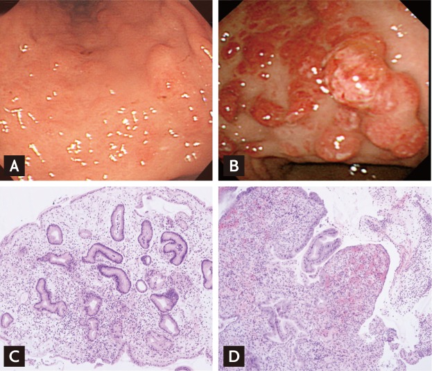
A 68-year-old woman, with hepatitis B virus cirrhosis, was hospitalized for melena. She had undergone regular esophagogastroduodenoscopy (EGD) examinations to follow-up esophageal variceal ligation. Fifteen months ago, EGD showed several raised erosions in the lower body (Fig. 1A) and grade 2/3 esophageal varices and small gastric varices. The laboratory examination revealed low hemoglobin (7.1 g/dL), mean corpuscular volume (75.8 fL), and mean corpuscular hemoglobin (24.6 pg). EGD revealed no bleeding from the esophageal or gastric varices. However, multiple polyps with superficial erosions or erythema were noted (Fig. 1B). The gastric polyps were biopsied and consisted of foveolar hyperplasia, edematous lamina propria, and ectatic capillaries (Fig. 1C). There was erosion of, and marked granulation tissue in, the lamina propria (Fig. 1D). She was diagnosed with gastric polyposis associated with portal hypertension.
 PDF Links
 PubReader
 ePub Link
 Full text via DOI
 Download Citation
 Print
Print


 |
 |