


To the Editor,
Epithelioid sarcoma (ES) is an uncommon mesenchymal tumor that most frequently occurs in the dermal or subcutaneous area of the distal extremities of young adults, mainly in men. ES has a poor prognosis owing to its aggressiveness, as it frequently recurs locally and can undergo metastasis mainly to the lymph nodes, soft tissues, bones, lungs, and brain. Unilateral or bilateral spontaneous pneumothorax with cystic lung metastasis has been reported [1-5]. To our knowledge, only five patients have been reported in the literature, none of whom had massive pleural effusion. We reviewed the existing literature on pneumothorax in ES cases and present herein the first reported case of spontaneous pneumothorax and massive pleural effusion in both lungs in ES.
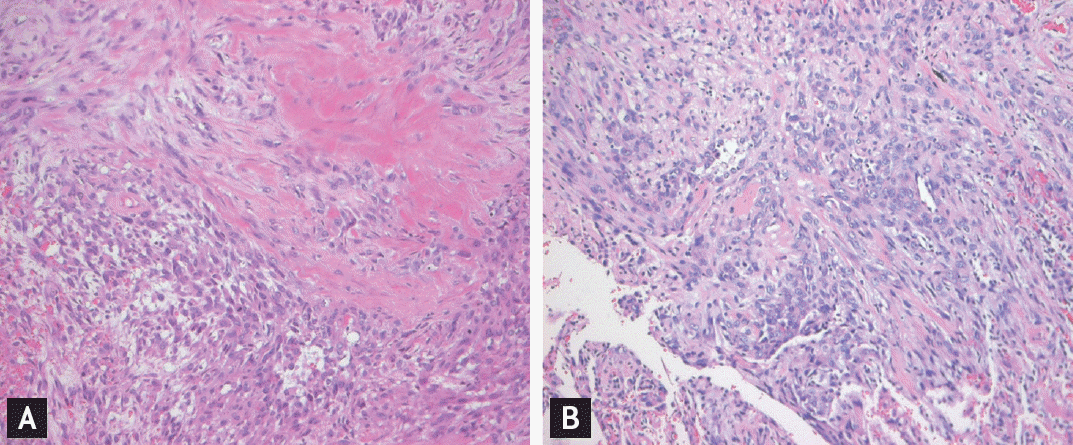
A 42-year-old man presented to Emergency Department of Chonbuk National University Hospital with a complaint of abrupt onset of left-sided pleuritic chest pain and dyspnea. Seven years prior, he was initially diagnosed with ES of the perianal area and underwent mass excision. A diagnostic biopsy revealed mild to moderate pleomorphic epithelioid and spindle cells arranged in nodular aggregates (Fig. 1A). The specimen was immunohistochemically positive for cytokeratin, epithelial membrane antigen, CD34, and vimentin, but negative for the smooth muscle actin, and S-100. Three years prior, the tumor metastasized to the right inguinal lymph node; therefore, he underwent right inguinal lymph node dissection.
Seven months prior to admission, the tumor recurred at the right inguinal lymph node and lung. He received three cycles of chemotherapy with ifosfamide, doxorubicin, and dacarbazine in combination for soft tissue sarcoma, resulting in progression with an enlarged inguinal lymph node and lung lesion.
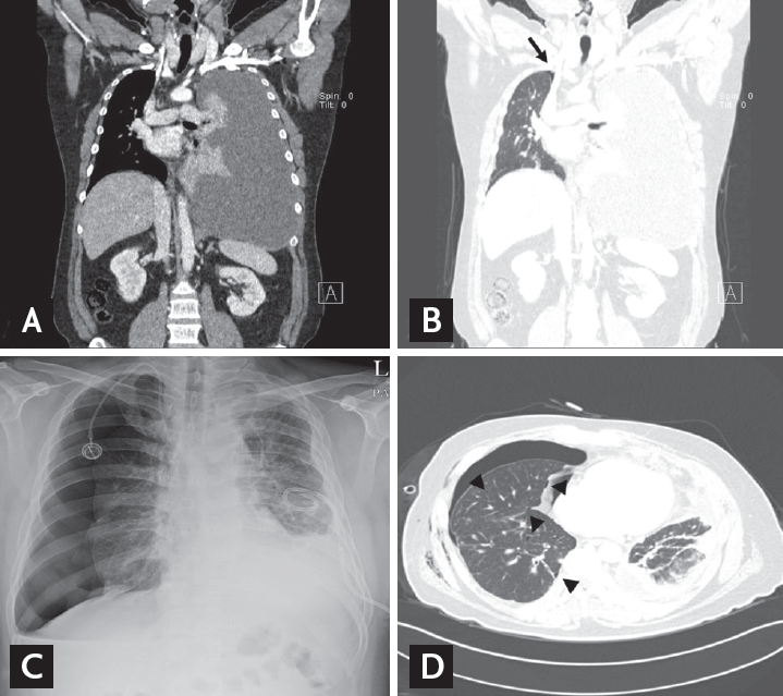
On examination, his vital signs were normal, but breath sounds from the left lung were decreased. Chest radiography revealed a left pleural effusion, and chest computed tomography (CT) showed a massive pleural effusion on the left lung and a small pneumothorax on the right lung (Fig. 2A and 2B).
Pig-tail catheter was inserted in the left lung. Pleural fluid analysis results were exudate: white blood cell count, 3,450/╬╝L (71% lymphocyte); total protein, 5.1 g/dL (serum value, 5.8 g/dL); lactate dehydrogenase, 980 IU/L (serum value, 349; normal range, 218 to 472); and adenosine deaminase, 9.51 IU/L (normal range, 4.3 to 20.3). In addition, pleural fluid cytology revealed a few atypical cells. The results of the mycobacterial and bacterial cultures were negative. Pleural fluid was drained approximately 1,000 mL for 5 days, which decreased the amount of effusion. And he was treated conservatively with high-flow oxygen due to small spontaneous pneumothorax. At 8 days after admission, severe dyspnea suddenly recurred. On examination, his breath sounds from the right lung was decreased. Chest radiography showed a right pneumothorax, and the chest CT scan demonstrated a right pneumothorax and multiple pulmonary cysts of various sizes (Fig. 2C and 2D). Thus, a chest tube was inserted in his right chest. After 11 days, his pneumothorax was not resolved and the air leakage continued. Therefore, he underwent wedge resection of bullae via video-assisted thoracoscopy. Operators found the ruptured bullae in the anterior segment of the right lung and multiple metastatic nodules in the visceral and parietal pleura of the entire right lung. His biopsy specimen are observed clusters of malignant epithelioid cells (Fig. 1B). On postoperative day 6, his symptom improved and the catheter was removed because drainage was less than 50 mL/day 3 more days consistently. However, his left lung was not fully expanded due to loculations and nodular thickening of pleural metastases. And he was discharged from the hospital. Four weeks later, he was readmitted aggravated pleural effusion, thickening and lung metastases and pig tail catheter was reinserted in the left lung. He died 1 month later owing to disease progression.
ES is a rare soft tissue sarcoma with a known high propensity for locoregional recurrence and distant metastases. Pulmonary metastasis of soft tissue sarcomas commonly manifest as solid nodules, but only few cases of cystic pulmonary metastasis have been described. Pneumothorax may have also been caused by distension of alveoli through the ball-valve effect, permitting passage of air along the interlobular septa to the pleura, where cysts may have formed and eventually ruptured [1].
In five patients with ES, spontaneous pneumothorax was reported (Table 1) [1-5]. The lung lesions were usually only cysts, except in one patient. None of the patients presented with pleural effusion. Our patient represents the first reported case of spontaneous pneumothorax and pleural effusion in both lungs in ES. The treatment of such cases is chest tube drainage, pleurodesis, and wedge resection because of air leakage. Three patients were treated with systemic chemotherapy with an anthracycline-based regimen but had a poor response. Patients had variable outcomes, with our patient surviving for just 3 months after pneumothorax. Because of the rarity of cases, prognostic factors of survival are unclear, but some cases progress slowly.
In conclusion, the coexistence of pneumothorax and pleural effusion in both lungs is an uncommon presentation of primary extremity sarcomas. Cystic metastasis with nodules in patients with sarcomas require attention because they can develop spontaneous pneumothorax and pleural effusion.
 PDF Links
 PubReader
 ePub Link
 Full text via DOI
 Download Citation
 Print
Print


