


To the Editor,
Although an association between pyogenic liver abscess and colon cancer has been suggested, liver abscess as a presenting sign of occult colorectal cancer has rarely been reported [1,2]. Destruction of the mucosal barrier by a colorectal neoplasm, followed by bacterial translocation into the portal venous system, might contribute to the formation of liver abscess [2]. We describe a case of recurrent pyogenic liver abscess at short intervals as the first manifestation of colorectal cancer in the absence of liver metastases. A healthy 66-year-old male visited the emergency room because of right upper abdominal pain associated with fever and chills over the past 5 days. He reported no history of cigarette smoking and alcohol consumption. Physical examination revealed tachycardia (pulse rate, 88 beats/min), a body temperature of 38.7°C, and tenderness of the right upper abdomen. Laboratory tests showed a white blood cell (WBC) count of 11,460/μL, with 83.9% neutrophils, and a hemoglobin level of 14.4 g/dL. The blood chemistry profile was as follows: fasting glucose, 106 mg/dL; total protein, 5.9 g/dL; albumin, 2.9 g/dL; total bilirubin, 0.9 mg/dL; aspartate aminotransferase, 107 U/L; and alanine aminotransferase, 195 U/L; alkaline phosphatase, 674 U/L; gamma-glutamyl transferase, 321 U/L. The C-reactive protein level was 12.9 mg/dL (normal range < 0.3). Serologic tests for hepatitis B and C were negative. Serologic tests for tumor markers revealed normal levels of α-fetoprotein (2.7 ng/mL), carbohydrate antigen 19-9 (9.1 U/mL), and carcinoembryonic antigen (0.7 ng/mL).
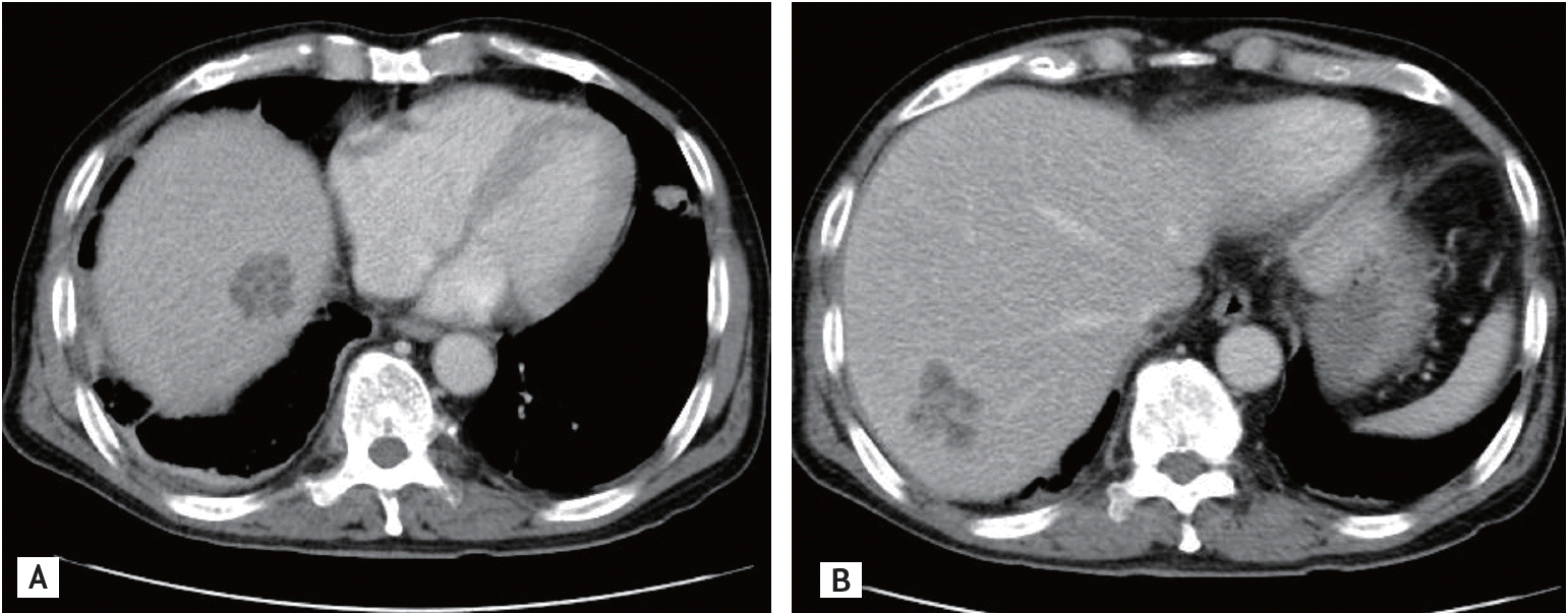
A computed tomography (CT) scan of the abdomen and pelvis demonstrated a multiseptated hypoechoic lesion in segment 6 of the liver, with a maximum diameter of 5.1 cm and poorly marginated margins (Fig. 1A). No other pathology was evident. A pyogenic liver abscess was suspected. Ultrasonography-guided percutaneous drainage and intravenous administration of a broad-spectrum antibiotic resulted in resolution of the fever and leukocytosis. Growth of Klebsiella pneumoniae was observed in pus cultures obtained from liver abscess aspirate and blood cultures. Cytology of liver abscess aspirate was negative for malignancy. The patient was discharged on oral antibiotic therapy after 4 weeks of hospitalization. A follow-up CT scan after 6 weeks of antibiotic therapy revealed regression of liver abscess (Fig. 1B).
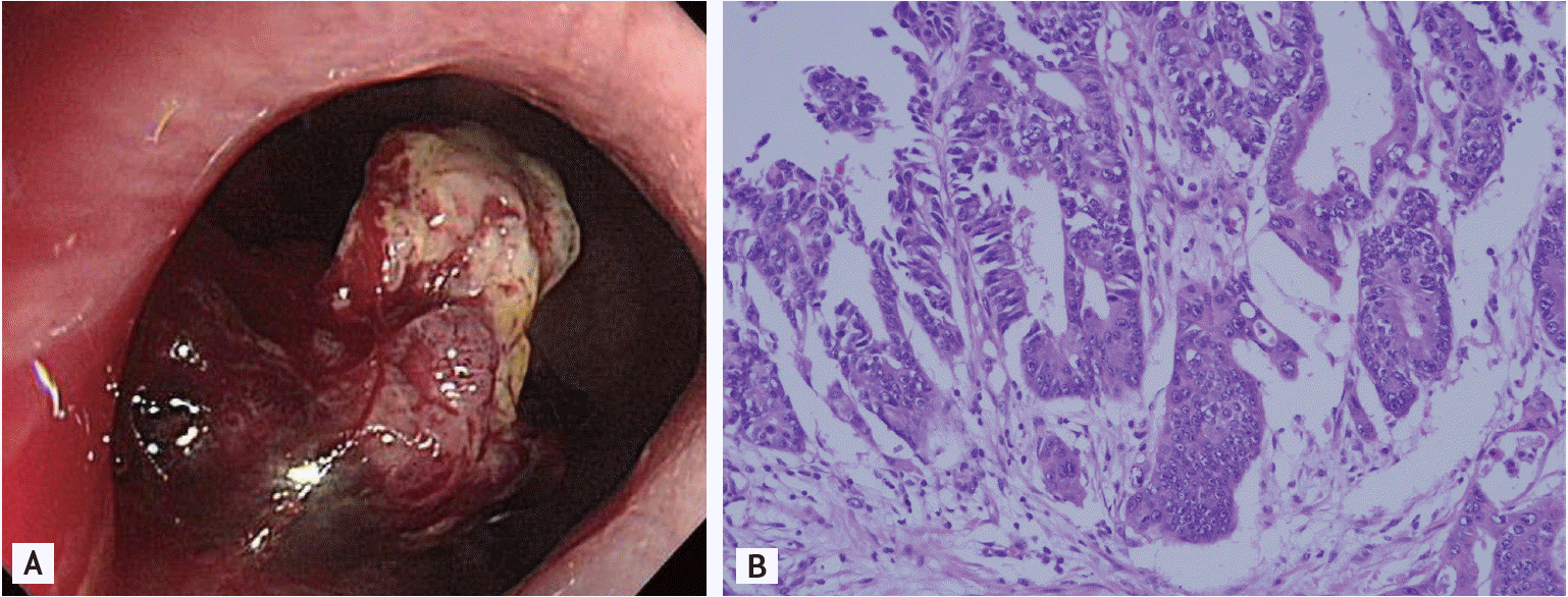
Two months following his initial presentation, the patient revisited the emergency room suffering from fever and chills. His WBC count was 25,660/μL, with 80.4% neutrophils, and the C-reactive protein level was 14.9 mg/dL. CT of the abdomen and pelvis revealed two newly developed hypodense lesions in segments 7/8 of the liver, suggestive of liver abscess (Fig. 2). Moreover, eccentric thickening of the rectal wall, which was not seen in previous CT scans, was noted. Intravenous administration of antibiotics and percutaneous drainage were repeated. Blood and aspirated abscess cultures again showed growth of K. pneumoniae. Colonoscopy revealed an about 2.5 cm fungating mass located 4.5 cm from the anal verge (Fig. 3A). Histologic examination of the biopsy specimen disclosed a well-differentiated adenocarcinoma (Fig. 3B). Six weeks later, the patient underwent concurrent chemoradiotherapy. Surgical resection has been scheduled.
A systematic review of colorectal cancer-associated pyogenic liver abscess cases reported worldwide was published recently. In this report, K. pneumoniae was the predominant organism isolated from pyogenic liver abscess (80%), and tumors were located mainly in the sigmoid colon and rectum (72.7%) in Eastern Asian patients [3]. Mucosal defects in the neoplastic lesions are believed to facilitate entry of microorganisms into the portal circulation, with subsequent hematogenous spread to the liver [2].
Despite the recent evidence of an association between pyogenic liver abscess and colorectal cancer, the need for colonoscopic investigation in patients with cryptogenic pyogenic liver abscess remains a matter of debate. In our case, colonoscopy was recommended based on accumulating evidence for a relationship between the pyogenic liver abscess and the underlying colorectal neoplasm, but the patient refused due to his general medical condition. Initially, CT of the abdomen and pelvis did not show any thickening of the colonic wall in this case. Hsu et al. [4] suggested that an abdominal CT scan inclusive of the pelvic area to evaluate liver abscess can assist in the diagnosis of colorectal cancer; however, the misdiagnosis of colorectal cancer in our case could be attributed to the collapsed bowel wall observed on the initial CT scan. Therefore, the possibility of colorectal neoplasm should be questioned in cases of cryptogenic pyogenic liver abscess, even if CT scans involving the pelvic area suggest no colonic pathology.
The patient did not complain of lower abdominal pain, changes in bowel habit, bloody stool, or tenesmus. Moreover, his carcinoembryonic antigen level was within normal limits. A recent review of pyogenic liver abscesses associated with colorectal cancers reported that only about 10% of the patients had colorectal cancer-associated symptoms. Moreover, colorectal cancer-related biomarkers were normal in most cases [3]. Therefore, the origin of pyogenic liver abscess should be identified in the absence of colorectal cancer-related symptoms or laboratory findings.
Recurrent liver abscess associated with colorectal neoplasm has been reported only rarely. The time period between two subsequent episodes of pyogenic liver abscess was less than 1 year in most of these reports [4,5]. To the best of our knowledge, the case reported herein represents the earliest recurrence, at 2 months after the first manifestation of pyogenic liver abscess. As a result, colorectal cancer was diagnosed because of recurrent pyogenic liver abscess. However, a delay in the diagnosis of underlying colorectal cancer in our case led to recurrent liver abscess, which resulted in deferred treatment for colorectal cancer. Early investigation of the underlying cause of cryptogenic pyogenic liver abscess is warranted.
In conclusion, a pyogenic liver abscess can be the sole initial manifestation of colorectal cancer without associated metastasis. Increasing awareness of this association emphasizes the importance of skepticism regarding the presence of colorectal cancer in cases of otherwise cryptogenic pyogenic liver abscess. A thorough investigation, including colonoscopy, is necessary to identify the origin of an unexplained liver abscess. Timely diagnosis of common colonic pathologies associated with pyogenic liver abscess can improve the outcome of colorectal neoplasm. The cost-effectiveness of a complete evaluation of the gastrointestinal tract in patients with cryptogenic pyogenic liver abscess has not yet been elucidated.

 PDF Links
 PubReader
 ePub Link
 Full text via DOI
 Download Citation
 Print
Print


