


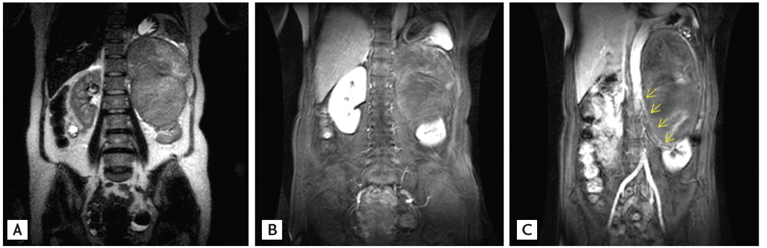
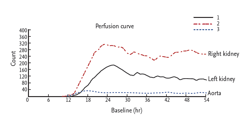
A 57-year-old woman was admitted to our hospital with abdominal pain in the left upper quadrant. She had left hemiplegia due to an intracranial hemorrhage 5 years previously, as well as hypertension and diabetes mellitus. Her blood pressure was 190/100 mmHg. Laboratory data revealed a serum creatinine level of 1.2 mg/dL and a serum potassium level of 3.4 mEq/L. Magnetic resonance imaging of the abdomen showed a large suprarenal mass containing hemorrhagic foci, and compressing the left kidney and left renal artery (Fig. 1). Plasma renin activity was 51.9 ng/mL/hr (normal range in the supine position, 0.15 to 2.33), and aldosterone level was 44.7 ng/dL (normal range in the supine position, 1.0 to 16). A 24-hour urine test showed the following results: vanillylmandelic acid, 82 mg/day (normal range, 0 to 8); metanephrine, 22.3 mg/day (normal range, 0 to 1.2); epinephrine, 59.4 pg/mL (normal range, 0 to 110); and norepinephrine, 29,658 pg/mL (normal range, 70 to 750). A technetium-99m diethylene triamine pentaacetic acid scan showed that perfusion of the left kidney was markedly lower than that of the right kidney, suggesting compression of the renal artery (Fig. 2).
After surgical resection of the mass, plasma renin activity, aldosterone, serum creatinine, and serum potassium levels normalized (to values of 0.1 ng/mL/hr, 1.0 ng/dL, 1.0 mg/dL, and 4.4 mEq/L, respectively), and blood pressure decreased to 140/90 mmHg. The left kidney was removed with the mass because of severe adhesion. The tumor measured 16 × 11 cm at its largest dimension, showed hemorrhage and necrosis at its center, and was composed of epithelioid to spindle-shaped cells without distinct sustentacular cells. Tumor cells stained positive for chromogranin A, synaptophysin, and CD56 (Fig. 3), but not for vimentin, S-100, or Melan-A; these findings were consistent with a pheochromocytoma.

 PDF Links
 PubReader
 ePub Link
 Full text via DOI
 Download Citation
 Print
Print


