


INTRODUCTION
Elevation of serum lipase activity is included in the diagnostic criteria for acute pancreatitis [1]. However, non-pancreatic elevations of serum lipase levels, such as increases due to renal insufficiency, bowel obstructions, diabetic ketoacidosis, inflammatory bowel disease, and intracranial hemorrhage and even idiopathic cases have been reported, and differential diagnosis is needed for clinical practice [2-5]. Because the interpretation of elevated serum lipase concentrations is complex, cross-sectional imaging studies, including computed tomography (CT) or magnetic resonance imaging, are usually recommended. However, these modalities have the limitations of radiation exposure or high expense.
Pancreatic lipase, also known as pancreatic triacylglycerol lipase, is secreted into the duodenum, and the serum level of this enzyme is low in patients without pancreatic diseases. However, under conditions of pancreatic injury, pancreatic autolysis induces elevation of the serum pancreatic lipase level. In the measurement of serum lipase levels, the triglyceride lipase gene subfamily, including lipoprotein lipase, hepatic lipase, and endothelial lipase, is also measured [6]. Thus, measurement of only serum pancreatic lipase concentrations is more specific and more rational for the differential diagnosis of serum lipase elevation. This study aimed to evaluate the clinical efficacy of serum lipase subtype analysis for the differential diagnosis of pancreatic and non-pancreatic serum lipase elevation.
METHODS
Ethics statement
This study was conducted according to the principles expressed in the Declaration of Helsinki. Voluntary participation was requested, and written informed consent was obtained from each participant. This study was approved by the Institutional Review Board of Hallym University Chuncheon Sacred Heart Hospital.
Study design
This study was conducted at Hallym University Chuncheon Sacred Heart Hospital, a teaching hospital in the Korea. From July 2012 through February 2014, consecutive patients who were referred to the pancreatobiliary department for serum lipase elevation were prospectively enrolled. Clinical findings and serum lipase subtypes (pancreatic, endothelial, lipoprotein, and hepatic lipase) were analyzed and compared after division into pancreatitis and non-pancreatitis groups.
The diagnosis of acute pancreatitis was made according to the revised definition of Atlanta 2012 ŌĆötwo of the following three features: (1) acute onset of persistent, severe, epigastric pain often radiating to the back; (2) serum lipase activity (or amylase activity) at least three times greater than the upper limit of normal; and (3) characteristic findings of acute pancreatitis on contrast-enhanced computed tomography (CECT) [1].
Clinical findings were recorded for the following variables: age, sex, alcohol use, smoking history, body mass index (BMI), presence of intracranial hemorrhage, and laboratory results, including serum amylase, lipase, aspartate aminotransferase (AST), alanine aminotransferase (ALT), alkaline phosphatase (ALP), gamma glutamyl transpeptidase (GGT), and C-reactive protein (CRP). Serum amylase and lipase levels were measured by enzymatic colorimetric assay (Cobas 8000 C702 Chemistry autoanalyzer, Roche-Hitachi Corp., Basel, Switzerland), which is a form of spectrophotometric assay. Among the serial laboratory outcomes, values at the time of the highest lipase level were selected and recorded. All of the laboratory variables were measured and reported by the Laboratory Medicine Department of Hallym University Chuncheon Sacred Heart Hospital (2013-82).
Enzyme-linked immunosorbent assay
Blood samples obtained from the enrolled patients were allowed to clot for 30 minutes at room temperature. After centrifugation, serum was collected and stored at ŌĆō80┬░C. The activity of serum lipase subtypes was measured using an enzyme-linked immunosorbent assay (ELISA) analysis kit (Cloud-Clone Corp., Houston, TX, USA) for pancreatic lipase, endothelial lipase, lipoprotein lipase, and hepatic lipase, according to the manufacturerŌĆÖs protocol. The minimum detectable doses the lipase subtypes were typically less than 0.239 ng/mL for pancreatic lipase, less than 27 pg/mL for endothelial lipase, less than 0.247 ng/mL for lipoprotein lipase, and less than 33 pg/mL for hepatic lipase. The coefficient of variation (CV) was calculated using the following equation, and the values of the intra- and interassay CVs were as follows; CV (%) = standard deviation / mean ├Ś 100; intra-assay, CV < 10%; inter-assay, CV < 12%. A biochemical analyzer assessed the lipase subtype levels in the serum. Absorbance (A) was detected at 450 nm. The content of each sample was estimated using a standard curve.
Statistical analysis
Continuous variables are expressed as the medians and interquartile ranges (IQRs) because they were not normally distributed. Categorical variables are expressed as numbers and percentages. The Mann-Whitney test and Fisher exact test were used to compare two variables. The diagnostic performance of serum lipase subtypes was assessed using the receiver-operating characteristic (ROC) curve, which plots sensitivity over 1-specificity. To detect the best cut-off value associated with serum lipase subtype analysis for the prediction of acute pancreatitis, a maximum of the Youden index was selected. Multivariate logistic regression analysis was performed to assess the independent risk factors associated with the detection of acute pancreatitis. A p < 0.05 (2-tailed) was adopted as the threshold of statistical significance for all of the tests. The analyses were performed using SPSS version 18.0 (SPSS Inc., Chicago, IL, USA), and Medcalc version 13.3.3 (Medcalc Software, Ostend, Belgium).
RESULTS
Characteristics of patients
Of the 34 eligible patients initially enrolled in this study, no patients were excluded due to refusal to participate; as a result, a total of 34 patients (18 male and 16 female) participated, and 12 patients (35.3%) were diagnosed with acute pancreatitis (CECT was performed for all of the enrolled patients). The characteristics of the enrolled patients are summarized in Table 1. The median age of enrolled patients was 55.5 years old (IQR, 45.5 to 69.5). The proportions of smokers and alcoholics were 32.4% (n = 11) and 47.1% (n = 16), respectively. The median BMI was 23.4 (IQR, 21.3 to 26.3). Among the enrolled population, the proportion of patients with intracranial hemorrhage was 17.6% (n = 6).
Laboratory values
The median levels of serum amylase and lipase were 448.5 IU/L (IQR, 240.8 to 930.7) and 2,214.1 IU/L (IQR, 1,063.8 to 4,375.3), respectively. In the subtype analysis of lipase, the median levels of serum lipase subtypes were as follows: pancreatic lipase (494.5 pg/mL; IQR, 271.4 to 902.9), endothelial lipase (268.3 pg/mL; IQR, 235 to 397), lipoprotein lipase (105.1 ng/mL; IQR, 98.8 to 109.5), and hepatic lipase (76,582.5 pg/mL; IQR, 41,438.5 to 123,801).
To determine the comparative activity of serum lipase subtypes, the fraction of each lipase in the total amount of lipase subtypes was calculated. The fraction of each lipase in the total amount of serum lipase subtypes were as follows: pancreatic lipase, median 0.003 (IQR, 0.001 to 0.005); endothelial lipase, median 0.002 (IQR, 0.001 to 0.003); lipoprotein lipase, median 0.560 (IQR, 0.458 to 0.721); and hepatic lipase, median 0.432 (IQR, 0.274 to 0.539). The detailed laboratory values of the enrolled patients are described in Table 1.
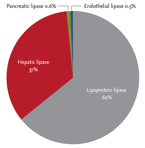
The distributions of each lipase in the total amount of serum lipase subtypes were as follows: lipoprotein lipase, 62%; hepatic lipase, 37%; pancreatic lipase, 0.6%; and endothelial lipase, 0.5% (the relative proportion of the mean value of each lipase subtype was calculated) (Fig. 1).
Univariate analysis for the associations of acute pancreatitis
Among the clinical variables, there were no statistically significant differences in age, sex, smoking history, BMI, presence of intracranial hemorrhage, or laboratory results, including serum amylase, lipase, AST, ALT, ALP, GGT, and CRP, between the pancreatitis and non-pancreatitis groups. Only alcohol consumption differed, being noted in nine of 12 patients (75%) in the acute pancreatitis group and seven of 12 (31.8%) in the non-pancreatitis group (p = 0.03).
Among the laboratory values, there were no statistically significant differences in serum amylase, lipase, pancreatic lipase, endothelial lipase, lipoprotein lipase, or hepatic lipase between the pancreatitis and non-pancreatitis groups. The level of serum pancreatic lipase in the patients with acute pancreatitis was higher than in the patients without acute pancreatitis, although the difference was statistically insignificant ([median, 626 pg/mL; IQR, 465 to 1,664.2] vs. [median, 407.8 pg/mL; IQR, 200 to 766.5], p = 0.17).
Among the fractions of each lipase in the total amount of lipase subtypes, there were no statistically significant differences in the fractions of endothelial lipase, lipoprotein lipase, or hepatic lipase. Only the fraction of pancreatic lipase in the total amount of serum lipase subtypes (FPL) showed a higher value in the acute pancreatitis group than in the non-pancreatitis group ([median, 0.004; IQR, 0.003 to 0.011] vs. [median, 0.002; IQR, 0.001 to 0.004], p = 0.04). The detailed content of the univariate analysis for the associations of acute pancreatitis is demonstrated in Table 2.
Prediction of acute pancreatitis
In the ROC curve analysis for the prediction of acute pancreatitis, FPL was the most valuable predictor (AUROC, 0.72; 95% confidence interval [CI], 0.54 to 0.86; p = 0.04). The best cut-off value associated with FPL was 0.0027. The sensitivity and specificity of this test using the cut-off value of 0.0027 were 83.3% (95% CI, 51.6 to 97.9) and 63.6% (95% CI, 40.7 to 82.8), respectively. The positive and negative likelihood ratios were 2.3 (95% CI, 1.3 to 4.2) and 0.3 (95% CI, 0.1 to 0.9), respectively. The positive and negative predictive values were 55.6% (95% CI, 30.8 to 78.5) and 87.5% (95% CI, 61.7 to 98.5), respectively. Other tests, including serum amylase, lipase, and lipase subtypes other than FPL showed, statistically non-significant results (Table 3, Fig. 2).
Multivariate analysis for the associations of acute pancreatitis
In multivariate analysis for the associations of acute pancreatitis, only FPL using a cut-off value of 0.0027 was associated with acute pancreatitis (OR, 8.3; 95% CI, 1.3 to 51.7; p = 0.02) (Table 4).
DISCUSSION
In the present study, the clinical efficacy of serum lipase subtype analysis for the differential diagnosis of pancreatic and non-pancreatic serum lipase elevations was assessed. Lipase and amylase are released from acinar cells in patients with acute pancreatitis, and the measurements of serum concentrations of these enzymes are used to confirm diagnosis [7]. Serum lipase measurement is generally recommended because of its greater specificity, sensitivity, and durability than serum amylase measurement [8-11]. However, non-pancreatic elevations of serum lipase levels make it difficult to diagnose acute pancreatitis, and radiologic imaging studies are inevitable. FPL showed adequate sensitivity (83.3%) and negative predictive value (87.5%) although relatively low specificity (63.6%) and positive predictive value (55.6%) in this study. However, considering that the total number of patients (n = 34) was small, and the prevalence of pancreatitis (32.4%) was low, these values could have been higher if applied in the larger cohort. The direct comparison of pancreatic lipase between patients with acute pancreatitis and those without showed statistically non-significant results, although the pancreatitis group showed higher levels (Table 2). The small sample size could be the reason for this study, which must be replicated with a larger sample size.
The sensitivity and specificity of serum lipase measurement for acute pancreatitis were reported as 55% to 100% and greater than 95%, respectively, at a cut-off value of 600 IU/L, showing greater specificity than FPL [7,10]. However, a major difference from our study was the target population. The reported diagnostic performance of serum lipase was based on a population with acute abdominal pain. However, this study included patients with serum lipase elevations regardless of symptoms. Thus, diagnostic performance could not be applied directly when comparing with serum lipase or amylase. Considering the radiation exposure and high expense of CT, FPL testing could be used for triage or as an add-on test in patients with serum lipase elevations and without definite symptoms suggesting acute pancreatitis. In particular, it could be useful in certain clinical situations, such as typical symptoms with a normal range of serum lipase or an elevated level of serum lipase without typical symptoms.
Another outcome was the distribution of each lipase subtype in serum. Among the isoforms of lipase, the majority portion of the serum concentration was lipoprotein lipase or hepatic lipase in this study, and this finding was consistent independent of acute pancreatitis (Tables 1 and 2, Fig. 1). Although we are not yet aware of the exact function of each lipase subtype, this result indicates the need for more specific measurement of lipase subtypes for different etiologies. Among the enrolled population, patients with lithium toxicity or hepatocellular carcinoma showed higher concentrations of hepatic lipase than pancreatic lipase compared to the patients with acute pancreatitis.
Despite the potential diagnostic performance of lipase subtype analysis, there are several concerns that need to be clarified for widespread application. First, there is no reference assay or reference concentration range for the measurement of serum pancreatic lipase [12-16]. In this study, the authors also could not enroll an asymptomatic control group with normal serum lipase levels. We assessed the enzymatic activity using the ELISA technique; however, it would be cumbersome to do so in clinical practice. Second, the reference diagnostic method was not perfect. We used the revised definition of Atlanta 2012 as the gold standard for the diagnosis of acute pancreatitis. Serum lipase activity (or amylase activity) at least three times greater than the upper limit of normal was generally accepted [1]. However, the diagnostic performance of the index test could change according to the diagnostic cut-off of the gold standard test and the study population [10]. Another issue was the lack of analysis of pancreatic lipase-related protein 2 (PLRP2). From the three different mRNAs encoding human pancreatic lipases, there are three expressions of human pancreatic lipase, PLRP1, and PLRP2, respectively [17,18]. PLRP1 is known to have no lipase activity, whereas PLRP2 is known to have lipase activity in vitro and is known to be reduced in patients with chronic calcifying pancreatitis, although statistically insignificantly [18,19]. Human gastric lipase, a member of the acid lipase gene family, is known to compensate partly for the loss of pancreatic lipase in situations of exocrine pancreatic insufficiency [20]. However, the activity of this enzyme was not measured in this study. Finally, the small sample size was also a limitation of this study.
There have been efforts to determine laboratory biomarkers for the diagnosis of acute pancreatitis to replace or compensate for traditional serum enzyme measurements, although measuring serum lipase activity remains the gold standard [10]. Non-pancreatic lipase elevation is complex with heterogeneous conditions to be clarified, and this study was the first clinical analysis of combined assessment of human pancreatic lipase isoforms. Through each of the etiological approaches to serum lipase subtypes, the characteristics and interactions of each lipase isoform could be elucidated. These results do not support that serum lipase subtype analysis could replace the standard lipase measurement for the diagnosis of acute pancreatitis. However, the test demonstrated adequate sensitivity to be used for triage or as an add-on test in serum lipase elevation, but these findings need to be replicated with a larger sample size.
KEY MESSAGE
1. The fraction of pancreatic lipase in the total amount of serum lipase subtypes was statistically higher in patients with pancreatitis.
2. Although serum lipase subtype analysis cannot replace the standard lipase measurement for the diagnosis of acute pancreatitis, the test demonstrated adequate sensitivity for use in triage or as an add-on test for serum lipase elevation.

 PDF Links
 PubReader
 ePub Link
 Full text via DOI
 Download Citation
 Print
Print


