


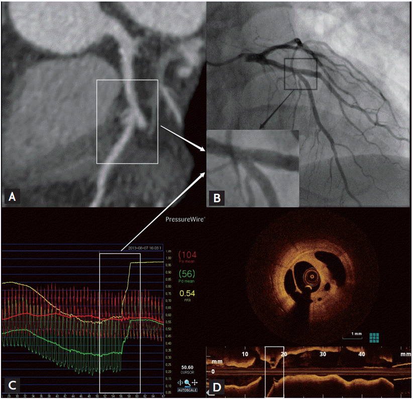
A 48-year-old man visited our hospital due to exertional chest pain. His resting electrocardiogram exhibited a T wave inversion in the inferior leads. A transthoracic echocardiogram showed a preserved left ventricular systolic function without any regional wall motion abnormalities. Multi-detector computed tomography was performed to evaluate the existence of any coronary artery disease and it revealed a significant stenosis in the right coronary artery; however, there was a minimal atherosclerotic lesion in the left coronary artery (Fig. 1A). The right coronary angiogram showed a significant stenosis in the mid portion of the right coronary artery and percutaneous coronary intervention for that lesion was performed successfully with a drug-eluting stent. The left coronary angiogram showed a focal minimal lesion in the mid left anterior descending artery, just proximal to the diagonal bifurcation (Fig. 1B). Contrary to our expectations, the fractional flow reserve (FFR) measured by a pressure wire at that lesion was significantly decreased at 0.54 (Fig. 1C). Intravascular optical coherence tomography (OCT) was performed to evaluate the cause of the discordance between the angiography and FFR. The OCT revealed a focal (2 mm length) lotus root-like lesion consisting of multiple cavities divided by septa within fibrous plaque (Fig. 1D). In spite of the variability in the size of the channels, microchannels were seen to communicate with each other. That was a suspicious finding of a recanalized intracoronary thrombus. That lesion was also successfully revascularized and the patientŌĆÖs symptoms no longer occurred after discharge.
Decision making for revascularization based on angiography alone without prior vessel specific stress testing can be difficult in patients with multi-vessel coronary disease including intermediate or ambiguous coronary lesions. Therefore, functional and physiological considerations of such coronary lesions should be warranted for the selection of the target vessel. This representative case well demonstrated a noteworthy risk of an underestimation using coronary angiography, which could be compensated by invasive functional or intravascular imaging procedures.
 PDF Links
 PubReader
 ePub Link
 Full text via DOI
 Download Citation
 Print
Print


