


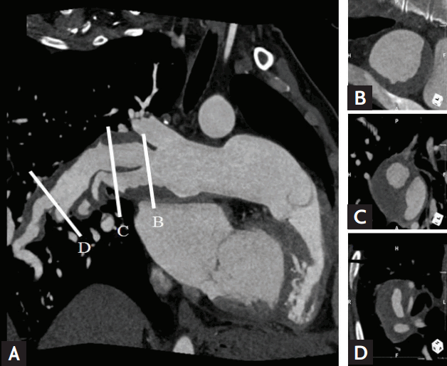
A 43-year-old woman was referred to our emergency department with acute onset of dyspnea, palpitation and chest discomfort. The patient did not have any pre-existing medical history nor any complaint of a particular symptom 1 day before the hospital visit. Physical examination revealed a pulse rate of 117 beats per minute, blood pressure of 100/60 mmHg, and pulse oximetry reading of 88%. A diagnosis of right ventricular hypertrophy and dilatation of the right ventricle (RV) along with severe RV deterioration was made on transthoracic echocardiography. Severe pulmonary hypertension was diagnosed with pulmonary artery (PA) pressure of approximately 90 mmHg by tricuspid regurgitation. An enlargement of the PA, an extensive intimal flap inside the proximal portion of the right lower lobe interlobar PA, and peripheral involvement of the superior and medial basal segmental arteries were revealed after an urgent multidetector computed tomography (MDCT) pulmonary angiography under the suspicion of pulmonary embolism (Fig. 1). Chronic thromboembolism within the right middle lobar PA and its segmental branches was accurately displayed on MDCT along with a large PDA. The patient was treated with oxygen and endothelin receptor antagonist, phosphodiesterase 5 inhibitors, and antibiotics for combined inflammation. A follow-up MDCT and cardiac magnetic resonance imaging showed progression of pulmonary hemorrhage and thrombosis in the PA up to its intersegmental branches (Fig. 2, Supplementary Video 1). An adjustment of medicine was priorly considered before the need for heart-lung transplantation. Fortunately, the patient was discharged in a clinically stable condition 1 month after proper medical treatment. After a 2-year follow-up period, she remained stable without any complications and blood examination was normalized including C-reactive protein.

 PDF Links
 PubReader
 ePub Link
 Full text via DOI
 Download Citation
 Supplement 1
 Print
Print


 |
 |