


A 68-year-old man with no risk factor of atherosclerosis presented to the cardiology division for ongoing dyspnea on exertion for 1 month. The chest computed tomography (CT) revealed a well-defined huge myocardial mass in the anterior wall of the left ventricle (LV) and multiple pathological lymphadenopathies at the supraclavicular, mediastinal, and right intrapulmonary stations. A diffuse myocardial invasion of a hypoechoic irregular mass was observed in the septum and the anterior portion of the LV (5.5 ├Ś 3.2 cm) on the transthoracic echocardiography (TTE). The latter showed severe hypokinesia from the mid-LV to the apex combined with a huge myocardial mass and enlarged cardiac chambers.
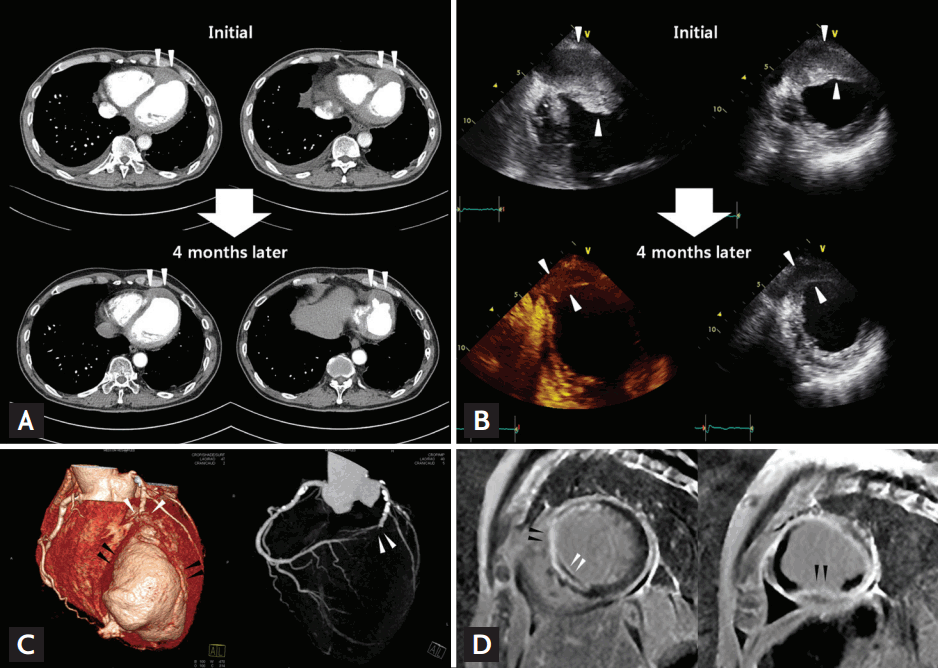
The excision of the cervical lymph node revealed a granulomatous inflammation suggesting tuberculosis or sarcoidosis. In consideration of the patientŌĆÖs cardiac mass and of his negative tuberculous polymerase chain reaction testing, cardiac sarcoidosis was suspected. The patient refused to undergo a myocardial biopsy or further treatment. After 4 months, a follow-up chest CT and TTE revealed a decrease in the size of the LV mass and a newly developed aneurysmal change in the apical portion of the LV (Fig. 1A and 1B, white arrowheads), with decreased mediastinal and cervical lymph nodes. The coronary CT showed an abrupt interruption of the left anterior descending coronary artery (white arrowheads) just before the protruding LV mass and aneurysm (black arrowheads) (Fig. 1C). Cardiac magnetic resonance imaging revealed apical aneurysm and both epicardial and endocardial involvement in the delayed hyperenhancement of the septum and the apical portion of the LV (Fig. 1D).
This case was significant in demonstrating that even self-regressing sarcoidosis can cause critical complications such as irreversible myocardial damage or consecutive fatal vascular injury. A multiple imaging approach can facilitate a more accurate and earlier diagnosis.
 PDF Links
 PubReader
 ePub Link
 Full text via DOI
 Download Citation
 Print
Print


