


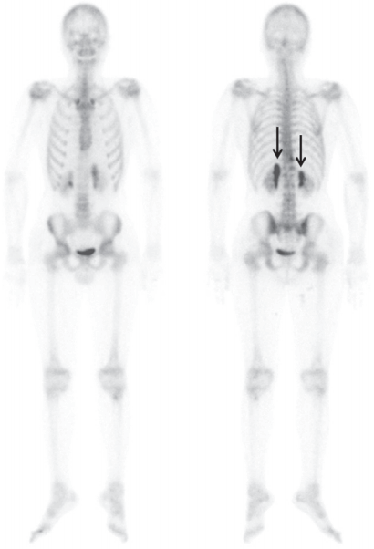
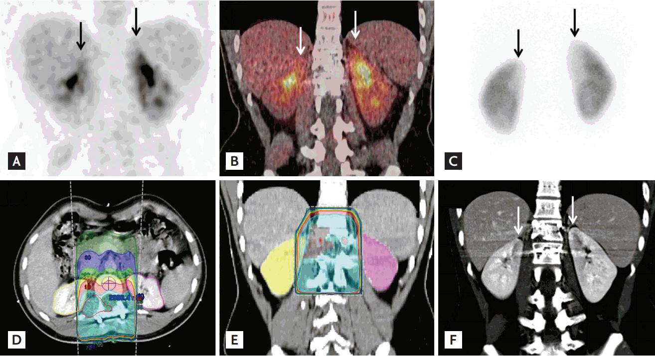
A 20-year-old male had EwingŌĆÖs sarcoma of the right paraspinal region (T12ŌĆōL2 vertebra level) and was treated with chemotherapy and radiation therapy (RT). 99mTc hydroxydiphosphonate bone scan was performed 4 months after the end of treatment. Intense uptake was noted in both kidneys (Fig. 1). On the fluorodeoxyglucose (FDG) positron emission tomography/computed tomography (PET/CT) for response evaluation, mild uptake was also seen in both kidneys (Fig. 2A and 2B). On 99mTc dimercaptosuccinic acid (DMSA) scan, photon defects were noted in both kidneys (Fig. 2C). When we reviewed the RT plan, the medial aspect of both kidneys was included in the radiation field (Fig. 2D and 2E). On contrast-enhanced CT, low-densities were noted in both kidneys (Fig. 2F). We thus diagnosed radiation nephritis (RN). On 6-month follow-up bone scan, the previously noted intensity on both kidneys was decreased.
RN is commonly seen following RT of intra-abdominal tumors or bony metastasis of spine. The histologic findings are vascular sclerosis with tubular ischemic change. Tubular dysfunction and ischemic change explain the photon defect on DMSA scan and the delayed enhancement and retention of contrast on CT. Delayed excretion of radiopharmaceuticals could cause increased uptake on bone scan. CT and bone scan findings were previously reported, but PET/CT findings were reported in only one case. Although the exact mechanism of increased activity on PET/CT remains uncertain, delayed tracer excretion via parenchymal injury could be a major cause. Radiation-induced inflammation can also cause. However, the lack of evidence of inflammation in other tissues included in the radiation field and the decreased blood flow to the kidneys on enhanced CT indicate that the inflammation was a minor contributor.
Focal renal activity on PET/CT could be misinterpreted as malignancy. Differential diagnostic criteria for distinguishing RN from malignancy include a RT history, a linear uptake pattern, and a sharp margin of uptake. Lesion locations were the medial or upper portion of the kidney. Clinicians need to exclude RN when linear increases in renal activity are incidentally detected on FDG PET/CT.
 PDF Links
 PubReader
 ePub Link
 Full text via DOI
 Download Citation
 Print
Print


