Korean J Intern Med > Volume 33(1); 2018 > Article
Park and Cheon: Updated treatment strategies for intestinal Behçet’s disease

Abstract

Behçet’s disease (BD) is a chronic, idiopathic, relapsing immune-mediated disease involving multiple organs, and is characterized by recurrent oral and genital ulcers, ocular disease, gastrointestinal ulcers, vascular diseases, and skin lesions. In particular, gastrointestinal involvement in BD is followed by severe complications, including massive bleeding, bowel perforation, and fistula, which can lead to significant morbidity and mortality. However, the management of intestinal BD has not yet been properly established. Intestinal BD patients with a severe clinical course experience frequent disease aggravations and often require recurrent corticosteroid and/or immunomodulatory therapies, or even surgery. However, a considerable number of patients with intestinal BD are often refractory to conventional therapies such as corticosteroids and immunomodulators. Recently, there has been a line of evidence suggesting that biologics such as infliximab and adalimumab are effective in treating intestinal BD. Moreover, new biologics targeting proteins other than tumor necrosis factor α are emerging and are under active investigation. Therefore, in this paper, we review the current therapeutic strategies and new clinical data for the treatment of intestinal BD.

INTRODUCTION

Behçet’s disease (BD) is a chronic, multi-systemic immune disorder with an unknown etiology, characterized by recurrent oral and genital aphthous ulcerations, arthritis, and skin manifestations, with ocular, vascular, neurological, and gastrointestinal (GI) involvement [1,2]. The diagnosis of intestinal BD is based on GI symptoms in BD patients and typical intestinal ulcerative lesions documented by objective measures [3]. Although the GI symptoms can range from asymptomatic or mild abdominal discomfort to severe abdominal pain, involvement of the GI tract often leads to severe morbidity and mortality [4]. Additionally, these patients can have further complications such as massive hemorrhage, fistula, or bowel perforation [3,5]. Similar to inflammatory bowel disease (IBD), intestinal BD is accompanied by different clinical courses [6] with chronic continuous symptoms or repeated episodes of relapse and remission [7,8]. Along with proper intervention with surgical management, the appropriate use of medical therapy given the life-long duration of the disease is of great importance in the management of intestinal BD [9,10]. Empirical medical therapies include 5-aminosalicylic acids (5-ASAs), corticosteroids, immunomodulators such as azathioprine and 6-mercaptopurine (6-MP), thalidomide, and anti-tumor necrosis factor α (TNF-α) agents. Therefore, we will cover and review the current optimal medical treatment options and recent data relevant to patients with intestinal BD. Furthermore, new treatment modalities that might have the potential to be developed as novel therapeutic agents in the near future will also be discussed.

PRINCIPLES OF APPROACH TO THE MANAGEMENT OF INTESTINAL BD

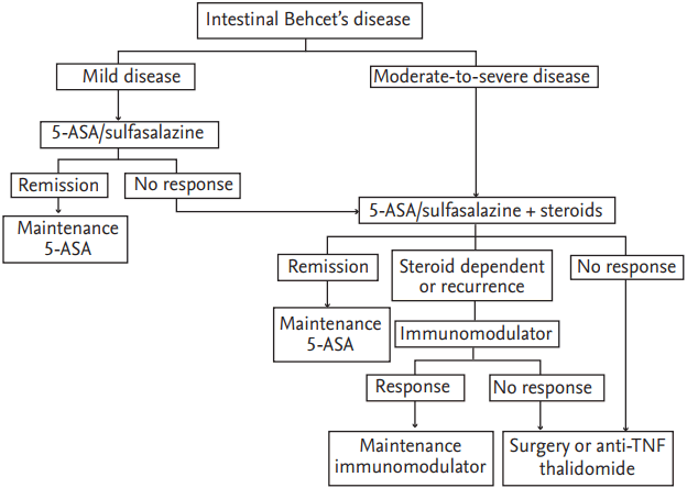
According to the 2008 European League Against Rheumatism (EULAR) guidelines, there are unfortunately no evidence-based treatment guidelines that can be recommended for the management of intestinal BD [11]. There are a few large, randomized and controlled trials of potential management for patients with intestinal BD. Therefore, the management of intestinal BD has been empirically based on the treatment guidelines for Crohn’s disease (CD) following opinions of expert physicians [2]. Correspondingly, according to the Japanese consensus of intestinal BD published in 2007, 5-ASAs, corticosteroids, immunomodulators, enteral nutrition, total parenteral nutrition, and surgical therapy were included in standard therapy for intestinal BD [12]. In addition, in 2014, the 2nd edition of consensus statements for intestinal BD suggested that anti-TNF-α agents such as adalimumab and infliximab should be considered as standard therapy for intestinal BD [13]. Cheon and Kim [2] and Lee et al. [14] proposed a treatment algorithm for intestinal BD based on expert opinion and accumulated published data (Fig. 1).

MEDICAL TREATMENT

5-Aminosalicylic acids

5-ASAs can reduce inflammation and have immunomodulatory actions in the intestine [15]. 5-ASAs widely used for IBD are sulfasalazine and mesalamine [15]. Although evidence of efficacy in intestinal BD is still insufficient, the Japanese consensus statements recommended 5-ASA as a first option for induction and maintenance therapy for mild or moderate intestinal BD [13]. 5-ASA is usually administered at a dose of 2 to 4 g/day [2]. Sulfasalazine, a colon-selective drug that contains a sulfa group, is usually administered 3 to 4 g/day in intestinal BD [2].
In 1997, a case study reported that sulfasalazine use was associated with clinical improvement in 79% (11 of 14, p < 0.005) of intestinal BD patients in Korea [16]. In contrast, Matsukawa et al. [17] reported the ineffectiveness of 5-ASA in recurrent intestinal ulcers. However, Jung et al. [18] showed that 5-ASA/sulfasalazine therapy has a positive effect on maintaining remission in Korean patients with intestinal BD, although younger age (< 35 years), higher C-reactive protein (CRP) level, and higher disease activity were associated with a poor response to 5-ASA/sulfasalazine. Another retrospective study on the effect of 5-ASA compounds reported that among 16 patients who were treated with 5-ASA compounds, 62.5% (n = 10) achieved clinical remission with a disease-free duration of 89.3 ± 64.5 months (Table 1) [13,16,18-34]. This discrepancy between studies possibly originates from differences in disease heterogeneity, especially disease severity.

Corticosteroids

Corticosteroids are fast-acting anti-inflammatory drugs that have been commonly used for patients with acutely moderate-to-severe and refractory intestinal BD [13,20-22,35]. However, evidence on the efficacy of corticosteroids in intestinal BD also remains insufficient, because prospective/randomized studies are lacking. Treatment with corticosteroids is considered the first-line therapy during the acute phase of the disease, initially at doses of 0.5 to 1.0 mg/kg/day of prednisolone for 1 to 2 weeks with tapering by 5 mg each week and stoppage within 3 months (Table 1) [2,36-38]. Prolonged use of prednisolone in excess of 10 mg/day is not recommended. Some case studies have reported the efficacy of an intravenous administration of a lipid emulsion of dexamethasone [20,21]. However, corticosteroid use was paradoxically associated with increased risk of GI bleeding and perforation [39]. Additionally, Park et al. [40] reported that corticosteroid use was significantly associated with re-bleeding in patients with intestinal BD. Despite these side effects, Park et al. [23] in 2010 performed a retrospective study of clinical outcomes after corticosteroid use in patients with moderate-to-severe intestinal BD. A total of 54 patients were enrolled and the median corticosteroid dosage was 0.58 mg/kg (interquartile range, 0.39 to 1.20). In this study, 46.3% of patients (n = 25) achieved complete remission after 1 month of treatment. Three months after treatment, 40.7% of patients (n = 22) showed treatment response. After 1 year, 48.1% (n = 26) remained responsive to treatment. However, after 1 year, 35.2% of the patients (n = 19) exhibited corticosteroid dependency, and 7.4% (n = 4) received surgery. Treatment response at three months was independently associated with a decreased risk of surgery in the long term (p = 0.009). Therefore, the authors suggested that short-term response rate to initial corticosteroid therapy was high in patients with intestinal BD [23]. Given this background, the Japanese consensus recommended that corticosteroids should be considered for induction therapy when patients have severe symptoms such as abdominal pain, diarrhea, or GI bleeding due to deep ulcers [13].

Immunomodulators

Immunomodulators included in this review are thiopurines (6-mercaptopurine, 6-MP, and its prodrug azathioprine), methotrexate, tacrolimus, interferon, cyclosporin, and intravenous immunoglobuilin [41].

Thiopurines

Thiopurines are the most commonly prescribed immunomodulators for patients with intestinal BD, especially those with moderate-to-severe disease and those who are corticosteroid-dependent/resistant or secondarily lose response to anti-TNF-α agents (Table 1) [1,2,13,24].
Thiopurine drugs such as azathioprine and 6-MP are also used in postoperative patients after intestinal resection to reduce the postoperative recurrence rate [25], and they were reported to be relatively useful in the maintenance of remission after surgery in intestinal BD patients [26]. The initial dose of azathioprine is 25 to 50 mg/day, and if the azathioprine therapy is tolerated without side effects such as leukopenia and liver dysfunction, the dose can be gradually increased every 2 to 3 weeks up to 2.0 to 2.5 mg/kg [13,26,42]. The dose for the initial use of 6-MP is 0.5 mg/kg, which can also be gradually increased up to 1.0 to 1.5 mg/kg in a similar manner to that of azathioprine [26]. Jung et al. [26] reported positive effects of thiopurine maintenance therapy in patients with intestinal BD in Korea. They retrospectively evaluated patients with intestinal BD who received prolonged thiopurine monotherapy for over 6 months in a tertiary single center [26]. Of the 272 patients with intestinal BD, 67 patients (24.6%) received thiopurine therapy, and 39 of these 67 patients (58.2%) maintained medically or surgically induced remission. However, a younger age at diagnosis and a lower hemoglobin level were significantly associated with a poor response to thiopurines in this study [26].
Choi et al. [24] and Lee et al. [25] independently reported the effects of postoperative thiopurine use in patients with intestinal BD (Table 1). Choi et al. [24] compared the reoperation rate between azathioprine users and non-users. Reoperation rates were significantly lower in azathioprine users (7%) than in non-users (25%) at 2 years, and 25% and 47% at 5 years, respectively [24]. In addition, Lee et al. [25] investigated the postoperative protective effect of thiopurine. In their study, the postoperative recurrence rate was significantly lower in patients who received postoperative thiopurines (p = 0.050) [25].
The most common side effect of thiopurine drugs was leukopenia, which is defined as a white blood cell (WBC) count lower than 3,000 or 4,000 cells/mm3 [26,27]. Following the American Gastroenterological Association recommendations, it is suggested that when patients are treated with azathioprine or 6-MP, routine laboratory monitoring, including complete blood count, should be frequently performed [43]. Park et al. [27] reported that lower WBC count during thiopurine maintenance therapy was associated with prolonged remission.

Methotrexate

The efficacy of other immunomodulators has also been reported. The use of methotrexate in patients with intestinal BD remains relatively poorly understood; however, Iwata et al. [44] reported the efficacy and safety of the combination of infliximab and methotrexate in 10 patients with refractory intestinal BD. All the 10 patients had improved symptoms and disease-related complications within 4 weeks. In addition, 50% of the patients (five of 10) showed disappearance of ileocecal valve ulcers at 6 months, and 90% of the patients (nine of 10) showed disappearance of the ulcers at 12 months (Table 2) [44-67]. Recently, Park et al. (unpublished data) also reported the efficacy of methotrexate in patients with intestinal BD as either mono- or combination therapy with adalimumab. In a retrospective review of 10 intestinal BD patients who received methotrexate treatment, four of the 10 patients received methotrexate monotherapy and the other six patients received combination therapy with adalimumab. In methotrexate monotherapy, three patients (30%) had steroid-free remission at 3 months, and four patients (50%) did at 6 months (unpublished data) (Table 1).

Tacrolimus

Oral tacrolimus is a macrolide antibiotic with potent immunosuppressive activity [68], and it is widely used for the prevention of allograft rejection in patients who underwent organ transplantation [28]. Matsumura et al. [28] reported improvement after using tacrolimus in a patient refractory to conventional therapeutic agents such as 5-ASA, corticosteroids, immunomodulators, and infliximab. In our experience at Severance Hospital, four patients underwent tacrolimus therapy after failure of conventional agents, but none had a response to that drug (unpublished data).

Interferon

In 1957, Isaacs and Lindenmann [69] first introduced interferon (IFN), a large family of glycoproteins known to produce cellular responses to various antigens such as microbes, viruses, and tumors [70,71]. Several studies reported the efficacy of IFN alfa-2a in the treatment of BD, especially in patients with mucocutaneous lesions, arthritis, and ocular manifestations [70,72,73]. Alpsoy et al. [70] reported a randomized, placebo-controlled, and double-blind study in 50 patients with BD. Of the 50 patients, 23 received IFN alfa-2a therapy, two of whom showed complete response while another 13 showed partial response. Eight patients showed no response. Grimbacher et al. [74] reported the efficacy of IFN alfa-2a in a patient who had eye and GI tract involvement. The patient experienced improved retinal infiltrates, skin lesions, and abdominal complaints within 2 weeks of treatment [74]. Monastirli et al. [75] presented a case demonstrating improvement of clinical symptoms in a patient who had intestinal BD and acute myelitis that were initially unresponsive to high-dose steroids; the patient remained drug-free until 12 months of follow-up. However, until now, there has been little research on the effect of IFN in patients with intestinal BD, and therefore it is difficult to clearly define the effect of IFN on intestinal BD.

Cyclosporin

Cyclosporin is one of the most potent immunosuppressive agents, which decreases T-cell activity and blocks the immune response of inflammatory cytokines [76]. Several studies reviewed the positive effect of cyclosporin on the ocular manifestations of BD [11,77,78]. Following the EULAR guidelines, cyclosporin should not be used in BD patients with central nervous system involvement [11]. With regard to intestinal BD, Bayraktar et al. [7] reported cyclosporin had no benefit on intestinal BD. Therefore, further study is needed to clarify the efficacy of cyclosporin in intestinal BD.

Intravenous immunoglobulin

Since 1952, immune globulin products from human plasma have been used to treat immune deficiency [29,79]. Intravenous immunoglobulin (IVIG) is an immunomodulating agent that has multiple activities and has been used for autoimmune and systemic inflammatory diseases [29]. There are a limited number of studies regarding the role of the IVIG in intestinal BD. Beales [30] reported a case of IVIG treatment without additional corticosteroids in a BD patient with colon involvement after initial failed treatment with corticosteroids and other immunomodulators. She significantly improved after IVIG initiation, and bowel lesions disappeared after 6 weeks. Cantarini et al. [29] reported successful treatment with IVIG in patients with severe and resistant BD. Of four patients, all had mucocutaneous involvement with BD and one patient had intestine involvement (Table 1) [29]. Therefore, the effect of IVIG on intestinal BD still warrants further study.

Thalidomide

Thalidomide, a synthetic derivative of glutamic acid, was first introduced in 1957 and used as a sedative agent with a teratogenic side effects; however, recent research has shown growing interest in its anti-inflammatory and immunomodulatory properties [2,31]. Therefore, there have been several studies of thalidomide treatment in pediatric patients with BD who were refractory to other immunomodulators, or those who developed undesirable side effects [80,81].
Sayarlioglu et al. [32] reported beneficial effects of thalidomide on recurrent perforating intestinal ulcers in an adult patient with BD. Additionally, Yasui et al. [31] reviewed a case of thalidomide for the treatment of juvenile-onset intestinal BD, which included seven patients with severe and recurrent intestinal involvement. The initial dose of thalidomide was 2 mg/kg/day, with dose adjustment according to response; if necessary, the dose of thalidomide was increased up to 3 mg/kg/day or decreased to 0.5 to 1 mg/kg/day [31]. All the seven patients achieved clinical improvement and were allowed to stop corticosteroid therapy [31]. In Korea, Lee et al. [33] also reported the efficacy of thalidomide in four patients who had chronic relapse of intestinal BD that repeatedly required corticosteroids and was refractory to conventional therapy such as 5-ASA and immunomodulators. Three of the four patients showed clinical symptom improvements; however, two of the four patients had to stop the therapy due to side effects such as general edema, leukopenia, and sepsis (Table 1).
It has been reported that thalidomide reduces TNF-α levels via degradation of its encoding messenger RNA, and thus shows immunomodulatory effects [82]. Hatemi et al. [83] investigated 13 patients with intestinal BD who were refractory to conventional therapy were treated with anti-TNF-α and/or thalidomide. Of those, 10 patients (75%) achieved clinical and endoscopic remission [83]. Therefore, thalidomide may become a therapeutic option for intestinal BD, but it should be carefully selected and monitored due to its teratogenicity and side effects such as edema, leukopenia, and sepsis. Further well-designed studies are necessary to conduct proper use.

Others

Colchicine

Colchicine is an anti-inflammatory drug that suppresses the secretion of cytokines and chemokines, and is used for the management of patients with gout and BD [35,84]. Some studies reported the improvement of arthralgia and erythema nodosum in patients with BD, but the effect on mucocutaneous manifestations was controversial in several studies [85-87]. Therefore, the EULAR recommended that colchicine is preferred when the dominant lesion is erythema nodosum [11]. However, currently, there is no evidence on the use of colchicine for intestinal BD. Choi et al. [24] suggested that there are no beneficial effects on intestinal lesions from their retrospective study. There is an insufficient number of studies to prove efficacy of colchicine in intestinal BD.

Stem cell transplantation and leukocytapheresis

Hematopoietic stem cell transplantation (HSCT) has been used to treat severe autoimmune and inflammatory conditions that are unresponsive to traditional therapies [38,88]. HSCT in patients with BD has been reported several times in forms of case reports either for refractory BD or patients with associated hematological conditions such as aplastic anemia or myelodysplastic syndrome (MDS) [88-90]. This treatment is based on the rationale that a vigorous immunoablative regimen can remove autoaggressive lymphocyte clones [2]. Yamato [89] and Rossi et al. [90] reported cases of successful stem cell transplantation for MDS with BD and severe/refractory BD. HSCT could be an alternative therapy in BD patients with severe organ involvement, including GI involvement, that is refractory to immunomodulators [88]. Allo-stem cell transplants have high transplant-related morbidity and mortality; therefore, autologous transplants have been performed more frequently in BD patients, and non-myeloablative regimens may be preferred over myeloablative ones [88].
According to the Japanese consensus statements for intestinal BD in 2014 [13], leukocytapheresis was classified into an experimental treatment to mechanically remove WBCs in patients with intestinal BD who are steroid-dependent or resistant [2]. This is because activated neutrophils in patients with severe intestinal BD increase leukocytosis or cytokines [91]. However, this procedure also warrants further well-designed studies [2].

Anti-TNF-α agents and biologics

Intestinal BD is associated with abnormal T-cell immune response and T helper type 1-associated cytokines such as TNF-α, IFN-γ, interleukin-12 (IL-12), and IL-18, which play a critical role in disease pathogenesis [92,93]. In patients with BD, the number of γδ cells producing TNF-α increases, TNF-α and its receptor levels are elevated in the blood, and the expression of TNF-α increases when the clinical course becomes worse [93]. Therefore, the importance of anti-TNF-α agents has been reported in several studies based on this background (Table 2) [45,46,67,94-97]. According to the Japanese consensus [13], anti-TNF-α mAbs (infliximab and adalimumab) should be considered as the standard therapy in patients with intestinal BD [2,13]. Infliximab is a chimeric monoclonal antibody against TNF-α [2] that can be considered for induction therapy with 5 mg/kg at 0, 2, and 6 weeks [13]. Responders should be administered maintenance therapy with infliximab every 8 weeks [13]. Adalimumab is a fully human anti-TNF-α monoclonal antibody [48], and can also be used for induction therapy with a dose of 160 mg at week 0 (baseline), 80 mg at week 2, and 40 mg at week 4 subcutaneously. Responders should be considered for maintenance therapy every other week at a dose of 40 mg [13]. In addition, several studies on the effects of various biologics such as etanercept, anakinra, canakinumab, and tocilizumab have been conducted [49-52].

Infliximab

Hassard et al. [53] reviewed a patient with chronically active, steroid-dependent intestinal BD who received 4 doses of infliximab during 6 months. The patient achieved remission, with a reduced Crohn’s Disease Activity Index score (from 270 to 13) at 2 weeks of infliximab use, and maintained remission after 10 weeks (Table 2) [53]. Naganuma et al. [54] also reported on the efficacy of infliximab induction and maintenance therapy. Four of six patients with intestinal BD achieved remission, and all four patients maintained remission with infliximab therapy (Table 2) [54]. In 2013, Lee et al. [45] reported a multicenter retrospective study of 28 patients with active moderate-to-severe intestinal BD who had been treated with infliximab at eight tertiary hospitals in Korea. At 2, 4, 30, and 54 weeks post-infliximab infusion, patients showed clinical response rates of 75%, 64.3%, 50%, and 39.1%, and clinical remission rates of 32.1%, 28.6%, 46.2%, and 39.1%, respectively. In addition, independent factors of maintenance response were older age at diagnosis (≥ 40 years), female sex, a longer disease duration (≥ 5 years), concomitant immunomodulator use, and achievement of remission at week 4 on multivariate analysis (Table 2) [45]. In a prospective open-label, single-arm phase 3 study in Japan, 18 patients with BD were enrolled, including 11 patients with intestinal BD, three patients with neurological BD, and four patients with vascular BD with poor response or resistance to conventional therapy [47]. Induction infliximab therapy was administered at 5 mg/kg at weeks 0, 2, and 6, with maintenance therapy every 8 weeks thereafter until week 46. Eleven of 18 of the patients (61%) were complete responders at weeks 14 and 30 and remained in remission until week 54. In addition, laboratory findings such as CRP levels were diminished, and 80% of patients showed healing or scarring ulceration at week 14. However, three patients with intestinal BD had to increase the dose of infliximab up to 10 mg/kg after week 30 due to loss of response, and two of three patients had symptoms that could not be controlled and worsened (Table 2) [47].
The efficacy of infliximab induction and maintenance therapy for intestinal BD has been widely accepted in many studies [13,47,55]. However, there is still insufficient evidence on the effect of combination therapy with immunomodulators as well as whether it can be used as a postoperative therapy in patients who underwent bowel resection. Byeon et al. [56] reviewed a patient with an unhealed postoperative anastomotic site and recurrent ulcers after a distal ileocecectomy, and reported endoscopic remission after 15 days of infliximab infusion. Iwata et al. [44] reported that concomitant therapy of infliximab with methotrexate was effective to achieve short-term remission and long-term response until 24 months. Well-designed further studies are needed.

Adalimumab

Adalimumab is a humanized anti-TNF-α monoclonal antibody [98] and is widely used in patients with rheumatoid arthritis (RA), psoriatic arthritis, ankylosing spondylitis, psoriasis, juvenile idiopathic arthritis, uveitis, and IBD [48,57,99]. In 2011, De Cassan et al. [58] first reported adalimumab efficacy in patients with intestinal BD with repeated steroid-dependent flares and failure of conventional therapy. After that, several studies showed the efficacy of adalimumab in patients with intestinal BD. Tanida et al. [48] performed a multicenter, open-label, uncontrolled study of adalimumab for the treatment of Japanese patients with intestinal BD who were refractory to conventional therapy, including corticosteroids and/or immunomodulators. A total of 20 patients with intestinal BD were treated with adalimumab with dose of 160 mg at induction, 80 mg 2 weeks later, and 40 mg every other week for 52 weeks; for some patients with incomplete response, the dose of adalimumab increased up to 80 mg every other week. After 24 weeks of adalimumab therapy, nine of 20 patients (45%) had alleviated GI symptoms and diminished endoscopic assessment scores to 1 or lower than at pre-treatment. In addition, 12 of 20 patients (60%) had reduced endoscopic assessment scores at week 52. Additionally, no newly discovered safety problems or deaths were reported (Table 2) [48]. Recently, Inoue et al. [46] published an open-label phase 3 study (NCT01243671) in Japan to evaluate the safety and efficacy of adalimumab for the treatment of the same patients included in the 52 week follow-up study from weeks 52 to 100. Long-term efficacy of adalimumab was assessed on the basis of marked improvement, defined as both a GI symptom score of ≤ 1 (did not affect patient’s daily life) and an endoscopic score of ≤ 1 (largest ulcer is ≤ 1/4 original size), and complete remission, defined as a GI symptom score of 0 (symptom-free) and an endoscopic score of 0 (complete ulcer healing). At weeks 52 and 100, 12 of 20 patients (60%) and eight of 20 patients (40%) showed marked improvement, while four of 20 patients (20%) and three of 20 patients (15%) showed complete remission, respectively (Table 2) [46]. In addition, Kimura et al. [95] reported that a patient with intestinal BD and MDS successfully improved in GI symptoms, CRP levels, leukocytopenia, and anemia 4 months after starting adalimumab.
Combination therapy with adalimumab and immunomodulators has not yet been established, but Vitale et al. [59] conducted a multicenter retrospective study with 100 BD patients for the comparison of adalimumab monotherapy and combination therapy with disease modifying anti-rheumatic drugs. Although the study was not limited to patients with intestinal BD, it did not show a significant difference in clinical outcomes between adalimumab monotherapy and combination therapy (Table 2) [59].

Etanercept

Etanercept is a dimeric human TNF receptor (TNFR) p75-Fc fusion protein that inhibits TNF-α activity [100,101]. Only a few studies have focused on etanercept as a treatment option for intestinal BD [49]. Ma et al. [49] reported on the outcomes of etanercept in the treatment of intestinal BD with 19 patients who were refractory to conventional therapy. Etanercept (25 mg twice a week for 3 months) was compared with conventional therapy, and the etanercept group showed significantly greater healing of ulceration, remission, and recovery of the erythrocyte sedimentation rate and CRP levels [49]. However, etanercept was not effective for the treatment of IBD, unlike other anti-TNF agents such as infliximab and adalimumab [102,103]. Sandborn et al. [102] conducted a randomized, double-blind, placebo-controlled trial in patients with active CD using etanercept. Forty-three patients were enrolled; however, 39% of the etanercept-treated patients had a clinical response compared with 45% of the placebo group (p = 0.763) at week 4 [102]. Furthermore, some recent studies have suggested a paradoxical development of IBD in patients receiving etanercept therapy. Forty-four (41 CD, three ulcerative colitis [UC]) of 443 cases (297 CD, 146 UC) were reported to develop de novo IBD after the initiation of etanercept, and 43 cases of flares of existing IBD patients were reported in association with etanercept therapy [104]. Therefore, the therapeutic efficacy of etanercept in patients with intestinal BD is also questionable.

Anakinra

Anakinra is a recombinant version of the IL-1 receptor antagonist (IL1-RA) and a biologic agent used to modify the immune response of IL-1 [105]. Although the exact etiology of BD is still unclear, it has been reported that IL-1 is a proinflammatory cytokine [106] which can be increased in patients with BD. Zou and Guan [106] reported increased susceptibility to BD due to polymorphisms in the IL-1-related gene. Cantarini et al. [50] reported on the effects of anakinra in patients with conventional therapy-resistant BD. Nine patients were refractory to anti-TNF agents and conventional therapy, and most of them received anakinra (100 mg daily) with low doses of corticosteroids. Eight of nine patients had symptom improvement after using anakinra; however, the eight had recurrence over time and 15% of patients experienced adverse events (AEs) [50]. Although the efficacy of anakinra has been shown in a few studies, the long-term effect is unclear and most of the studies were not limited to patients with intestinal BD; therefore, more studies are required to clarify the relationship between recurrence and dosage, and to determine the exact efficacy in intestinal BD.

Canakinumab

Canakinumab is a fully human anti-IL-1β antibody which could also be considered as a therapeutic option for resistant or refractory BD [51,60]. Vitale et al. [51] reported successful use of canakinumab in three patients with BD. Two patients had GI involvement and had failed to respond to conventional therapy such as sulfasalazine, methotrexate, cyclosporin, azathioprine, corticosteroids, anti-TNF agents, and anakinra (100 mg/day). Therefore, canakinumab was used at a dose of 150 mg every 6 (case 2) and/or 8 weeks (case 1) by subcutaneous injection, which led to clinical improvement and achieved clinical remission [51]. Recently, Emmi et al. [60] reported outcomes on the use of IL-inhibitors including canakinumab in patients with BD. Three patients used canakinumab (150 mg every 6 to 8 weeks for 12 months) and achieved complete remission at 12 months with no serious AEs (Table 2) [60]. However, the study was not limited to intestinal BD. Therefore, further studies are required.

Tocilizumab

Tocilizumab is a recombinant-humanized anti-human IL-6 receptor monoclonal antibody [107]. IL-6 is a cytokine that plays an important role in immune function, and some studies have shown that circulating IL-6 level is elevated in patients with BD [52,108]. Deroux et al. [52] reported four experienced cases using tociliziumab and reviewed the cases in the literature to evaluate the safety and efficacy of tocilizumab in patients with refractory BD. Three of the four patients were BD patients with GI involvement and all patients were previously unresponsive to conventional therapy. All patients received tocilizumab (8 mg/kg every 4 weeks) and experienced significantly decreased disease activity [52]. However, tocilizumab failed in clinical trials for patients with CD because of serious adverse effects such as bowel perforation and abscess [109,110]. This is because IL-6 is known to play an important role in stimulating intestinal epithelial proliferation and repairing intestinal injury [111]. Therefore, we should pay attention to the use of tocilizumab in patients with intestinal BD in whom intestinal perforation is one of the important complications, and further studies are required.

CONCLUSIONS

Although intestinal BD is not common, GI manifestation occurs more frequently in East Asia, including Korea, than in the Mediterranean region [2,36,112,113]. In addition, complications such as intestinal perforation and bleeding considerably increase morbidity and mortality [2,3]; therefore, it is important to manage and treat patients properly. Appropriate medical therapy in intestinal BD is important to prevent poor outcomes such as frequently recurred ulcer, surgery, complications, and mortality. 5-ASA, corticosteroids, immunomodulators, and anti-TNF agents have been proposed as standard therapy in intestinal BD. However, there is still no definite guideline for established medical therapy because of its rarity and lack of clinical data. Studies on the pathogenesis of BD have been carried out, and studies on new therapies such as the therapeutic effects of related biological agents have been actively under investigation. We anticipate more advanced treatment strategies and established treatment guidelines in intestinal BD in the near future.

Conflict of Interest

No potential conflict of interest relevant to this article was reported.

Acknowledgments

This study was supported by a grant (NRF-2017R1A1A1A05001011) from the Basic Science Research Program through the National Research Foundation of Korea, which is funded by the Ministry of Science, ICT, and Future Planning; and a faculty research grant (2012-31-0477) from the Department of Internal Medicine, Yonsei University College of Medicine.

Figure 1.
A proposed algorithm for the treatment of intestinal Behçet’s disease. Adapted from Lee et al. [14. 5-ASA, 5-aminosalicylic acid; anti-TNF, anti-tumor necrosis factor.
kjim-2017-377f1.gif
Table 1.
Studies of medical treatments of intestinal BD: 5-ASA, corticosteroids, immunomodulators, thalidomide, IVIG
Drugs Study Type of study No. of patients or articles Dose of medication Indication/Outcomes
5-ASA Yoo et al. (1997) [16] Case report Total: 31 Sulfasalazine user: 14 1 g/day → 2–4 g/day → 1–2 g/day (maintenance) Symptom improved: 79%
Sonta et al. (2000) [34] Case report Total: 1 1,200 mg/day Lesion: esophageal ulcers
Positive clinical efficacy
Jung et al. (2012) [18] Retrospective cohort study Total: 292 - Maintaining remission; mild-to-moderate intestinal BD
Clinical remission: 143
Hisamatsu et al. (2014) [13] Japanese consensus statements Relevant articles: 15 5-ASA: 2.25–3.00 g/day Indication: maintenance therapy
Sulfasalazine: 3–4 g/day
Hatemi et al. (2016) [19] Retrospective cohort study Total: 60 - Remission: 10 of 16 patients (62.5%)
5-ASA user: 16 Disease free duration: 89.3 ± 64.5 months
Corticosteroids Nakase et al. (2001) [20] Case report Total: 2 Case 1: PD 30 mg → 40 mg → no improved: IV 2.5 mg dexamethasone every 2 weeks Case 1: after IV corticosteroid use, oral, abdominal pain improved & inflammatory parameters: normalized
Case 2: PD 40 mg → 25 mg/ day → recur: IV 2.5 mg limethasone every 2 weeks Case 2: after IV corticosteroid use, abdominal symptoms disappeared & inflammatory parameters: normalized
Toda et al. (2002) [21] Case report Total: 1 1 g/day of IV methylpred- nisolone, 3 days → IV PD 40 mg/day → 20 mg PD IA injection Disease remission
Yasuo et al. (2003) [22] Case report Total: 1 PD 0.5 mg/kg daily Lesion: esophageal ulcer
Symptom improved & esophagoscopy after 4 weeks: mucosa healing
Park et al. (2010) [23] Retrospective cohort study Total: 54 Median dosage: 0.58 mg/kg (range, 0.39–1.20) Clinical remission: 25 (46.3%)
1 Year treatment response: 26 (48.1%)
Cumulative probability of surgery: 17.5% at 1 year; 49.1% at 3 years
Hisamatsu et al. (2014) [13] Japanese consensus statements Relevant articles: 15 The initial dose of corticosteroids: 0.5–1 mg/kg/day for 1–2 weeks Indication: induction therapy
Clinical improvement → tapered by 5 mg every week & stopped Severe symptoms (i.e., abdominal pain, diarrhea, GI bleeding)
Immunomodulators Choi et al. (2000) [24] Retrospective cohort study Total: 43 AZA: 50 mg/day Re-operation rates: decreased in AZA user group (7% vs. 25% at 2 years; 25% vs. 47% at 5 years) maintenance ther- apy in patients who require surgery
Remission: 16 (38%)
Matsumura et al. (2010) [28] Case report Total: 1 Oral tacrolimus: 10–12 ng/mL (induction) → 5–10 ng/mL (maintenance) Refractory case to 5-ASA, steroid, immunomodulators, thalidomide, infliximab
Remission: 33 months after starting tacrolimus
Jung et al. (2012) [26] Retrospective cohort study Total: 272 AZA: Initial dose 25 or 50 mg daily → 2.0–2.5 mg/kg (gradually increased every 2–4 weeks) Indication: maintenance therapy
AZA user: 67 (24.6%) Remission: 39 (58.2%) of 67 (maintaining medically or surgically induced remission)
6-MP: Initial dose 0.5 → 1.0–1.5 mg/kg (gradually increased) Clinical relapse: 13 (33.3%) of 39
Relapse risk factors: younger age (< 25 years) at diagnosis, lower hemoglobin level (< 11 g/dL)
Hisamatsu et al. (2014) [13] Japanese consensus statements Relevant articles: 15 AZA: Initial dose 25–50 mg/day Indication: corticosteroid- dependent, corticosteroid- resistant, or anti-TNF-α mAb-resistant
Lee et al. (2015) [25] Retrospective cohort study Total: 77 AZA: 2–2.5 mg/kg/day Postoperative recurrence rate: lower in patients who received post-operative thiopurines than 5-ASA user (p = 0.050)
Thiopurine user: 27 (35.1%) 6-MP: 1–1.5 mg/kg/day
Park et al. (2015) [27] Retrospective cohort study Total: 196 (IBD) AZA: 2–2.5 mg/kg/day Cumulative relapse-free survival rate: higher in the leukopenic group than in the non-leukopenic group (p < 0.001)
Intestinal BD: 83 6-MP: converted to an equivalent AZA dose by multiplying by 2.08
Hatemi et al. (2016) [19] Retrospective cohort study Total: 44 AZA Remission and no relapse: 24 (65%)
Mean follow-up duration: 68.6 ± 43.6 months
Park et al. (2017) (unpublished data) Retrospective cohort study Total: 10 MTX: various dose (subcutaneously & oral) Steroid free remission: 3 patients (30%) at 3 months, 4 patients (50%) at 6 months
MTX monotherapy: 4 MTX + ADA: 6 ADA: 160 mg in week 0, 80 mg in week 2, and 40 mg every other week CRP levels: significantly decreased at 6 months com- pared with the baseline (p = 0.039).
Thalidomide Sayarlioglu et al. (2004) [32] Case report Total: 1 100 mg/day Lesion: recurrent intestinal perforation cecum, transverse colon, terminal ileum
Remission: 4 months follow-up
Yasui et al. (2008) [31] Case report Total: 7 Initial dose: 2 mg/kg/day → if necessary, 3 mg/kg/day (increase) or 1–0.5 mg/kg/ day (decreased) Inclusion case: conventional therapy failure, severe steroid toxicity
Clinical improvement: all patients
Lee et al. (2010) [33] Case report Total: 4 Case 1: 200 mg/day Inclusion case: refractory to steroid, 5-ASA, immunomodulators
Case 2: 100 mg/day Clinical improvement: 3 of 4
Case 3: 100 mg/day
Case 4: 50 → 100 mg/day
IVIG Beales (1998) [30] Case report (letters) Total: 1 400 mg/kg/day for 5 days Lesion: oral, genital ulcer, uveitis, acneiform rash, arthralgia, intestine
Previous therapy: cyclosporine, thalidomide maintenance → IV methyl PD, oral mesala- mine add → no effect
Clinical improvement: after 30 months
Cantarini et al. (2016) [29] Case report Total: 4 Case 3: 400 mg/kg/day for 5 days per month Lesion: mucocutaneous, ocular, neurological, GI
GI BD: 1 Previous therapy: prednisone, cyclosporine, AZA, mesalazine
Clinical improvement: after 14 months

BD, Behçet’s disease; 5-ASA, 5-aminosalicylic acid; IVIG, intravenous immunoglobulin; PD, prednisolone; IV, intravenous; IA, intra-arterial; GI, gastrointestinal; AZA, azathioprine; 6-MP, 6-mercaptopurine; TNF-α, tumor necrosis factor α; mAb, monoclonal antibody; IBD, inflammatory bowel disease; MTX, methotrexate; ADA, adalimumab; CRP, C-reactive protein.

Table 2.
Studies of medical treatment of intestinal BD: anti-TNF agents and biologics
Drugs Study Type of study No. of patients Dose of medication Outcomes
Infliximab Hassard et al. (2001) [53] Case report Total: 1 5 mg/kg, at 0, 2, 7, and 23 weeks Disease activity: CDAI 270 (baseline) → 13 (at 2 weeks)
Remission: after 10 weeks
Travis et al. (2001) [55] Case report Total: 2 Case 1: 3 mg/kg (dose reduced because of recent sepsis) Case 1: CRP 72 mg/L → 45 (on steroids) → 70 (sepsis) → 29 (on infliximab) → 12 (12 weeks later)
Case 2: 5 mg/kg Case 2: CRP 109 mg/L → 25 (on steroids) → 73 (recur) → 53→ < 6 (on infliximab)
Kram et al. (2003) [61] Case report Total: 1 5 mg/kg dose over a 6-week period (3 infusions) CRP 18.7 → normal (on infliximab) → 22.5 mg/mL (relapse) → normal (on infliximab + MTX)
Lee et al. (2007) [67] Case report Total: 1 5 mg/kg CRP: 70 → 6.7 mg/L (on infliximab) CDAI: 183 → 88 (on infliximab)
HBI: 10 → 2 (on infliximab)
Byeon et al. (2007) [56] Case report Total: 1 5 mg/kg Lesion: unhealed anastomosis site, early recurrent ulcers after a distal ileocecectomy
Remission: on endoscopy at 15 days after the infliximab infusion
Ugras et al. (2008) [62] Case report Total: 1 5 mg/kg at weeks 0, 2, and 6 Remission: 7 months
Naganuma et al. (2008) [54] Case report Total: 6 5 mg/kg at 0, 2, 6 weeks (induction) & every 8 weeks (maintain) Clinical remission: 4 of 6 (induction), 4 of 4 (maintain)
Surgery: 2 of 6 (case 5-ileum ulceration, case 6-ileum ulceration with internal fistula)
Maruyama et al. (2012) [63] Case report Total: 1 5 mg/kg at 0, 2, and 6 weeks (induction) & every 8 weeks (maintain) Remission: 6 years
Iwata et al. (2011) [44] Retrospective study Total: 10 Infliximab (3 mg/kg at 0, 2, and 6 weeks & every 8 weeks) + MTX (3 mg/kg to 200 mg) Short-term response: improved clinical symptoms & decreased ESR, CRP within 4 weeks
Long-term response: remain remission at 6, 12, and 24 months
Lee et al. (2013) [45] Retrospective multicenter study Total: 28 5 mg/kg at weeks 0,2, and 6 & every 8 weeks Moderate to severe disease
Clinical response: 2 weeks (75%), 4 (64.3%), 30 (50%), 54 (39.1%)
Clinical remission: 2 (32.1%), 4 (28.6%), 30 (46.2%), 54 (39.1%)
Biological response rate: 2 (82.1%), 4 (57.1%), 30 (53.8%), 54 (43.5%)
Sustained response: 15 (53.6%)
Kinoshita et al. (2013) [64] Retrospective study Total: 15 5 mg/kg at 0, 2 and 6 weeks & every 8 weeks Short-term response at week 10: 12 of 15 (80%)
Clinical remission: 8 of 15 (53%)
Clinical response: 4 of 15 (27%)
Long-term response after 12 months: 7 of 11 (64%)
Clinical remission: 3 of 11 (27%)
Clinical response: 4 of 11 (36%)
After 24 months: 4 of 8 (50%)
Clinical remission & response: 3 of 8 (38%)
Ideguchi et al. (2014) [65] Retrospective study Total: 43 Infliximab user: 7 - 1 of 7: remission
2 of 7: insufficient efficacy
1 of 7: AE, sepsis
2 of 7: unchanged
1 of 7: for concurrent rheumatoid arthritis
Hibi et al. (2016) [47] Prospective, open-label, single-arm phase 3 study Total: 18 Intestinal BD (11), NBD (3), VBD (4) 5 mg/kg at weeks 0, 2, 6 (induction) & every 8 weeks (maintain) until week 46 Complete responder: 61% (11 of 18)
10 mg/kg (patients who showed inad- equate responses after week 30) Intestinal BD: improvement in clinical symptoms & decrease in CRP levels after week 2
ADA De Cassan et al. (2011) [58] Case report Total: 2 Induction dose of 160 mg subcutaneously 80 mg 2 weeks later Intestinal BD
Maintenance: 40 mg every other week Repeated steroid-dependent flares and failure of maintenance
Clinical response, remission: 1-year follow-up
Shimizu et al. (2012) [66] Case report Total: 1 40 mg every other week Lesion: marginal ulcer after surgery (total gastrectomy) in stomach
Improvement: after 3 months
Tanida et al. (2015) [48] Multicenter, open-label, uncontrolled study Total: 20 160 mg at the start Intestinal BD
80 mg at 2 weeks Symptom relief & endoscopic assessment scores of 1 or lower at week 24 of treatment: 9 of 20 (45%)
40 mg, every other week & 80 mg for inadequate response/flare for 52 weeks At week 52: 12 patients (60%)
Tanida et al. (2016) [57] Case report Total: 8 ADA: 160 mg in week 0, 80 mg in week 2, 40 mg every other week over 52 weeks Refractory intestinal BD (failed to respond to conventional treatments)
Primary outcome
MI: at 10 weeks; 5 of 8 (62.5%); at 52 weeks; 6 of 8 (75%)
Improvement of Global GI symptoms to score 0: at 10 weeks; 3 of 8 (37.5%); at 52 weeks; 4 of 8 (50%)
Vitale et al. (2017) [59] Multicenter retrospective observational study Total: 100 40 mg subcutane- ously every 14 days (5 patients: initially treated with 40 mg every 7 days) GI involvement: 47 of 100
At 12 weeks: ADA induced clinical efficacy in 81 patients
At 24 months: 67 of 100 patients were still on ADA therapy
Combination therapy with DMARDs: no significantly superior to monotherapy
Inoue et al. (2017) [46] Open label study following a phase 3 clinical trial Total: 20 Initiated at 160 mg; 2 weeks, 80 mg, followed by 40 mg every other week until the study end Intestinal BD
Study completion: 15 Incidence of AEs through week 100; 544.4 events/100 person-years
MI at weeks 52: 60.0%
MI at weeks 100: 40.0%
CR at weeks 52: 20.0%
CR at weeks 100: 15.0%
Etanercept Ma et al. (2014) [49] Retrospective comparative study based on observation Total: 54 Etanercept, subcutaneously at the dose of 25 mg twice a week Intestinal BD
Conventional therapy: 35 Primary outcome: 4 criteria for diagnosis of BD
Etanercept: 19 Remission rate, ulcer healing, recovery rate of ESR and CRP: significantly higher in Etanercept group than conventional group.
Less adverse reactions in intestinal BD: etanercept group
ANA/CAN Cantarini et al. (2015) [50] Case report Total: 9 ANA, 100 or 150 mg/day GI involvement BD: 3 of 9
With prednisone (5–25 mg/day) On refractory to TNF blockers, standardized therapies
Response to ANA: 8 of 9
Relapse: 8 of 9
Remain completely under control on ANA monotherapy: 1 patient
Emmi et al. (2016) [60] Retrospective study Total: 30 ANA, 100 mg/day, subcutaneously GI involvement BD: 13% (4 of 30)
ANA: 27 CAN, 150 mg, subcutaneously every 6–8 weeks CR at 12 months: 13 of 27
CAN: 3 Maintain therapy: 6 of 13
Maintained the same drug: 1 of 6
Shifted from ANA to CAN: 6
AE: ANA, 4 of 27 (15%); CAN, 0 of 27
Vitale et al. (2014) [51] Case report Total: 3 CAN, 150 mg every 6 weeks Case 1: GI involve (+), symptom-free, 6-month follow-up
Case 2: GI involve (+), symptom-free, 12-month follow-up
Case 3: GI involve (–), symptom-free, 6-month follow-up
Tocilizumab Deroux et al. (2015) [52] Case report & literature review Total: 15 8 mg/kg every 4 weeks or 2 weeks Refractory BD
Case: 4 GI involvement: 3 of 4 (case)/1 of 11 (literature)
Literature review related case: 11 In 4 cases: BD activity decreased significantly
In literature review (11 previous cases): 8 of 11, improvement; 3 of 11, without efficacy

BD, Behçet’s disease; TNF, tumor necrosis factor; CDAI, Crohn’s Disease Activity Index; CRP, C-reactive protein; MTX, methotrexate; HBI, Harvey-Bradshaw Index; ESR, erythrocyte sedimentation rate; AE, adverse event; BD, Behçet’s disease; NBD, neurological Behçet’s disease; VBD, vascular Behçet’s disease; ADA, adalimumab; GI, gastrointestinal; DMARD, disease modifying anti-rheumatic drug; MI, marked improvement; CR, complete remission; ANA, anakinra; CAN, canakinumab.

REFERENCES

1. Hisamatsu T, Hayashida M. Treatment and outcomes: medical and surgical treatment for intestinal Behcet’s disease. Intest Res 2017;15:318–327.
crossref pmid pmc
2. Cheon JH, Kim WH. An update on the diagnosis, treatment, and prognosis of intestinal Behcet’s disease. Curr Opin Rheumatol 2015;27:24–31.
crossref pmid
3. Lee HJ, Cheon JH. Optimal diagnosis and disease activity monitoring of intestinal Behcet’s disease. Intest Res 2017;15:311–317.
crossref pmid pmc
4. Kim DH, Cheon JH. Intestinal Behcet’s disease: a true inflammatory bowel disease or merely an intestinal complication of systemic vasculitis? Yonsei Med J 2016;57:22–32.
crossref pmid pmc
5. Park JJ, Kim WH, Cheon JH. Outcome predictors for intestinal Behcet’s disease. Yonsei Med J 2013;54:1084–1090.
crossref pmid pmc
6. Jung YS, Cheon JH, Park SJ, Hong SP, Kim TI, Kim WH. Clinical course of intestinal Behcet’s disease during the first five years. Dig Dis Sci 2013;58:496–503.
crossref pmid
7. Bayraktar Y, Ozaslan E, Van Thiel DH. Gastrointestinal manifestations of Behcet’s disease. J Clin Gastroenterol 2000;30:144–154.
crossref pmid
8. Grigg EL, Kane S, Katz S. Mimicry and deception in inflammatory bowel disease and intestinal Behcet disease. Gastroenterol Hepatol (N Y) 2012;8:103–112.
pmid pmc
9. Jung YS, Yoon JY, Lee JH, et al. Prognostic factors and long-term clinical outcomes for surgical patients with intestinal Behcet’s disease. Inflamm Bowel Dis 2011;17:1594–1602.
crossref pmid
10. Baek SJ, Kim CW, Cho MS, et al. Surgical treatment and outcomes in patients with intestinal Behcet disease: long-term experience of a single large-volume center. Dis Colon Rectum 2015;58:575–581.
crossref pmid
11. Hatemi G, Silman A, Bang D, et al. EULAR recommendations for the management of Behcet disease. Ann Rheum Dis 2008;67:1656–1662.
crossref pmid
12. Kobayashi K, Ueno F, Bito S, et al. Development of consensus statements for the diagnosis and management of intestinal Behcet’s disease using a modified Delphi approach. J Gastroenterol 2007;42:737–745.
crossref pmid
13. Hisamatsu T, Ueno F, Matsumoto T, et al. The 2nd edition of consensus statements for the diagnosis and management of intestinal Behcet’s disease: indication of anti-TNFα monoclonal antibodies. J Gastroenterol 2014;49:156–162.
crossref pmid pmc
14. Lee HW, Kim WH, Cheon JH. The medical treatments of intestinal Behcet’s disease: an update. Intest Res 2013;11:155–160.
crossref
15. Lim WC, Wang Y, MacDonald JK, Hanauer S. Aminosalicylates for induction of remission or response in Crohn’s disease. Cochrane Database Syst Rev 2016;7:CD008870.
crossref pmid
16. Yoo HM, Han KH, Kim PS, et al. Clinical features of intestinal Behet’s disease and therapeutic effects of sulfasalazine. Korean J Gastroenterol 1997;29:465–472.

17. Matsukawa M, Yamasaki T, Kouda T, Kurihara M. Endoscopic therapy with absolute ethanol for postoperative recurrent ulcers in intestinal Behcet’s disease, and simple ulcers. J Gastroenterol 2001;36:255–258.
crossref pmid
18. Jung YS, Hong SP, Kim TI, Kim WH, Cheon JH. Long-term clinical outcomes and factors predictive of relapse after 5-aminosalicylate or sulfasalazine therapy in patients with intestinal Behcet disease. J Clin Gastroenterol 2012;46:e38–e45.
crossref pmid
19. Hatemi I, Esatoglu SN, Hatemi G, Erzin Y, Yazici H, Celik AF. Characteristics, treatment, and long-term outcome of gastrointestinal involvement in Behcet’s syndrome: a strobe-compliant observational study from a dedicated multidisciplinary center. Medicine (Baltimore) 2016;95:e3348.
crossref pmid pmc
20. Nakase H, Okazaki K, Kawanami C, et al. Therapeutic effects on intestinal Behcet’s disease of an intravenous drug delivery system using dexamethasone incorporated in lipid emulsion. J Gastroenterol Hepatol 2001;16:1306–1308.
crossref pmid
21. Toda K, Shiratori Y, Yasuda M, et al. Therapeutic effect of intraarterial prednisolone injection in severe intestinal Behcet’s disease. J Gastroenterol 2002;37:844–848.
crossref pmid
22. Yasuo M, Miyabayashi H, Okano T, Aoki H, Ichikawa K, Hirose Y. Successful treatment with corticosteroid in a case of Behcet’s syndrome with multiple esophageal ulcerations. Intern Med 2003;42:696–699.
crossref pmid
23. Park JJ, Cheon JH, Moon CM, et al. W1321 long-term clinical outcomes after the first course of corticosteroid therapy in patients with moderate to severe intestinal Behcet’s disease. Gastroenterology 2010;138(5 Suppl 1):S698–S699.

24. Choi IJ, Kim JS, Cha SD, et al. Long-term clinical course and prognostic factors in intestinal Behcet’s disease. Dis Colon Rectum 2000;43:692–700.
crossref pmid
25. Lee HW, Cheon JH, Lee HJ, et al. Postoperative effects of thiopurines in patients with intestinal Behcet’s disease. Dig Dis Sci 2015;60:3721–3727.
crossref pmid
26. Jung YS, Cheon JH, Hong SP, Kim TI, Kim WH. Clinical outcomes and prognostic factors for thiopurine maintenance therapy in patients with intestinal Behcet’s disease. Inflamm Bowel Dis 2012;18:750–757.
crossref pmid
27. Park MS, Kim DH, Kim DH, et al. Leukopenia predicts remission in patients with inflammatory bowel disease and Behcet’s disease on thiopurine maintenance. Dig Dis Sci 2015;60:195–204.
crossref pmid
28. Matsumura K, Nakase H, Chiba T. Efficacy of oral tacrolimus on intestinal Behcet’s disease. Inflamm Bowel Dis 2010;16:188–189.
crossref pmid
29. Cantarini L, Stromillo ML, Vitale A, et al. Efficacy and safety of intravenous immunoglobulin treatment in refractory Behcet’s disease with different organ involvement: a case series. Isr Med Assoc J 2016;18:238–242.
pmid
30. Beales IL. Gastrointestinal involvement in Behcet’s syndrome. Am J Gastroenterol 1998;93:2633.
crossref
31. Yasui K, Uchida N, Akazawa Y, et al. Thalidomide for treatment of intestinal involvement of juvenile-onset Behcet disease. Inflamm Bowel Dis 2008;14:396–400.
crossref pmid
32. Sayarlioglu M, Kotan MC, Topcu N, Bayram I, Arslanturk H, Gul A. Treatment of recurrent perforating intestinal ulcers with thalidomide in Behcet’s disease. Ann Pharmacother 2004;38:808–811.
crossref pmid
33. Lee HJ, Cheon JH, Lee KJ, et al. Clinical experience of thalidomide in the treatment of Korean patients with intestinal Behcet’s disease: pilot experience in a single center. Intest Res 2010;8:63–69.
crossref
34. Sonta T, Araki Y, Koubokawa M, et al. The beneficial effect of mesalazine on esophageal ulcers in intestinal Behcet’s disease. J Clin Gastroenterol 2000;30:195–199.
crossref pmid
35. Saleh Z, Arayssi T. Update on the therapy of Behcet disease. Ther Adv Chronic Dis 2014;5:112–134.
crossref pmid pmc
36. Kaklamani VG, Vaiopoulos G, Kaklamanis PG. Behcet’s disease. Semin Arthritis Rheum 1998;27:197–217.
crossref pmid
37. Sakane T, Takeno M, Suzuki N, Inaba G. Behcet’s disease. N Engl J Med 1999;341:1284–1291.
crossref pmid
38. Lopalco G, Rigante D, Venerito V, et al. Update on the medical management of gastrointestinal Behcet’s disease. Mediators Inflamm 2017;2017:1460491.
pmid pmc
39. Narum S, Westergren T, Klemp M. Corticosteroids and risk of gastrointestinal bleeding: a systematic review and meta-analysis. BMJ Open 2014;4:e004587.
crossref pmid pmc
40. Park J, Cheon JH, Park YE, et al. Risk factors and outcomes of acute lower gastrointestinal bleeding in intestinal Behcet’s disease. Int J Colorectal Dis 2017;32:745–751.
crossref pmid pdf
41. Sharara AI. When to start immunomodulators in inflammatory bowel disease? Dig Dis 2016;34:125–131.
crossref pmid
42. Kim B, Park SJ, Hong SP, Cheon JH, Kim TI, Kim WH. Overlooked management and risk factors for anemia in patients with intestinal Behcet’s disease in actual clinical practice. Gut Liver 2015;9:750–5.
crossref pmid pmc pdf
43. Feuerstein JD, Nguyen GC, Kupfer SS, Falck-Ytter Y, Singh S, American Gastroenterological Association Institute Clinical Guidelines Committee. American Gastroenterological Association Institute Guideline on therapeutic drug monitoring in inflammatory bowel disease. Gastroenterology 2017;153:827–834.
crossref pmid
44. Iwata S, Saito K, Yamaoka K, et al. Efficacy of combination therapy of anti-TNF-α antibody infliximab and methotrexate in refractory entero-Behcet’s disease. Mod Rheumatol 2011;21:184–191.
crossref pmid
45. Lee JH, Cheon JH, Jeon SW, et al. Efficacy of infliximab in intestinal Behcet’s disease: a Korean multicenter retrospective study. Inflamm Bowel Dis 2013;19:1833–1838.
pmid
46. Inoue N, Kobayashi K, Naganuma M, et al. Long-term safety and efficacy of adalimumab for intestinal Behcet’s disease in the open label study following a phase 3 clinical trial. Intest Res 2017;15:395–401.
crossref pmid pmc
47. Hibi T, Hirohata S, Kikuchi H, et al. Infliximab therapy for intestinal, neurological, and vascular involvement in Behcet disease: efficacy, safety, and pharmacokinetics in a multicenter, prospective, open-label, single-arm phase 3 study. Medicine (Baltimore) 2016;95:e3863.
crossref pmid pmc
48. Tanida S, Inoue N, Kobayashi K, et al. Adalimumab for the treatment of Japanese patients with intestinal Behcet’s disease. Clin Gastroenterol Hepatol 2015;13:940–948.
crossref pmid
49. Ma D, Zhang CJ, Wang RP, Wang L, Yang H. Etanercept in the treatment of intestinal Behcet’s disease. Cell Biochem Biophys 2014;69:735–739.
crossref pmid
50. Cantarini L, Vitale A, Scalini P, et al. Anakinra treatment in drug-resistant Behcet’s disease: a case series. Clin Rheumatol 2015;34:1293–1301.
crossref pmid
51. Vitale A, Rigante D, Caso F, et al. Inhibition of interleukin-1 by canakinumab as a successful mono-drug strategy for the treatment of refractory Behcet’s disease: a case series. Dermatology 2014;228:211–214.
crossref pmid
52. Deroux A, Chiquet C, Bouillet L. Tocilizumab in severe and refractory Behcet’s disease: four cases and literature review. Semin Arthritis Rheum 2016;45:733–737.
crossref pmid
53. Hassard PV, Binder SW, Nelson V, Vasiliauskas EA. Anti-tumor necrosis factor monoclonal antibody therapy for gastrointestinal Behcet’s disease: a case report. Gastroenterology 2001;120:995–999.
crossref pmid
54. Naganuma M, Sakuraba A, Hisamatsu T, et al. Efficacy of infliximab for induction and maintenance of remission in intestinal Behcet’s disease. Inflamm Bowel Dis 2008;14:1259–1264.
crossref pmid
55. Travis SP, Czajkowski M, McGovern DP, Watson RG, Bell AL. Treatment of intestinal Behcet’s syndrome with chimeric tumour necrosis factor alpha antibody. Gut 2001;49:725–728.
crossref pmid pmc
56. Byeon JS, Choi EK, Heo NY, et al. Antitumor necrosis factor-alpha therapy for early postoperative recurrence of gastrointestinal Behcet’s disease: report of a case. Dis Colon Rectum 2007;50:672–676.
crossref pmid
57. Tanida S, Mizoshita T, Nishie H, et al. Long-term efficacy of adalimumab in patients with intestinal Behcet’s disease: eight consecutive cases. J Clin Med Res 2016;8:334–337.
crossref pmid pmc
58. De Cassan C, De Vroey B, Dussault C, Hachulla E, Buche S, Colombel JF. Successful treatment with adalimumab in a familial case of gastrointestinal Behcet’s disease. J Crohns Colitis 2011;5:364–368.
crossref pmid pdf
59. Vitale A, Emmi G, Lopalco G, et al. Adalimumab effectiveness in Behcet’s disease: short and long-term data from a multicenter retrospective observational study. Clin Rheumatol 2017;36:451–455.
crossref pmid pdf
60. Emmi G, Talarico R, Lopalco G, et al. Efficacy and safety profile of anti-interleukin-1 treatment in Behcet’s disease: a multicenter retrospective study. Clin Rheumatol 2016;35:1281–1286.
crossref pmid pdf
61. Kram MT, May LD, Goodman S, Molinas S. Behcet’s ileocolitis: successful treatment with tumor necrosis factor-alpha antibody (infliximab) therapy. Report of a case. Dis Colon Rectum 2003;46:118–121.
crossref pmid
62. Ugras M, Ertem D, Celikel C, Pehlivanoglu E. Infliximab as an alternative treatment for Behcet disease when other therapies fail. J Pediatr Gastroenterol Nutr 2008;46:212–215.
crossref pmid
63. Maruyama Y, Hisamatsu T, Matsuoka K, et al. A case of intestinal Behcet’s disease treated with infliximab monotherapy who successfully maintained clinical remission and complete mucosal healing for six years. Intern Med 2012;51:2125–2129.
crossref pmid
64. Kinoshita H, Kunisaki R, Yamamoto H, et al. Efficacy of infliximab in patients with intestinal Behcet’s disease refractory to conventional medication. Intern Med 2013;52:1855–1862.
crossref pmid
65. Ideguchi H, Suda A, Takeno M, et al. Gastrointestinal manifestations of Behcet’s disease in Japan: a study of 43 patients. Rheumatol Int 2014;34:851–856.
crossref pmid
66. Shimizu Y, Takeda T, Matsumoto R, et al. Clinical efficacy of adalimumab for a postoperative marginal ulcer in gastrointestinal Behcet disease. Nihon Shokakibyo Gakkai Zasshi 2012;109:774–780.
pmid
67. Lee JH, Kim TN, Choi ST, et al. Remission of intestinal Behcet’s disease treated with anti-tumor necrosis factor alpha monoclonal antibody (Infliximab). Korean J Intern Med 2007;22:24–27.
crossref pmid pmc
68. Matsuoka K, Saito E, Fujii T, et al. Tacrolimus for the treatment of ulcerative colitis. Intest Res 2015;13:219–226.
crossref pmid pmc
69. Isaacs A, Lindenmann J. Virus interference. I. The interferon. Proc R Soc Lond B Biol Sci 1957;147:258–267.
crossref pmid
70. Alpsoy E, Durusoy C, Yilmaz E, et al. Interferon alfa-2a in the treatment of Behcet disease: a randomized placebo-controlled and double-blind study. Arch Dermatol 2002;138:467–471.
crossref pmid
71. Kotter I, Gunaydin I, Zierhut M, Stubiger N. The use of interferon alpha in Behcet disease: review of the literature. Semin Arthritis Rheum 2004;33:320–335.
crossref pmid
72. Zouboulis CC, Orfanos CE. Treatment of Adamantiades-Behcet disease with systemic interferon alfa. Arch Dermatol 1998;134:1010–1016.
crossref pmid
73. Tsambaos D, Eichelberg D, Goos M. Behcet’s syndrome: treatment with recombinant leukocyte alpha-interferon. Arch Dermatol Res 1986;278:335–336.
crossref pmid
74. Grimbacher B, Wenger B, Deibert P, Ness T, Koetter I, Peter HH. Loss of vision and diarrhoea. Lancet 1997;350:1818.
crossref pmid
75. Monastirli A, Chroni E, Georgiou S, et al. Interferon-α treatment for acute myelitis and intestinal involvement in severe Behcet’s disease. QJM 2010;103:787–790.
crossref pmid pdf
76. Matsuda S, Koyasu S. Mechanisms of action of cyclosporine. Immunopharmacology 2000;47:119–125.
crossref pmid
77. Ozdal PC, Ortac S, Taskintuna I, Firat E. Long-term therapy with low dose cyclosporin A in ocular Behcet’s disease. Doc Ophthalmol 2002;105:301–312.
crossref pmid
78. Ozyazgan Y, Yurdakul S, Yazici H, et al. Low dose cyclosporin A versus pulsed cyclophosphamide in Behcet’s syndrome: a single masked trial. Br J Ophthalmol 1992;76:241–243.
crossref pmid pmc
79. Hooper JA. Intravenous immunoglobulins: evolution of commercial IVIG preparations. Immunol Allergy Clin North Am 2008;28:765–778.
crossref pmid
80. Shek LP, Lee YS, Lee BW, Lehman TJ. Thalidomide responsiveness in an infant with Behcet’s syndrome. Pediatrics 1999;103(6 Pt 1):1295–1297.
crossref pmid
81. Kari JA, Shah V, Dillon MJ. Behcet’s disease in UK children: clinical features and treatment including thalidomide. Rheumatology (Oxford) 2001;40:933–938.
crossref pmid pdf
82. Moreira AL, Sampaio EP, Zmuidzinas A, Frindt P, Smith KA, Kaplan G. Thalidomide exerts its inhibitory action on tumor necrosis factor alpha by enhancing mRNA degradation. J Exp Med 1993;177:1675–1680.
crossref pmid pmc
83. Hatemi I, Hatemi G, Pamuk ON, Erzin Y, Celik AF. TNF-alpha antagonists and thalidomide for the management of gastrointestinal Behcet’s syndrome refractory to the conventional treatment modalities: a case series and review of the literature. Clin Exp Rheumatol 2015;33(6 Suppl 94):S129–S137.
pmid
84. Atas H, Cemil BC, Canpolat F, Gonul M. The effect of colchicine on mean platelet volume in Behcet’s disease. Ann Clin Lab Sci 2015;45:545–549.
pmid
85. Yurdakul S, Mat C, Tuzun Y, et al. A double-blind trial of colchicine in Behcet’s syndrome. Arthritis Rheum 2001;44:2686–2692.
crossref pmid
86. Aktulga E, Altac M, Muftuoglu A, et al. A double blind study of colchicine in Behcet’s disease. Haematologica 1980;65:399–402.
pmid
87. Hatemi G, Silman A, Bang D, et al. Management of Behcet disease: a systematic literature review for the European League Against Rheumatism evidence-based recommendations for the management of Behcet disease. Ann Rheum Dis 2009;68:1528–1534.
crossref pmid
88. Soysal T, Salihoglu A, Esatoglu SN, et al. Bone marrow transplantation for Behcet’s disease: a case report and systematic review of the literature. Rheumatology (Oxford) 2014;53:1136–1141.
crossref pmid pdf
89. Yamato K. Successful cord blood stem cell transplantation for myelodysplastic syndrome with Behcet disease. Int J Hematol 2003;77:82–85.
crossref pmid pdf
90. Rossi G, Moretta A, Locatelli F. Autologous hematopoietic stem cell transplantation for severe/refractory intestinal Behcet disease. Blood 2004;103:748–750.
crossref pmid
91. Namba K, Sonoda KH, Kitamei H, et al. Granulocytapheresis in patients with refractory ocular Behcet’s disease. J Clin Apher 2006;21:121–128.
crossref pmid
92. Gul A. Behcet’s disease: an update on the pathogenesis. Clin Exp Rheumatol 2001;19(5 Suppl 24):S6–S12.
pmid
93. Lee CK, Kim HJ. Pathogenesis and treatment of intestinal Behcet’s disease. Korean J Gastroenterol 2007;50:3–8.
pmid
94. Vallet H, Riviere S, Sanna A, et al. Efficacy of anti-TNF alpha in severe and/or refractory Behcet’s disease: multicenter study of 124 patients. J Autoimmun 2015;62:67–74.
crossref pmid
95. Kimura M, Tsuji Y, Iwai M, et al. Usefulness of adalimumab for treating a case of intestinal Behcet’s disease with trisomy 8 myelodysplastic syndrome. Intest Res 2015;13:166–169.
crossref pmid pmc
96. Ishioka M, Onochi K, Suzuki Y, et al. Successful treatment with infliximab for refractory para-ileostomal ulceration in a patient with Behcet’s disease. Clin J Gastroenterol 2015;8:193–196.
crossref pmid
97. Zou J, Ji DN, Cai JF, Guan JL, Bao ZJ. Long-term outcomes and predictors of sustained response in patients with intestinal Behcet’s disease treated with infliximab. Dig Dis Sci 2017;62:441–447.
crossref pmid pdf
98. Park J, Cheon JH. Could adalimumab be used safely and effectively in intestinal Behcet’s disease refractory to conventional therapy? Intest Res 2017;15:263–265.
crossref pmid pmc
99. Wu KC, Ran ZH, Gao X, et al. Adalimumab induction and maintenance therapy achieve clinical remission and response in Chinese patients with Crohn’s disease. Intest Res 2016;14:152–163.
crossref pmid pmc
100. Haraoui B, Bykerk V. Etanercept in the treatment of rheumatoid arthritis. Ther Clin Risk Manag 2007;3:99–105.
crossref pmid pmc
101. Cantarini L, Tinazzi I, Caramaschi P, Bellisai F, Brogna A, Galeazzi M. Safety and efficacy of etanercept in children with juvenile-onset Behcets disease. Int J Immunopathol Pharmacol 2009;22:551–555.
crossref pmid
102. Sandborn WJ, Hanauer SB, Katz S, et al. Etanercept for active Crohn’s disease: a randomized, double-blind, placebo-controlled trial. Gastroenterology 2001;121:1088–1094.
crossref pmid
103. Van den Brande JM, Braat H, van den Brink GR, et al. Infliximab but not etanercept induces apoptosis in lamina propria T-lymphocytes from patients with Crohn’s disease. Gastroenterology 2003;124:1774–1785.
crossref pmid
104. O’Toole A, Lucci M, Korzenik J. Inflammatory bowel disease provoked by etanercept: report of 443 possible cases combined from an IBD referral center and the FDA. Dig Dis Sci 2016;61:1772–1774.
crossref pmid pdf
105. Mertens M, Singh JA. Anakinra for rheumatoid arthritis. Cochrane Database Syst Rev 2009;(1):CD005121.
crossref pmid
106. Zou J, Guan JL. Interleukin-1-related genes polymorphisms in Turkish patients with Behcet disease: a meta-analysis. Mod Rheumatol 2014;24:321–326.
crossref pmid
107. Koryurek OM, Kalkan G. A new alternative therapy in dermatology: tocilizumab. Cutan Ocul Toxicol 2016;35:145–152.
crossref pmid
108. Hamzaoui K, Hamzaoui A, Guemira F, Bessioud M, Hamza M, Ayed K. Cytokine profile in Behcet’s disease patients. Relationship with disease activity. Scand J Rheumatol 2002;31:205–210.
crossref pmid
109. Coskun M, Vermeire S, Nielsen OH. Novel targeted therapies for inflammatory bowel disease. Trends Pharmacol Sci 2017;38:127–142.
crossref pmid
110. Danese S, Vermeire S, Hellstern P, et al. 764 Results of andante, a randomized clinical study with an anti-IL6 antibody (PF-04236921) in subjects with Crohn’s disease who are anti-TNF inadequate responders. Gastroenterology 2016;150(4 Suppl 1):S155.
crossref
111. Kuhn KA, Manieri NA, Liu TC, Stappenbeck TS. IL-6 stimulates intestinal epithelial proliferation and repair after injury. PLoS One 2014;9:e114195.
crossref pmid pmc
112. Yazici Y, Yurdakul S, Yazici H. Behcet’s syndrome. Curr Rheumatol Rep 2010;12:429–435.
crossref pmid
113. Yi SW, Cheon JH, Kim JH, et al. The prevalence and clinical characteristics of esophageal involvement in patients with Behcet’s disease: a single center experience in Korea. J Korean Med Sci 2009;24:52–56.
crossref pmid pmc

Editorial Office
101-2501, Lotte Castle President, 109 Mapo-daero, Mapo-gu, Seoul 04146, Korea
Tel: +82-2-2271-6792    Fax: +82-2-790-0993    E-mail: kaim@kams.or.kr                

Copyright © 2024 by Korean Association of Internal Medicine.

Close layer
prev next