Korean J Intern Med > Volume 35(3); 2020 > Article
Kim and Park: Heart failure with preserved ejection fraction: insights from recent clinical researches
This article has been corrected. See Korean J Intern Med. 2020 Jul 01; 35(4): 1026.

Abstract

Heart failure (HF) with preserved ejection fraction (HFpEF) accounts for nearly half of the cases of HF and its incidence might be increasing with the aging society. Patients with HFpEF present with significant symptoms, including exercise intolerance, impaired quality of life, and have a poor prognosis as well as frequent hospitalization and increased mortality compared with HF with reduced ejection fraction. The concept of HFpEF is still evolving and may be a virtual complex rather than a real systemic disorder. Thus, beyond solely targeting cardiac abnormalities management strategies need to be extended, such as left ventricular diastolic dysfunction. In this review, we examine new diagnostic algorithms, pathophysiology, current management status, and ongoing trials based on heterogeneous pathophysiology and etiology in HFpEF.

INTRODUCTION

Heart failure with preserved ejection fraction (HFpEF) is the most common form of heart failure (HF) in the elderly, which accounts for about 50% of HF [1]. With the aging of the population and the increasing burden of comorbidities, the prevalence of HFpEF is steadily increasing [2], whereas its prognosis is not improving. The mortality and acute decompensation rate of HFpEF are similar to that of heart failure with reduced ejection fraction (HFrEF) [3]. Moreover, the rehospitalization rate of HFpEF is as high as that of HFrEF [3,4]. Additionally, patients with HFpEF have a similar or poorer quality of life (QOL) than that of patients with HFrEF [5]. Despite the health and economic importance of HFpEF, optimal medical therapy remains unclear. The medical management of HFpEF is challenging because of the diverse phenotypes of HFpEF [5] and there are few therapies that are proven to be effective for HFpEF, regarding the improvement of mortality or HF hospitalization [6]. Here, we summarize the clinical management of HFpEF and review recent clinical trials, and then provide a therapeutic clue for HFpEF.

DIAGNOSIS OF HFpEF

HF is a clinical syndrome that results from a structural or functional impairment of contraction or filling of the heart [7]. Currently, the diagnostic process of HFpEF includes typical symptoms and signs of HF and natriuretic peptides, after that, it is categorized by left ventricular ejection fraction (LVEF) [7-9]. The cut-off LVEF for HFpEF varies between 40%, 45%, and 50% in clinical trials. In current guidelines [7-9], HFpEF is diagnosed when the patient presents with evidence of increased vascular volume (i.e., elevated natriuretic peptides) or myocardial abnormality to implicate the symptoms and signs of HF with LVEF ≥ 50%. The diagnostic criteria of the current guidelines are summarized in Table 1.
However, the diagnosis of HFpEF remains challenging. The gold standard test for confirming HFpEF is a demonstration of elevated left ventricular filling pressure (LVFP): elevated pulmonary capillary wedge pressure (PCWP) at rest ≥ 15 mmHg or during exercise ≥ 25 mmHg by right catheterization. Although the current guidelines recommend right catheterization in patients with an intermediate pretest probability of HFpEF, the performance of right catheterization is limited in routine clinical practice due to complex technique, cost, and invasiveness. Uncertainty exists in the diagnostic criteria of HFpEF in the current guidelines. The diagnosis of HFpEF depends on the level of natriuretic peptides and echocardiographic data, but the sensitivities of both are quite low [10-12]. In particular, natriuretic peptides might have limited value in evaluating HFpEF [11]. A considerable portion of HFpEF patients with clinical, echocardiographic, and hemodynamic evidence of HF had a normal range of natriuretic peptides [13]. Furthermore, as HFpEF is regarded to have several distinct phenotypes with different pathophysiology, uniform diagnostic criteria of current guidelines could be a major limitation in providing proper treatment of HFpEF.
Therefore, new diagnostic algorithms for HFpEF have been published [14,15]. Reddy et al. [14] reported an H2FPEF score based on six variables, which were important comorbidities and etiologies of HFpEF (Table 2) and demonstrated that the H2FPEF score was superior to the current algorithm (increase in area under the curve of 0.169; 95% confidence interval [CI], 0.12 to 0.22l; p < 0.0001). A higher H2FPEF score was significantly related to future cardiovascular (CV) or HF-related events [16,17].
The HF association of the European Society of Cardiology reported a new diagnostic algorithm for HFpEF and the Heart Failure Association (HFA)-PEFF diagnostic algorithm in the past year [15]. This algorithm is composed of four steps. First, in step P as a pre-test assessment: medical history, electrocardiogram, laboratory tests including natriuretic peptides, and echocardiography should be performed to exclude other causes of dyspnea in all patients with symptoms and signs of HF. In the next step (step E), the HFA-PEFF score is calculated for each patient (Table 3). If the HFA-PEFF score is ≥ 5, HFpEF is diagnosed and if the score is ≤ 1, HFpEF could be excluded. If the HFA-PEFF score is 2–4, which is the intermediate probability of HFpEF, a functional test is recommended in the third step (step F1). Exercise stress echocardiography and invasive hemodynamic test during exercise are common functional tests for HFpEF. Lastly, the specific etiologies of HFpEF should be evaluated for advanced targeted therapy of HFpEF (step F2). The validation of HFA-PEFF was evaluated in a small cohort, and it is useful to diagnose HFpEF [18].
The H2FPEF score is relatively simple and easy to apply. Contrarily, the HFA-PEFF score is complex and costly, but it is sophisticated and helps in finding the etiology of HFpEF. Moreover, no study has validated both scores in the same study population that could elucidate which score is more accurate in diagnosing HFpEF. The diagnosis of HFpEF is still challenging, despite the use of new diagnostic algorithms. Nevertheless, the insights from these new scores are changing the diagnostic paradigm of HFpEF, in which comorbidity, etiology, and phenotype of HFpEF are considered and evaluated in the process of diagnosis.

PATHOPHYSIOLOGY AND ETIOLOGY OF HFpEF

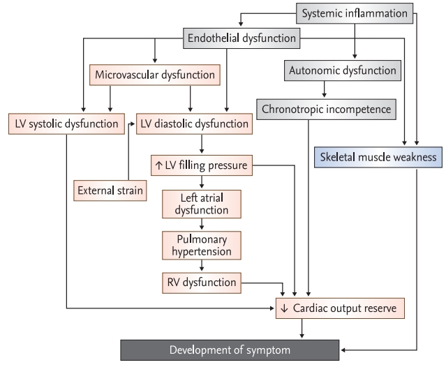
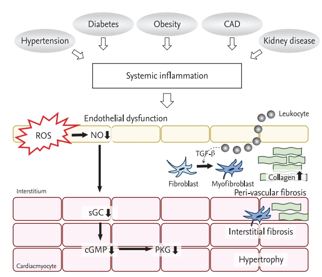
The pathophysiology of HFpEF is intricate and is not yet well understood. Patients with HFpEF are older, predominantly women, and have multiple comorbidities including hypertension, obesity, coronary artery disease (CAD), diabetes, anemia, atrial fibrillation (AF), renal insufficiency, and sleep apnea [13]. These comorbidities affect ventricular and vascular remodeling and are essential for the development of HFpEF (Fig. 1) [19,20]. These comorbidities induce a low-grade systemic inflammation, which induces endothelial dysfunction of systemic and coronary microvasculature [21,22]. The production and bioavailability of nitric oxide (NO) in the endothelium are impaired in HFpEF. Abnormalities in NO-cyclic guanosine monophosphate (cGMP) signaling, including soluble guanylate cyclase (sGC) activity and reduced protein kinase G (PKG) activity in cardiac myocytes, promote myocardial hypertrophy, increased stiffness of the myocardium, and interstitial fibrosis (Fig. 2) [21,23,24]. These result in left ventricular diastolic dysfunction (LVDD) and increased arterial stiffness.

Left ventricular diastolic dysfunction

LVDD is well-known pathophysiology of HFpEF. LVDD is caused by myocardial structural changes, microvascular dysfunction, systematic inflammation, and increased passive chamber stiffness [25]. The increased LVFP, the fundamental and pathologic hemodynamics of HFpEF, predominantly results from LVDD. The passive chamber stiffness is rapidly increased and the prolonged relaxation time is not shortened during exercise and these result in an increase of LVFP in HFpEF [26]. In the earlier stage, LVFP is normal but markedly increases during exercise, while in an advanced stage it increases continuously even at rest [27]. Elevated LVFP leads to elevation of left atrial (LA) pressure (passive LA hypertension) and contributes to an increase in pulmonary capillary hydrostatic pressure and an increase in vascular permeability, resulting in the development of interstitial edema [28]. Increased LVFP is closely related to the development of typical symptoms of HFpEF such as exercise intolerance and exertional dyspnea, symptom severity, and prognosis of HFpEF [29,30].

Left atrial dysfunction

Under the condition of prolonged increased LVFP, secondary structural and functional remodeling of the LA develops. The preservation of LA function might be crucial to the adaptation of HFpEF, which prevents pulmonary congestion and right ventricular (RV) dysfunction [31]. Impaired LA function is related to exacerbation of pulmonary congestion, a change in lung function, and the development and worsening of pulmonary venous hypertension [31-33]. In a recent meta-analysis, it was proven that impaired LA function is valuable for diagnosis as well as for estimating the prognosis of HFpEF [34]. Especially in HFpEF patients with AF, LA structural and functional remodeling is exacerbated, which is a major contributor to disease progression, such as the development of pulmonary hypertension (PH), and RV dysfunction [35].

Pulmonary hypertension and right ventricular dysfunction

The presence of PH in HFpEF patients is associated with poor prognosis [21,36]. PH not only develops from passive LA hypertension but also from increased pulmonary vascular resistance, which is derived from pulmonary vascular disorders such as pulmonary vasoconstriction and pulmonary vascular remodeling [36-38]. A considerable number of HFpEF patients had pulmonary vascular disorders and increased mortality compared to patients with pure passive PH [36]. Regardless of the type of PH, persisting PH eventually induces RV dysfunction. The development of RV dysfunction is associated with a markedly increased risk of mortality [39].

Extrinsic restraint

The extrinsic restraint on the heart by pericardium and epicardial fat could lead to an increase in LVFP. Epicardial fat, as an external restraint, induces an increase in intracavitary pressure [40] that could promote inflammation, fibrosis, and coronary microvascular dysfunction (CMD) [20].

Left ventricular systolic dysfunction

The left ventricular systolic dysfunction evaluated by tissue deformation imaging analysis is present at rest and worsens during exercise in HFpEF [41-43]. Abnormal ventricular-vascular coupling by arterial stiffening is regarded as the cause of subtle left ventricular systolic dysfunction. It results in a significantly impaired proportional increase in stroke volume output in response to exercise, which is one of the major causes of exercise intolerance in HFpEF. The increased left ventricular strain is closely associated with common comorbidities of HFpEF [44], and it has a prognostic value for CV mortality and HF hospitalization in HFpEF [42].

Coronary microvascular dysfunction

A considerable portion of patients presenting with symptoms of ischemia referred for coronary angiography are found to have evidence of ischemia but no significant stenosis of the coronary artery [45]. Most of these patients who have ischemia and no obstructive coronary artery disease (INOCA) have CMD [45]. In epidemiologic studies, INOCA was more prevalent in women and in patients with multiple comorbidities, which are similar to HFpEF [46]. Furthermore, patients with INOCA are likely to have preserved LVEF and a higher incidence of HF hospitalization [47]. Therefore, a link between CMD and HFpEF was strongly suggested [46], and the interaction between LVDD and CMD was clearly associated with an increased risk of HFpEF [48- 50]. CMD has become one of the major phenotypes and pathogenic cause of HFpEF.

Chronotropic incompetence

Many patients with HFpEF display impaired cardiac output reserve during exercise, despite normal values at rest [43]. Not only left ventricular systolic dysfunction, as mentioned above, but also chronotropic incompetence results in a reduced cardiac output reserve [43].

Skeletal muscle weakness

Recent studies have identified an important role of the skeletal muscle in HFpEF. In patients with HFpEF, skeletal muscle mass is decreased [51], the composition of skeletal muscle changes with increased fatty infiltration [52] and the capillary density within the muscle is reduced [53]. The oxidative metabolism of skeletal muscle fiber changes to a slow type and a decreased mitochondrial content and abnormal fusion of mitochondria in skeletal muscle are observed [54]. These changes reduce peak O2 uptake and result in exercise intolerance in patients with HFpEF.

TREATMENT

Currently, there are limited data on disease-modifying agents available for HFpEF, which improve clinical outcomes in randomized controlled trials (RCTs). Therefore, treatment guidelines, focused on optimal volume control using diuretics and proper management of risk factors and comorbidities are required. However, with increasing insight in the pathophysiology of HFpEF, various trials in treatment of HFpEF in accordance with the phenotype, have been published. The important trials of HFpEF treatment are described in Table 4.

TRADITIONAL PHARMACOLOGICAL STRATEGIES OF HF: BLOCKADE OF THE ACTIVATED NEUROHORMONAL SYSTEM

Renin-angiotensin-aldosterone system

Inappropriate activation of the renin-angiotensin-aldosterone system (RAAS) occurs in diseases that are associated with the development and progression of HFpEF, in common with HFrEF [55-58]. RAAS promotes the increase of arterial stiffness and myocardial stiffness and causes LVDD and LVH in HFpEF [55]. Therefore, several randomized clinical trials have been conducted to evaluate the prognostic value of RAAS blockade.
The angiotensin-converting enzyme inhibitor or angiotensin receptor blocker was not efficient for CV mortality and HF hospitalization in three large trials (CHARM-preserved [Candesartan in Heart Failure: Assessment of Reduction in Mortality and Morbidity], PEP-CHF [Perindopril in Elderly people with Chronic Heart Failure], and I-PRESERVED [Irbesartan in Heart Failure With Preserved Ejection Fraction]) [59-61]. They did not improve CV mortality, and only candesartan reduced HF hospitalization slightly [59]. A detailed summary of these trials is described in Table 4.
The Treatment of Preserved Cardiac Function Heart Failure With an Aldosterone Antagonist Trial (TOPCAT) is a large RCT to investigate the efficacy of aldosterone antagonists in symptomatic patients with HFpEF (LVEF ≥ 45%) [62]. The differences in the primary endpoint between spironolactone and placebo failed to reach a statistical significance; however, HF hospitalization was modestly decreased by spironolactone. Interestingly, in the subgroup analysis, spironolactone reduced primary outcome in patients with elevated natriuretic peptide levels at enrollment [62] in the Americas (hazard ratio [HR], 0.82; 95% CI, 0.69 to 0.98; p = 0.026) [63]. In Russia and Georgia, patients were enrolled by clinical judgment rather than having increased natriuretic peptides, and were relatively healthy and had lower compliance with study medication than in the Americas [64]. The result obtained in Russia/Georgia would not reflect a true response to spironolactone. For this reason, re-evaluation of the clinical efficacy of spironolactone in HFpEF is necessary. There is currently an ongoing trial in progress: Spironolactone Initiation Registry Randomized Interventional Trial in Heart Failure with Preserved Ejection Fraction (SPIRRIT, NCT 02901184), and their results are expected.
The angiotensin receptor-neprilysin inhibitor (ARNI) sacubitril-valsartan is a combination of inhibitors of neurohormonal activation and up-regulation of the adaptive natriuretic peptide pathway. In a phase-II study, (PARAMOUNT [65]) sacubitril-valsartan induced a greater decrease in N-terminal pro-B type natriuretic peptide (NT-proBNP), a larger reduction in LA size, and a greater improvement of symptoms than valsartan in patients with HFpEF. Therefore, the outcome trial, Efficacy and Safety of LCZ696 Compared to Valsartan, on Morbidity and Mortality in Heart Failure Patients With Preserved Ejection Fraction (PARAGON-HF) trial [66] was conducted in symptomatic HFpEF patients with increased natriuretic peptides. Despite a numerically lower event rate, the efficacy for HF hospitalization and CV death by sacubitril-valsartan approached but did not achieve a statistical significance (HR, 0.87; 95% CI, 0.75 to 1.01; p = 0.06). In post hoc analysis [13], the absolute risk reduction of sacubitril-valsartan was greatest in patients who were recently hospitalized within 1 month (approximately 25% to 30% risk reduction) and it gradually decreased with an increased interval from hospitalization. The sacubitril-valsartan might have alleviated the remaining neurohormonal activation after discharge. These data could provide clues for the initiation or switching time to sacubitril-valsartan in patients with HFpEF. The pre-specified analysis of outcomes by gender in the PARAGON-HF trial reported that the beneficial effect of sacubitril-valsartan was greater in women than in men (rate ratio [RR], 0.73, [95% CI, 0.59 to 0.90] in women vs. RR, 1.03, [95% CI, 0.84 to 1.25] in men, p interaction = 0.017) [67]. The possible reasons were further myocardial remodeling even in the same LVEF, more prominent age-related arterial stiffening in female patients with HFpEF, and differences in the signaling of natriuretic peptide [67]. Recently, Solomon et al. [68] reported the results of a pooled analysis of combined data from the Prospective Comparison of ARNI With ACEI to Determine Impact on Global Mortality and Morbidity in Heart Failure (PARADIGM-HF) [69] and PARAGON-HF [66] trials. The overall treatment benefit was at LVEF ≤ 42.5% and was maximized at lower ejection fraction. The sacubitril-valsartan was valuable in women with LVEF ≤ 60%, contrarily the threshold of LVEF at which efficacy of sacubitril-valsartan was highest was 45% to 50% in men. Therefore, it could be presumed that sacubitril-valsartan is effective for all patients with middle-ranged ejection fraction (HFmrEF). This result was consistent with the clinical characteristics of HFrEF and HFmrEF, which were similar and different from those of HFpEF [70].
There should be a careful application of RAAS blockade to patients with HFpEF because the phenotypes of patients in the real world are different from the inclusion criteria of trials. As the LVEF cutoff of trials varied, a considerable portion of registered patients to these trials might belong to HFmrEF. Neurohormonal activation is less prominent in HFpEF, rather than in HFmrEF or HFrEF. Furthermore, a large proportion of trial patients had already taken RAAS blocker (20% to 86% of the study population) and beta-blocker (55% to 80% of the study population) at enrollment [71], because of their comorbidities including CAD and arrhythmia and their neurohormonal system might be stabilized. Thus, it might leave a little room for the additional benefit from another RAAS inhibitor. Therefore, the messages of clinical trials should be interpreted and accepted judiciously.

Beta-adrenergic signaling

Beta-blockers are the cornerstone of the management of HFrEF [8] because of a significant improvement in mortality and morbidity [72]. Sympathetic activation induces an increment of HR and leads to a shortening of the left ventricular diastolic filling time. Therefore, slowing HR and reversal of sympathetic overactivation could provide a benefit in HFpEF. A lower heart rate at discharge was closely related to a survival benefit in patients with HFpEF [73]. Several observational studies reported a modest benefit of beta-blockers for survival in HFpEF [74-76]. However, in pre-specified sub-analysis of the Study of Effects of Nebivolol Intervention on Outcomes and Rehospitalization in Seniors With Heart Failure (SENIORS) [77] the effect of beta-blockers nebivolol was similar between patients with HFrEF (LVEF ≤ 35%) and patients with HFpEF (LVEF > 35%), although the efficacy of nebivolol to reduce mortality was not statistically significant in both the patient groups. In the Japanese Diastolic Heart Failure Study (J-DHF) [78], an RCT to evaluate the effect of carvedilol in HFpEF (LVEF > 40%), it did not decrease CV death and HF hospitalization. Even in several meta-analyses, the efficacy of beta-blockers for mortality as well as HF hospitalization, in HFpEF is still controversial [79-81]. Recently, a pre-specified TOPCAT trial reported that the use of beta-blockers was related to an increase in HF hospitalization and was not associated with CV mortality in HFpEF [82]. The explained mechanism of ineffectiveness, even harm, was due to an increase in central blood pressure (BP) and myocardial wall stress due to increment of left ventricular volume and pressure by prolonged diastolic filling time [83,84]. However, recently reported Korean registry data showed that the use of beta-blockers reduced all-cause mortality in patients with HFpEF (LVEF ≥ 40%) and acute exacerbation of HF [85]. This benefit existed in patients with global longitudinal strain (GLS) < 14%, but not in patients with GLS ≥ 14% [86]. The conflicting results regarding the efficacy of beta-blockers are still unknown. This is due to the difference in the definition of HFpEF, especially LVEF cut-off value, heart rate at enrollment, and their changes after medication and pre-existing comorbidities, such as AF and CAD [87]. Despite these, beta-blockers might be beneficial to patients with HFpEF, especially in patients with myocardial remodeling with reduced GLS.

DISEASE-MODIFYING PHARMACOLOGICAL STRATEGIES

As mentioned above, the deregulation of the NO-sGCcGMP-PKG pathway is a potentially key mechanism of HFpEF. Therefore, therapeutic approaches have been tried to use medications that act on this pathway, including nitrates, phosphodiesterase-5 inhibitors, and sCG stimulators. Several trials have evaluated the role of these agents in the pathogenesis and treatment of HFpEF, with limited success.

Organic and inorganic nitrates

In altered endothelium-cardiomyocyte signaling by inflammation, NO is absent in cardiomyocytes. Therefore, organic NO supplementation was supposed to be helpful for HFpEF due to the restoration of myocardial NO content as well as alleviation of increased arterial load [5]. In the Nitrate’s Effect on Activity Tolerance in Heart Failure with Preserved Ejection Fraction (NEAT-HFpEF) trial [88], organic nitrate isosorbide mononitrate did not improve QOL and NT-proBNP, even if the daily activity level was reduced and HF symptoms worsened in the isosorbide mononitrate group. The possible causes were excessive hypotension and decreased cardiac output due to preload reduction [89], the rapid development of tolerance, and endothelial dysfunction by organic nitrates [90]. In contrast, inorganic nitrate (NO3) has a different NO metabolism: the nitrate-nitrite pathway. The nitrate-nitrite pathway could be an important route to restore NO in HFpEF, especially in the presence of hypoxia and acidosis such as skeletal muscle during exercise [91]. In small trials, the delivery of inorganic nitration via NO3--rich beetroot juice improved the exercise capacity of HFpEF patients [92,93]. The administration of sodium nitrite via infusion or inhalation improved cardiac output reserve, ventricular filling pressure, and pulmonary artery pressure during exercise in HFpEF. However, Inorganic Nitrite Delivery to Improve Exercise Capacity in HFpEF (INDIE-HFpEF) trial [94] showed that inhaled inorganic nitrite for 4 weeks failed to improve exercise capacity and QOL. The nitrite inhalation with its short-acting nature could be the cause of this result, as other administrations of inorganic nitrite, which provide a persistent and higher level of NO, achieved positive results. It is necessary to confirm the effectiveness of NO3 in larger and long-term trials.

sGC stimulators

The sGC stimulators (riociguat and vericiguat) enhance cGMP production by acting on the NO receptor of sGC and activating sGC to generate cGMP. In Acute Hemodynamic Effects of Riociguat in Patients with Pulmonary Hypertension Associated with Diastolic HF (DILATE-1) [95] and Soluble guanylate Cyclase stimulatoR in heArT failurE patients with PRESERVED EF (SOCRATES-PRESERVED) [96] trials, sGC stimulators showed limited improvement in hemodynamic and echocardiographic parameters.

Sildenafil

Despite the possible positive effect of sildenafil in HFrEF [97], two separate studies for evaluating the efficacy of sildenafil in HFpEF with PH or RV dysfunction failed to show improvement in exercise capacity and QOL, and sildenafil was associated with impairment of renal function and increment of neurohormone level [98,99].

Other PKG-stimulating drugs, sacubitril

PKG is an intrinsic suppressor of ventricular hypertrophy and interstitial fibrosis [5]; therefore, stimulation of PKG is a potential therapy in HFpEF. cGMP is mandatory to stimulate PKG. As mentioned above, cGMP is synthesized by sGC via the NO-sGC-cGMP-PKG pathway and is generated by receptor guanyl cyclase linked to natriuretic peptide receptors. Therefore, the sacubitril, a neprilysin inhibitor, stimulates PKG by elevating the level of natriuretic peptides. The efficacy of ARNI in HFpEF is described above.

NON-PHARMACOLOGICAL THERAPY

Devices targeted to high LVFP

Pressure monitoring

As mentioned above, increased LVFP is a hallmark of the pathophysiology of HFpEF. PCWP is a hemodynamic parameter that reflects LA pressure and LVFP. A therapeutic strategy of hemodynamic monitoring accompanied by early therapeutic intervention might improve the clinical outcome of HFpEF patients. The CardioMEMS Heart Sensor Allows Monitoring of Pressure to Improve Outcomes in NYHA Class III Heart Failure Patients (CHAMPION) trial demonstrated that active reduction of LVFP guided by an assessment of central hemodynamics in HF patients regardless of LVEF significantly decreased HF hospitalization [100]. This finding persisted in additional analysis in HFpEF patients (LVEF ≥ 50%) [101] and real-word data [102].

Interatrial septal shunt

The other device targeted to high LVFP is an interatrial septal shunt to reduce LA pressure. In the Reduce Elevated Left Atrial Pressure in Patients with Heart Failure (REDUCE LAP-HF) study, an interatrial shunt was efficient in decreasing LAP and improving functional capacity [103] and these efficacies were confirmed in a long-term study with the improvement of hemodynamic data during exercise without significant complication [104,105]. The interatrial shunt might be helpful in the management of HFpEF, especially high LA filling pressure due to LA dysfunction, even though further studies to evaluate the long-term improvement are required.

Pacing

Chronotropic incompetence contributes to impaired cardiac output reserve, as described above. The restored normal heart rate response during exercise by pacemaker might be beneficial to HFpEF with chronotropic incompetence. The Rate-Adaptive Atrial Pacing In Diastolic Heart Failure (RAPID-HF) trial is ongoing to evaluate the impact of rate-adaptive atrial pacing on exercise capacity (NCT 02145351).

PREVENTIVE STRATEGIES FOR HFPEF: MANAGEMENT OF COMORBIDITIES

Hypertension

Hypertension is the most common comorbidity of HFpEF patients. In many previous RCTs of antihypertensive medication, optimal treatment of hypertension reduced the incidence of HF [106]. In HFpEF, the additional benefit of lowering BP is uncertain. As mentioned above, there was discordance between BP lowering and clinical outcome in large trials with neurohormonal inhibitors in HFpEF, even though neurohormonal inhibition had a favorable effect on ventricular hypertrophy, interstitial fibrosis, and myocardial stiffness [5]. Nevertheless, a recent meta-analysis showed that BP lowering was closely related to the reduction in HF hospitalization rather than all-cause mortality in HFpEF patients [107]. Furthermore, optimal BP control is important to prevent other major CV outcomes, including stroke and CAD, even in HFpEF patients.

Sodium-glucose cotransporter-2 inhibitors

The diabetes is prevalent in 20% to 40% of HFpEF patients [21]. Three RCTs on sodium-glucose cotransporter-2 (SGLT2) inhibitors in type 2 diabetes with high CV risk or established CV disease demonstrated a valuable effect of SGLT2 inhibitors [108]. McMurray et al. [109] reported that dapagliflozin significantly reduced worsening HF or CV death in HFrEF patients. The benefit of dapagliflozin on HF was similar between the patients with and without diabetes. The potential mechanism of the beneficial effect of SGLT2 inhibitors is not only to decrease intravascular volume via osmotic diuresis and natriuresis but also to reduce neurohormonal activation [110]. SGLT2 inhibitors increase metabolic efficiency and myocardial energy supply [110]. Furthermore, SGLT2 inhibitors induce a decrease in oxidative stress and fibrosis, an increase of endothelial function, and vascular compliance, which may be favorable in HFpEF [110,111]. The diastolic parameter was improved by the administration of SGLT2 inhibitors in patients with type 2 diabetes patients [112,113]. The RCTs for the efficacy of SGLT2 inhibitor in HFpEF (EMPagliflozin outcomE tRial in Patients With chrOnic heaRt Failure With Preserved Ejection Fraction [EMPEROR-Preserved; NCT 03057951], Effect of EMpaglifozin on ExeRcise ability and heart failure symptoms, In patients with chronic heArt faiLure with preserved ejection fraction [EMPERIAL-preserved; NCT 03448406], Dapagliflozin Evaluation to Improve the LIVEs of Patients With PReserved Ejection Fraction Heart Failure [DELIVER; NCT 03619213], dapagliflozin in PRESERVED Ejection Fraction Heart Failure [PRESERVED-HF; NCT 03030235]) are ongoing and we look forward to the results of these studies shortly.

Lifestyle modification

Lifestyle modifications, including exercise, weight reduction, and dietary composition, are beneficial in preventing and treating HF [114]. However, the potential benefit of lifestyle modification has not been fully elucidated in RCT or cohort studies. Nevertheless, the efficacy of exercise was reported in several studies that included endurance and resistance training, improved exercise capacity, and QOL in HFpEF [115,116]. About half of HFpEF patients have obesity, and an increase in body adiposity triggers systemic inflammation and impairment of cardiac, vascular, and skeletal muscle function [5]. Kitzman et al. [117] demonstrated that caloric reduction during 20 weeks significantly improved peak O2 consumption, symptoms, and QOL in older and obese patients. The additive benefit was derived by a combination of caloric restriction and exercise. Lifestyle modification might be important for the management of HFpEF as well as a cardiometabolic syndrome, representative comorbidity of HFpEF. Further investigation to prove the beneficial effect of lifestyle modification in HFpEF is required.

CONCLUSIONS

HFpEF is the most common form of HF with an increase in the elderly population, and its prognosis has not yet improved. The trials of medications have been neutral or less effective in terms of their primary outcomes. The possible explanations for this result are incomplete understanding of the pathophysiology, heterogeneity of the study populations, lack of universal diagnostic criteria of HFpEF, unconnected pathophysiological mechanisms related to treatment, and suboptimal designs for statistical power of the trials. Currently, the concept of HFpEF is evolving; HFpEF is a multifaceted syndrome. The pathophysiology of HFpEF is multifactorial, with several mechanisms and comorbidities involved, and different from that of HFrEF. HFpEF results from a complex interaction of heart, vasculature, and peripheries: LVDD, systolic functional reserve, autonomic imbalance, and macro- and microvasculature response to increased oxygen demand. Substantial heterogeneity exists in these interactions and dominance in each patient with HFpEF. Therefore, the definition of HFpEF by LVEF is bound to be limited. Recently, new diagnostic strategies have been proposed that appear promising; however, further validation is required. Treatment of HFpEF needs to be approached differently according to various phenotypes of HFpEF, and several treatment trials are on the way as per different therapeutic approaches (summarized in Table 5). In our daily clinical practice, we need to understand the nature of the diversity of HFpEF and approach each patient with HFpEF individually.

Conflict of Interest

No potential conflict of interest relevant to this article was reported.

Figure 1.
Schematic pathophysiology of heart failure with preserved ejection fraction. LV, left ventricular; RV, right ventricular.
kjim-2020-104f1.jpg
Figure 2.
Cellular mechanism of heart failure with preserved ejection fraction. CAD, coronary artery disease; ROS, reactive oxygen species; NO, nitric oxide; TGF-β transforming growth factor-β; sGC, soluble guanylate cyclase; cGMP, cyclic quanosine monophosphate; PKG, protein kinase G.
kjim-2020-104f2.jpg
Table 1.
Diagnostic of HFpEF [7-9]
Current guideline of HFpEF
2016 KSHF guideline [7] 2016 ESC guideline [8] 2013 AHA guideline [9]
Clinical manifestation Symptoms and signs of HF Symptoms and signs of HF Symptoms and signs of HF
LVEF ≥ 50% ≥ 50% ≥ 50%
Natriuretic peptides BNP ≥ 35 pg/mL or NT-proBNP ≥ 125 pg/mL BNP > 35 pg/mL or NT-proBNP > 125 pg/mL
Imaging Abnormal LVDD Relevant structural heart disease (LVH and/or LAE) Abnormal LVDD
LVDD
-LAVI > 34 mL/m2
-LVMI ≥ 115/95 g/m2 (M/W)
-E/e’ ≥ 13
-Mean e’< 9 cm/sec

HFpEF, heart failure with preserved ejection fraction; KSHF, Korean Society of Heart Failure; ESC, European Society of Cardiology; AHA, American Heart Association; HF, heart failure; LVEF, left ventricular ejection fraction; BNP, B-type natriuretic peptide; NT-proBNP, N-terminal pro-B type natriuretic peptide; LVDD, left ventricular diastolic dysfunction; LVH, left ventricular hypertrophy; LAE, left atrial enlargement; LAVI, left atrial volume index; LVMI, left ventricular mass index; M/W, men/women.

Table 2.
Diagnostic of H2FpEF [14]
Clinical variable H2FpEF score [14] Point
H2 Heavy BMI > 30 kg/m2 2
Hypertension Antihypertensive medication ≥ 2 1
F Atrial fibrillation Paroxysmal or persistent 3
P Pulmonary hypertension Doppler echocardiographic estimated PASP > 35 mmHg 1
E Elder Age > 60 years 1
F Filling pressure Doppler echocardiographic E/e’ > 9 1
Sum 0–9

BMI, body mass index; PASP, pulmonary artery systolic pressure.

Table 3.
Diagnostic of HFA-PEFF [15]
HFA-PEFF score [15]
Major
Minor
Value Point Value Point
Functional Septal e’ < 7 cm/sec or lateral e’ < 10 cm/sec 2 Avergaed E/e’ 9–14 1
or or
Averaged E/e’ ≥ 15 GLS < 16%
or
TR Vmax > 2.8 m/sec (PASP > 35 mmHg)
Morphological LAVI > 34 mL/m2 2 LAVI 29–34 mL/m2 1
or or
LVMI ≥ 149/122 g/m2 (M/W) ± RWT > 0.42 LVMI ≥ 115/95 m2 (M/W)
or
RWT > 0.42
or
LV wall thickness ≥ 12 mm
Biomarker (SR) NT-proBNP > 220 pg/mL 2 NT-proBNP > 660 pg/mL 1
or or
BNP > 80 pg/mL BNP > 240 pg/mL
(AF) NT-proBNP 125–220 pg/mL 2 NT-proBNP 365–660 pg/mL 1
or or
BNP 35–80 pg/mL BNP 105–240 pg/mL

≥ 5 points: heart failure with preserved ejection fraction; 2–4 points: exercise stress test or invasive hymodynamic measurement.

HFA-PEFF, heart Failure Association-PEFF; TR, tricuspid regurgitation; PASP, pulmonary artery systolic pressure; GLS, global longitudinal strain; LAVI, left atrial volume index; LVMI, left ventricular mass index; M/W, men/women; RWT, regional wall thickness; LV, left ventricular; SR, sinus rhythm; NT-proBNP, N-terminal pro-B type natriuretic peptide; BNP, B type natriuretic peptide; AF, atrial fibrillation.

Table 4.
Clinical trials of pharmacological and non-phamacological therapy in HFpEF
Trials, year (no. of patients) Interventions Inclusion criteria Characteristics of study population Primary endpoint Trial result
Renin-angiotensin-aldosterone system
CHARM-preserved 2003 (n = 2,023) [58] Candesartan NYHA II–IV HF with prior cardiac hospitalization Mean age: 67 ± 11 years, 40% female 65% hypertension, 28% diabetes CV death or HF hospitalization No benefit for primary endpoint
Mean LVEF: 54% ± 9.4% Reduction of HF hospitalization
LVEF ≥ 40% Median FU: 36.6 months
PEP-CHF 2006 (n = 850) [59] Perindopril Age ≥ 70 years Mean age: 76 ± 5 years, 55% female 79% hypertension, 20% diabetes All cause mortality or HF hospitalization No benefit for primary endpoint
Diastolic heart failure Mean LVEF: 64% Reduction of HF hospitalization
Median FU: 2.1 years Improvement of symptom and exercise capacity
I-PRESERVED 2008 (n = 4,128) [60] Irbesartan Age ≥ 60 years Mean age: 72 ± 7 years, 60% female 88% hypertension, 27% diabetes Death from any cause or hospitalization for CV cause No benefit for primary endpoint
NYHA II–IV Mean LVEF: 60% ± 9% No improvement of QOL
LVEF ≥ 45% Mean FU: 49.5 months
TOPCAT 2014 (n = 3,445) [63] Spironolactone HF with history of HF hospitalization within 12month and elevated BNP within 60 days Median age: 69 years, 52% female 91% hypertension, 32% diabetes CV death or cardiac arrest or HF hospitalization No benefit for primary endpoint
Median LVEF: 56% Reduction of HF hospitalization
LVEF ≥ 45% Mean FU: 3.3 years
PARAGON-HF 2019 (n = 4,822) [66] Sacubitrilvalsartan NYHA II–IV Mean age: 73 ± 8 years, 52% female 96% hypertension, 43% diabetes HF hospitalization or CV death No benefit for primary endpoint
LVEF ≥ 45% Mean LVEF: 60% ± 9% Reduce primary endpoint in subgroup (patients wuth LVEF below median [≤ 57%] and women)
Elevated NPs Total FU time up to 57 months
Beta-blocker
SENIORS 2009 (n = 752) [80] Nebivolol CHF history Mean age: 76 ± 5 years, 51% female 78% hypertension, 24% diabetes All cause mortality or CV hospitalization No benefit for primary endpoint
HF hospitalization within 12month Mean LVEF: 49% ± 10%
FU: 21 months
J-DHF 2013 (n = 245) [81] Carvedilol LVEF ≥ 40% Mean age: 76 ± 5 years, 55% female 80% hypertension, 33% diabetes CV death or HF hospi talization No benefit for primary endpoint
Mean LVEF: 63% ± 11% Reduce primary endpoints in patients with higher dose
Median FU: 3.2 years
Disease-modifying agents
NEAT-HFpEF 2015 (n = 110) [88] Isosorbide mononitrate (organic nitrate) LVEF ≥ 50% Mean age: 69 ± 9 years, 49% female 88% hypertension, 43% diabetes Daily activity level Decreased activity and worsen QOL
HF with objective evidence (≥ 1) Mean LVEF: 62% ± 8%
- HF hospitalization with congestion , increased LVEDP or PCWP, elevated NP, LVDD on ECHO Drug intervention for 6 weeks
INDIE-HFpEF 2018 (n = 105) [94] Nebulized inorganic nitrate Age ≥ 40 years Mean age: 68 ± 9 years, 68% female 81% hypertension, 38% diabetes Peak O2 consumption No improvement in exercise capacity
LVEF ≥ 50% Mean LVEF: 61% ± 5%
HF with objective evidence (≥ 1) Drug intervention for 4 weeks
- HF hospitalization with congestion, increased LVEDP or PCWP, elevated NP, LVDD on ECHO
DILATE-1 2014 (n = 21) [95] Riociguat (sGC stimulator) HFpEF with PH Mean age: 68 ± 9 years, 68% female 81% hypertension, 44% diabetes Peak decrease in mPAP No significant effect on mPAP
LVEF ≥ 50%
mPAP ≥ 25 mmHg Mean LVEF: 62% ± 7%
PAWP > 15 mmHg at rest
SOCRATES-PRESERVED 2017 (n = 477) [96] Vericiguat (sGC stimulator) NYHA II–IV Mean age: 73 ± 10 years, 48% female 49% diabetes Change of NT-proBNP and LAV No signigicant change of NT-pro BNP and LAV
LVEF ≥ 45% Mean LVEF: 57%
Elevated NPs Drug intervention for 4 weeks
History of HF hospitalization or IV diuretics within 4 weeks
RELAX 2016 (n = 216) [98] Sildenafil HFpEF with RVD and RV-RA coupling Mean age: 68 ± 9 years, 68% female 81% hypertension, 38% diabetes Peak oxygen uptake No improvement of RV function, exercise capacity and ventilatory efficiency
NYHA II–IV
LVEF ≥ 50% Mean LVEF: 61% ± 5%
Evidence of HF (≥ 1) Drug intervention for 4 weeks
- HF hospitalization, elevated LVFP and LAE
Non-pharmacological therapy
CHAMPION 2011 (n = 119) [101] Wireless implantable hemodynamic monitoring HF with NYHA III Mean age: 66 ± 12 years, 40% female 82% hypertension, 58% diabetes HF hospitalization Reduction of HF hospitalization
LVEF ≥ 40% Drug intervention for 6 months
REDUCE LAP-HF 2016 (n = 66) [103] Interatrial shunt device Age ≥ 40 years Mean age: 69 ± 8 years, 65% female 81% hypertension, 33% diabetes Successful device implantation Safe
LVEF ≥ 40% Drug intervention for 6 months Reduction of PCWP Reduction of LAP during exercise during exercise
Exercise PCWP ≥ 25 mmHg, PCWP-RAP gradient ≥ 5 mmHg
REDUCE LAP-HF I 2018 (n = 94) [104] Interatrial shunt device NYHA III, IV Mean age: 70 ± 9 years, 50% female 82% hypertension, 55% diabetes Exercise PCWP Reduction of PCWP during exercise
LVEF ≥ 40%
Exercise PCWP ≥ 25 mmHg, PCWP-RAP gradient ≥ 5 mmHg Drug intervention for 1 months
Manage of comorbidities
Ex-DHF 2011 (n = 64) [115] Endurance/resistance training Age ≥ 45 years Mean age: 65 ± 7 years, 56% female 86% hypertension, 14% diabetes Peak VO2 Improvement of exercise capacity and QOL
NYHA II–III
LVEF ≥ 50% Drug intervention for 3 months
CV risk factor (overweight, diabetes, hypertension, hyperlipidemia, smoking)
SECRET-1 2016 (n = 200) [117] Caloric restriction HF with obesity Mean age: 61 ± 5 years, 56% female 95% hypertension, 35% diabetes Peak VO2 Increased of peak oxygen uptake both caloric restriction and aerobic exercise training with addictive value
Age ≥ 60 years Disease-specific QOL
Aerobic exercise training BMI ≥ 30 Intervention for 3 months
LVEF ≥ 50%

HFpEF, heart failure with preserved ejection fraction; CHARM, Candesartan in Heart Failure: Assessment of Reduction in Mortality and Morbidity; NYHA, New York Heart Association; HF, heart failure; LVEF, left ventricular ejection fraction; FU, follow-up; CV, cardiovascular; PEP-CHF, Perindopril in Elderly people with Chronic Heart Failure; I-PRESERVED, Irbesartan in Heart Failure With Preserved Ejection Fraction; QOL, quality of life; TOPCAT, Treatment of Preserved Cardiac Function Heart Failure With an Aldosterone Antagonist Trial; BNP, brain natriuretic peptide; PARAGON-HF, Efficacy and Safety of LCZ696 Compared to Valsartan, on Morbidity and Mortality in Heart Failure Patients With Preserved Ejection Fraction; NP, natriuretic peptide; SENIORS, Study of Effects of Nebivolol Intervention on Outcomes and Rehospitalization in Seniors With Heart Failure; CHF, congestive heart failure; J-DHF, Japanese Diastolic Heart Failure Study; NEAT-HFpEF, Nitrate’s Effect on Activity Tolerance in Heart Failure with Preserved Ejection Fraction; LVEDP, left ventricular end diastolic pressure; PCWP, pulmonary capillary wedge pressure; LVDD, left ventricular diastolic dysfunction; ECHO, echocardiography; INDIE-HFpEF, Inorganic Nitrite Delivery to Improve Exercise Capacity in HFpEF; DILATE-1, Acute Hemodynamic Effects of Riociguat in Patients with Pulmonary Hypertension Associated with Diastolic HF; sGC, soluble guanylate cyclase; PH, pulmonary hypertension; mPAP, mean pulmonary artery pressure; PAWP, pulmonary arterial wedge pressure; SOCRATES-PRESERVED, Soluble guanylate Cyclase stimulatoR in heArT failurE patients with PRESERVED EF; NT-proBNP, N-terminal pro-B-type natriuretic peptide; LAV, left atrial volume; RELAX, phosphodiesterase-5 inhibition to improve clinical status and exercise capacity in diastolic heart failure; RVD, right ventricular dysfunction; RV-RA, right ventricular-right atrial; LVFP, left ventricular filling pressure; LAE, left atrial enlargement; CHAMPION, CardioMEMS Heart Sensor Allows Monitoring of Pressure to Improve Outcomes in NYHA Class III Heart Failure Patients; REDUCE LAP-HF, Reduce Elevated Left Atrial Pressure in Patients with Heart Failure; RAP, right atrial pressure; Ex-DHF, enhancement of physical activity in elderly patients with diastolic heart failure; VO2, oxygen uptake; SECRET-1, Study of the Effect of Caloric Restriction and Exercise Training in patients with heart failure and a normal ejection fraction-1; BMI, body mass index.

Table 5.
Ongoing trials in HFpEF
Name ClinicalTrials.gov identifier Intervention Inclusion criteria Primary endpoint Estimated completion
SPIRRIT-HFPEF NCT 02901184 Spironolactone Age ≥ 50 years Time to CV death or first HF hospitalization. 6/2,022
LVEF ≥ 40% in last 12 months
Stable HF defined by symptoms ans signs
NT-proBNP > 300 ng/L in SR or > 750 ng/L in AF
PARAGLIDE-HF NCT 03988634 Sacubitril/valsartan Age ≥ 40 years Proportional change in NT-proBNP at weeks 4 and 8 9/2,021
LVEF ≥ 40%
Elevated NT-proBNP
Currently hospitalized for or within 30 days following discharge of an acute decompensated HFpEF admission
PRISTINE-HF NCT 04128891 Sacubitril/valsartan Age ≥ 40 years Improvement in microvascular function and ischaemia, as assessed by OS-CMR 2/2,024
LVEF ≥ 45%
NYHA II–III
Structural heart disease: LAE, LVH
Elevated NT-proBNP
KNO3CK OUT HFPEF NCT 02840799 Potassium nitrate (KNO3) Age 18–90 years Peak VO2 12/2,020
LVEF ≥ 50%
Potassium chloride (KCl) NYHA II–III
Elevated filling pressure on echocardiography
CAPACITY-HFpEF NCT 03254485 IW-1973 (sGC stimulator) Age ≥ 45 years Treatment-emergent adverse event Complete
LVEF ≥ 40% Peak VO2 at week 12
Peak VO2 < 80% of age-and sex-adjusted normal value
Medical history of HF
At least 2 of comorbidities (diabetes, hypertension, BMI > 30 kg/m2, age ≥ 70 years)
SATELLITE NCT 03756285 AZD4831 (myeloperoxidase inhibitor) Age 45–85 years Change of myeloperoxidase activity 10/2,020
LVEF ≥ 40%
NYHA II–IV
Elevated NT-proBNP
One of the following:
- HF hospitalization within 12 months
- Structural heart disease on echocardiography
- PCWP at rest > 15 or > 25 mmHg at exercise
- E/e’ ratio ≥ 13 at rest
NCT 03611153 Oral myeloperoxidase inhibitor Age ≥ 30 years Exercise PCWP 1/2,021
LVEF ≥ 50%
NYHA II–III
Evated filling pressures at rest (PCWP ≥ 15) or with exercise (PCWP ≥ 25)
DELIVER NCT 03619213 Dapaglifqlozin Age ≥ 40 years Time to the first occurrence of any of the components of this composite: (1) CV death; (2) hospitalisation for HF; (3) urgent HF visit 6/2,021
NYHA II–IV
LVEF > 40% and evidence of structural heart disease
DETERMINE-preserved NCT 03877224 Dapagliflozin Age ≥ 40 years Change of 6MWD at week 16 7/2,020
NYHA II–IV Exercise capacity
LVEF > 40% and evidence of structural heart disease HF symptom relieve
Elevated NT-proBNP
EMPEROR-Preserved NCT 03057951 Empagliflozin Age ≥ 18 years Time to first event of adjudicated CV death or adjudicated HF hospitalization 11/2,020
NYHA II–IV
LVEF > 40% and evidence of structural heart disease
Elevated NT-proBNP
FAIR-HFpEF NCT 03074591 Ferric carboxymaltose (IV iron) Age ≥ 18 years Exercise capacity-change of 6MWT at week 52 7/2,021
NYHA II–III
LVEF > 45%
One of the following
- Elevated NT-proBNP or previous hospitalization
Evidence of LVDD
ID (Hb 9–14 g/dL) with ferritin < 100 ng/mL or ferritin 100–299 with transferrin saturation < 20%
INABLE-Training NCT 02713126 Exercise training Age ≥ 40 years Change of peak VO2 at week12 12/2,020
Oral sodium nitrite NYHA II–IV
LVEF ≥ 50%
One of the following:
- Previous HF hospitalization with radiographic evidence
- Catheterization documented elevated filling pressures
- Elevated natruretic peptide
- Echo evidence of LVDD/elevated filling pressures
RAPID-HF NCT 02145351 Rate-adaptive atrial pacing Age ≥ 18 years Change of peak VO2 at week 4 5/2,021
NYHA II–III
LVEF > 40%
Baseline sinus rhythm
Documented chronotropic incompetence
CCM-HFpEF NCT 03240237 Cardiac contractility modulation Age 40–80 years Mean change from baseline to 24 weeks in Kansas City Cardiomyopathy questionnaire 12/2,023
LVEF ≥ 50%
NYHA II–III
Elevated NTproBNP
Has the following:
- LAVI ≥ 34 mL/m² or LVH > 12mm and either
- E/e’ ≥ 13 OR or septal e’ < 7 cm/sec (or lateral e’ < 10 cm/sec)
NCT 00327649 Low level transcutaneous vagus nerve stimulation HFpEF, defined as signs and symptoms of heart failure Diastolic dysfunction 8/2,020
Plus 2 of the following 4 comorbidities
- Age ≥ 65years, diabetes, hypertension, obesity
NCT 02499601 CORolla™ TAA device Age ≥ 18 years Number of participants with all-cause mortality and serious adverse events 9/2,020
NYHA III, IV
LVEF > 50%
PCWP > 15 mmHg by RHC
Echocardiographic criteria:
- LVEDVI < 97 mL/m2, E/e’ > 12, LAVI > 29 mL/m2

HFpEF, heart failure with preserved ejection fraction; SPIRRIT-HFPEF, Spironolactone Initiation Registry Randomized Interventional Trial in Heart Failure with Preserved Ejection Fraction ; LVEF, left ventricular ejection fraction; HF, heart failure; NT-proBNP, N-terminal pro-B-type natriuretic peptide; SR, sinus rhythm; AF, atrial fibrillation; CV, cardiovascular; PARAGLIDE-HF, A Multicenter, Randomized, Double-blind, Double-dummy, Parallel Group, Active Controlled Study to Evaluate the Effect of Sacubitril/Valsartan (LCZ696) Versus Valsartan on Changes in NT-proBNP and Outcomes, Safety, and Tolerability in HFpEF Patients With Acute Decompensated Heart Failure (ADHF) Who Have Been Stabilized During Hospitalization and Initiated In-hospital or Within 30 Days Post-discharge; PRISTINE-HF, PRospectIve Study of Sacubitril/ValsarTan on MyocardIal OxygenatioN and Fibrosis in PatiEnts With Heart Failure and Preserved Ejection Fraction; NYHA, New York Heart Association; LAE, left atrial enlargement; LVH, left ventricular hypertrophy; OS-CMR, oxygenation sensitive cardiac magnetic resoance; KNO3CK OUT HFPEF, Effect of KNO3 Compared to KCl on Oxygen UpTake in Heart Failure With Preserved Ejection Fraction; VO2, oxygen uptake; CAPACITY-HFpEF, A Multicenter, Randomized, Double-blind, Placebo-controlled, Phase 2 Study Evaluating the Safety and Efficacy of Different Doses of IW-1973 Over 12 Weeks in Patients With Heart Failure With Preserved Ejection Fraction; sGC, soluble guanylate cyclase; BMI, body mass index; SATELLITE, Safety and Tolerability Study of AZD4831 in Patients With Heart Failure; PCWP, pulmonary capillary wedge pressure; DELIVER, Dapagliflozin Evaluation to Improve the LIVEs of Patients With PReserved Ejection Fraction Heart Failure; DETERMINE-preserved, Dapagliflozin Effect on Exercise Capacity Using a 6-minute Walk Test in Patients With Heart Failure With Preserved Ejection Fraction; 6MWD, 6-minute walk distane; EMPEROR-Preserved, EMPagliflozin outcomE tRial in Patients With chrOnic heaRt Failure With Preserved Ejection Fraction; FAIR-HFpEF, Effect of IV Iron in Patients With Heart Failure With Preserved Ejection Fraction; IV, intravenous; LVDD, left ventricular diastolic dysfunction; Hb, hemoglobin; 6MWT, 6-minute walk teast; INABLE-traing, Inorganic Nitrite to Amplify the Benefits and Tolerability of Exercise Training in Heart Failure With Preserved Ejection Fraction; RAPID-HF, Rate-Adaptive Atrial Pacing In Diastolic Heart Failure; CCM-HFpEF, Cardiac Contractility Modulation Therapy in Subjects With Heart Failure With Preserved Ejection Fraction; RHC, right heart catheterization; LAVI, left atrial volume index; LVEDVI, left ventricular end-diastolic volume index.

REFERENCES

1. Kitzman DW, Gardin JM, Gottdiener JS, et al. Importance of heart failure with preserved systolic function in patients > or = 65 years of age. CHS Research Group. Cardiovascular Health Study. Am J Cardiol 2001;87:413–419.
crossref pmid
2. Heidenreich PA, Albert NM, Allen LA, et al. Forecasting the impact of heart failure in the United States: a policy statement from the American Heart Association. Circ Heart Fail 2013;6:606–619.
crossref pmid pmc
3. Chamberlain AM, Dunlay SM, Gerber Y, et al. Burden and timing of hospitalizations in heart failure: a community study. Mayo Clin Proc 2017;92:184–192.
crossref pmid pmc
4. Fonarow GC, Stough WG, Abraham WT, et al. Characteristics, treatments, and outcomes of patients with preserved systolic function hospitalized for heart failure: a report from the OPTIMIZE-HF Registry. J Am Coll Cardiol 2007;50:768–777.
crossref pmid
5. Shah SJ, Kitzman DW, Borlaug BA, et al. Phenotype-specific treatment of heart failure with preserved ejection fraction: a multiorgan roadmap. Circulation 2016;134:73–90.
crossref pmid pmc
6. Upadhya B, Kitzman DW. Heart failure with preserved ejection fraction: new approaches to diagnosis and management. Clin Cardiol 2020;43:145–155.
crossref pmid
7. Kim MS, Lee JH, Kim EJ, et al. Korean guidelines for diagnosis and management of chronic heart failure. Korean Circ J 2017;47:555–643.
crossref pmid pmc
8. Ponikowski P, Voors AA, Anker SD, et al. 2016 ESC guidelines for the diagnosis and treatment of acute and chronic heart failure: the task force for the diagnosis and treatment of acute and chronic heart failure of the European Society of Cardiology (ESC). Developed with the special contribution of the Heart Failure Association (HFA) of the ESC. Eur J Heart Fail 2016;18:891–975.
crossref pmid
9. Yancy CW, Jessup M, Bozkurt B, et al. 2013 ACCF/AHA guideline for the management of heart failure: a report of the American College of Cardiology Foundation/American Heart Association Task Force on Practice Guidelines. J Am Coll Cardiol 2013;62:e147–e239.
crossref pmid
10. Michaud M, Maurin V, Simon M, Chauvel C, Bogino E, Abergel E. Patients with high left ventricular filling pressure may be missed applying 2016 echo guidelines: a pilot study. Int J Cardiovasc Imaging 2019;35:2157–2166.
crossref pmid pdf
11. Richards AM. N-terminal B-type natriuretic peptide in heart failure. Heart Fail Clin 2018;14:27–39.
crossref pmid
12. Abergel E, Lafitte S, Mansencal N. Evaluation of left ventricular filling pressure: Updated recommendations lack new evidence and have severe interpretation issues. Arch Cardiovasc Dis 2018;111:707–711.
crossref pmid
13. Vaduganathan M, Claggett BL, Desai AS, et al. Prior heart failure hospitalization, clinical outcomes, and response to sacubitril/valsartan compared with valsartan in HFpEF. J Am Coll Cardiol 2020;75:245–254.
crossref pmid
14. Reddy YNV, Carter RE, Obokata M, Redfield MM, Borlaug BA. A simple, evidence-based approach to help guide diagnosis of heart failure with preserved ejection fraction. Circulation 2018;138:861–870.
crossref pmid pmc
15. Pieske B, Tschope C, de Boer RA, et al. How to diagnose heart failure with preserved ejection fraction: the HFAPEFF diagnostic algorithm: a consensus recommendation from the Heart Failure Association (HFA) of the European Society of Cardiology (ESC). Eur Heart J 2019;40:3297–3317.
crossref pdf
16. Segar MW, Patel KV, Berry JD, Grodin JL, Pandey A. Generalizability and implications of the H<sub>2</sub>FPEF score in a cohort of patients with heart failure with preserved ejection fraction. Circulation 2019;139:1851–1853.
crossref pmid
17. Sueta D, Yamamoto E, Nishihara T, et al. H2FPEF score as a prognostic value in HFpEF patients. Am J Hypertens 2019;32:1082–1090.
crossref pmid pdf
18. Barandiaran Aizpurua A, Sanders-van Wijk S, Brunner-La Rocca HP, et al. Validation of the HFA-PEFF score for the diagnosis of heart failure with preserved ejection fraction. Eur J Heart Fail 2020;22:413–421.
crossref pmid
19. Mohammed SF, Borlaug BA, Roger VL, et al. Comorbidity and ventricular and vascular structure and function in heart failure with preserved ejection fraction: a community-based study. Circ Heart Fail 2012;5:710–719.
crossref pmid pmc
20. Zakeri R, Cowie MR. Heart failure with preserved ejection fraction: controversies, challenges and future directions. Heart 2018;104:377–384.
crossref pmid
21. Pfeffer MA, Shah AM, Borlaug BA. Heart failure with preserved ejection fraction in perspective. Circ Res 2019;124:1598–1617.
crossref pmid
22. Gevaert AB, Boen JRA, Segers VF, Van Craenenbroeck EM. Heart failure with preserved ejection fraction: a review of cardiac and noncardiac pathophysiology. Front Physiol 2019;10:638.
crossref pmid pmc
23. Franssen C, Chen S, Unger A, et al. Myocardial microvascular inflammatory endothelial activation in heart failure with preserved ejection fraction. JACC Heart Fail 2016;4:312–324.
crossref pmid
24. Zile MR, Baicu CF, Ikonomidis JS, et al. Myocardial stiffness in patients with heart failure and a preserved ejection fraction: contributions of collagen and titin. Circulation 2015;131:1247–1259.
crossref pmid pmc
25. Westermann D, Kasner M, Steendijk P, et al. Role of left ventricular stiffness in heart failure with normal ejection fraction. Circulation 2008;117:2051–2060.
crossref pmid
26. Borlaug BA, Jaber WA, Ommen SR, Lam CS, Redfield MM, Nishimura RA. Diastolic relaxation and compliance reserve during dynamic exercise in heart failure with preserved ejection fraction. Heart 2011;97:964–969.
crossref pmid pmc
27. Borlaug BA, Kane GC, Melenovsky V, Olson TP. Abnormal right ventricular-pulmonary artery coupling with exercise in heart failure with preserved ejection fraction. Eur Heart J 2016;37:3293–3302.
crossref pdf
28. Melenovsky V, Andersen MJ, Andress K, Reddy YN, Borlaug BA. Lung congestion in chronic heart failure: haemodynamic, clinical, and prognostic implications. Eur J Heart Fail 2015;17:1161–1671.
crossref pmid
29. Obokata M, Olson TP, Reddy YNV, Melenovsky V, Kane GC, Borlaug BA. Haemodynamics, dyspnoea, and pulmonary reserve in heart failure with preserved ejection fraction. Eur Heart J 2018;39:2810–2821.
crossref pmid pmc pdf
30. Dorfs S, Zeh W, Hochholzer W, et al. Pulmonary capillary wedge pressure during exercise and long-term mortality in patients with suspected heart failure with preserved ejection fraction. Eur Heart J 2014;35:3103–3112.
crossref pmid pdf
31. Melenovsky V, Hwang SJ, Redfield MM, Zakeri R, Lin G, Borlaug BA. Left atrial remodeling and function in advanced heart failure with preserved or reduced ejection fraction. Circ Heart Fail 2015;8:295–303.
crossref pmid
32. Reddy YNV, Obokata M, Wiley B, et al. The haemodynamic basis of lung congestion during exercise in heart failure with preserved ejection fraction. Eur Heart J 2019;40:3721–3730.
crossref pmid pdf
33. Telles F, Nanayakkara S, Evans S, et al. Impaired left atrial strain predicts abnormal exercise haemodynamics in heart failure with preserved ejection fraction. Eur J Heart Fail 2019;21:495–505.
crossref pmid
34. Khan MS, Memon MM, Murad MH, et al. Left atrial function in heart failure with preserved ejection fraction: a systematic review and meta-analysis. Eur J Heart Fail 2020;22:472–485.
crossref pmid
35. Patel RB, Shah SJ. Therapeutic targeting of left atrial myopathy in atrial fibrillation and heart failure with preserved ejection fraction. JAMA Cardiol 2020;Mar. 4. [Epub]. https://doi.org/10.1001/jamacardio.2020.0136.
crossref pdf
36. Vanderpool RR, Saul M, Nouraie M, Gladwin MT, Simon MA. Association between hemodynamic markers of pulmonary hypertension and outcomes in heart failure with preserved ejection fraction. JAMA Cardiol 2018;3:298–306.
crossref pmid pmc
37. Andersen MJ, Hwang SJ, Kane GC, et al. Enhanced pulmonary vasodilator reserve and abnormal right ventricular: pulmonary artery coupling in heart failure with preserved ejection fraction. Circ Heart Fail 2015;8:542–550.
crossref pmid
38. Gorter TM, Obokata M, Reddy YNV, Melenovsky V, Borlaug BA. Exercise unmasks distinct pathophysiologic features in heart failure with preserved ejection fraction and pulmonary vascular disease. Eur Heart J 2018;39:2825–2835.
crossref pmid pmc pdf
39. Obokata M, Reddy YNV, Melenovsky V, Pislaru S, Borlaug BA. Deterioration in right ventricular structure and function over time in patients with heart failure and preserved ejection fraction. Eur Heart J 2019;40:689–697.
crossref pmid pdf
40. Obokata M, Reddy YNV, Pislaru SV, Melenovsky V, Borlaug BA. Evidence supporting the existence of a distinct obese phenotype of heart failure with preserved ejection fraction. Circulation 2017;136:6–19.
crossref pmid pmc
41. Tan YT, Wenzelburger F, Lee E, et al. The pathophysiology of heart failure with normal ejection fraction: exercise echocardiography reveals complex abnormalities of both systolic and diastolic ventricular function involving torsion, untwist, and longitudinal motion. J Am Coll Cardiol 2009;54:36–46.
crossref pmid
42. Shah AM, Claggett B, Sweitzer NK, et al. Prognostic importance of impaired systolic function in heart failure with preserved ejection fraction and the impact of spironolactone. Circulation 2015;132:402–414.
crossref pmid pmc
43. Abudiab MM, Redfield MM, Melenovsky V, et al. Cardiac output response to exercise in relation to metabolic demand in heart failure with preserved ejection fraction. Eur J Heart Fail 2013;15:776–785.
crossref pmid pmc
44. Potter E, Marwick TH. Assessment of left ventricular function by echocardiography: the case for routinely adding global longitudinal strain to ejection fraction. JACC Cardiovasc Imaging 2018;11:260–274.
crossref pmid
45. Herscovici R, Sedlak T, Wei J, Pepine CJ, Handberg E, Bairey Merz CN. Ischemia and no obstructive coronary artery disease (INOCA): what is the risk? J Am Heart Assoc 2018;7:e008868.
crossref pmid pmc
46. Elgendy IY, Pepine CJ. Heart failure with preserved ejection fraction: is ischemia due to coronary microvascular dysfunction a mechanistic factor? Am J Med 2019;132:692–697.
crossref pmid
47. Murthy VL, Naya M, Taqueti VR, et al. Effects of sex on coronary microvascular dysfunction and cardiac outcomes. Circulation 2014;129:2518–2527.
crossref pmid pmc
48. Taqueti VR, Solomon SD, Shah AM, et al. Coronary microvascular dysfunction and future risk of heart failure with preserved ejection fraction. Eur Heart J 2018;39:840–849.
crossref pmid pdf
49. Shah SJ, Lam CSP, Svedlund S, et al. Prevalence and correlates of coronary microvascular dysfunction in heart failure with preserved ejection fraction: PROMIS-HFpEF. Eur Heart J 2018;39:3439–3450.
crossref pmid pmc pdf
50. Park SM, Wei J, Cook-Wiens G, et al. Left ventricular concentric remodelling and functional impairment in women with ischaemia with no obstructive coronary artery disease and intermediate coronary flow reserve: a report from the WISE-CVD study. Eur Heart J Cardiovasc Imaging 2019;20:875–882.
crossref pmid pdf
51. Haykowsky MJ, Brubaker PH, Morgan TM, Kritchevsky S, Eggebeen J, Kitzman DW. Impaired aerobic capacity and physical functional performance in older heart failure patients with preserved ejection fraction: role of lean body mass. J Gerontol A Biol Sci Med Sci 2013;68:968–975.
crossref pmid pmc pdf
52. Heinonen I, Bucci M, Kemppainen J, et al. Regulation of subcutaneous adipose tissue blood flow during exercise in humans. J Appl Physiol (1985) 2012;112:1059–1063.
crossref pmid
53. Kitzman DW, Nicklas B, Kraus WE, et al. Skeletal muscle abnormalities and exercise intolerance in older patients with heart failure and preserved ejection fraction. Am J Physiol Heart Circ Physiol 2014;306:H1364–H1370.
crossref pmid pmc
54. Molina AJ, Bharadwaj MS, Van Horn C, et al. Skeletal muscle mitochondrial content, oxidative capacity, and Mfn2 expression are reduced in older patients with heart failure and preserved ejection fraction and are related to exercise intolerance. JACC Heart Fail 2016;4:636–645.
crossref pmid pmc
55. Jia G, Aroor AR, Hill MA, Sowers JR. Role of renin-angiotensin-aldosterone system activation in promoting cardiovascular fibrosis and stiffness. Hypertension 2018;72:537–548.
crossref pmid pmc
56. Pugliese NR, Masi S, Taddei S. The renin-angiotensin-aldosterone system: a crossroad from arterial hypertension to heart failure. Heart Fail Rev 2020;25:31–42.
crossref pmid pdf
57. Cabandugama PK, Gardner MJ, Sowers JR. The renin angiotensin aldosterone system in obesity and hypertension: roles in the cardiorenal metabolic syndrome. Med Clin North Am 2017;101:129–137.
crossref
58. Seccia TM, Caroccia B, Muiesan ML, Rossi GP. Atrial fibrillation and arterial hypertension: a common duet with dangerous consequences where the renin angiotensin-aldosterone system plays an important role. Int J Cardiol 2016;206:71–76.
crossref pmid
59. Yusuf S, Pfeffer MA, Swedberg K, et al. Effects of candesartan in patients with chronic heart failure and preserved left-ventricular ejection fraction: the CHARM-Preserved Trial. Lancet 2003;362:777–781.
crossref
60. Cleland JG, Tendera M, Adamus J, et al. The perindopril in elderly people with chronic heart failure (PEP-CHF) study. Eur Heart J 2006;27:2338–2345.
crossref
61. Massie BM, Carson PE, McMurray JJ, et al. Irbesartan in patients with heart failure and preserved ejection fraction. N Engl J Med 2008;359:2456–2467.
crossref pmid
62. Pitt B, Pfeffer MA, Assmann SF, et al. Spironolactone for heart failure with preserved ejection fraction. N Engl J Med 2014;370:1383–1392.
crossref
63. Pfeffer MA, Claggett B, Assmann SF, et al. Regional variation in patients and outcomes in the Treatment of Preserved Cardiac Function Heart Failure With an Aldosterone Antagonist (TOPCAT) trial. Circulation 2015;131:34–42.
crossref pmid
64. de Denus S, O’Meara E, Desai AS, et al. Spironolactone metabolites in TOPCAT: new insights into regional variation. N Engl J Med 2017;376:1690–1692.
crossref
65. Solomon SD, Zile M, Pieske B, et al. The angiotensin receptor neprilysin inhibitor LCZ696 in heart failure with preserved ejection fraction: a phase 2 double-blind randomised controlled trial. Lancet 2012;380:1387–1395.
crossref pmid
66. Solomon SD, McMurray JJV, Anand IS, et al. Angiotensin-neprilysin inhibition in heart failure with preserved ejection fraction. N Engl J Med 2019;381:1609–1620.
crossref pmid
67. McMurray JJV, Jackson AM, Lam CSP, et al. Effects of sacubitril-valsartan versus valsartan in women compared with men with heart failure and preserved ejection fraction: insights from PARAGON-HF. Circulation 2020;141:338–351.
crossref pmid
68. Solomon SD, Vaduganathan M, Claggett BL, et al. Sacubitril/valsartan across the spectrum of ejection fraction in heart failure. Circulation 2020;141:352–361.
crossref pmid
69. McMurray JJ, Packer M, Desai AS, et al. Angiotensin-neprilysin inhibition versus enalapril in heart failure. N Engl J Med 2014;371:993–1004.
crossref pmid
70. Koh AS, Tay WT, Teng THK, et al. A comprehensive population-based characterization of heart failure with midrange ejection fraction. Eur J Heart Fail 2017;19:1624–1634.
crossref pmid
71. Kuno T, Ueyama H, Fujisaki T, Briasouli A, Takagi H, Briasoulis A. Meta-analysis evaluating the effects of renin-angiotensin-aldosterone system blockade on outcomes of heart failure with preserved ejection fraction. Am J Cardiol 2020;125:1187–1193.
crossref pmid
72. Chatterjee S, Biondi-Zoccai G, Abbate A, et al. Benefits of β blockers in patients with heart failure and reduced ejection fraction: network meta-analysis. Version 2. BMJ 2013;346:f55.
crossref pmid pmc
73. Lam PH, Dooley DJ, Deedwania P, et al. Heart rate and outcomes in hospitalized patients with heart failure with preserved ejection fraction. J Am Coll Cardiol 2017;70:1861–1871.
crossref pmid
74. Shah R, Wang Y, Foody JM. Effect of statins, angiotensin-converting enzyme inhibitors, and beta blockers on survival in patients >or=65 years of age with heart failure and preserved left ventricular systolic function. Am J Cardiol 2008;101:217–222.
crossref pmid
75. Gomez-Soto FM, Romero SP, Bernal JA, et al. Mortality and morbidity of newly diagnosed heart failure with preserved systolic function treated with β-blockers: a propensity-adjusted case-control populational study. Int J Cardiol 2011;146:51–55.
crossref pmid
76. Yanagihara K, Kinugasa Y, Sugihara S, et al. Discharge use of carvedilol is associated with higher survival in Japanese elderly patients with heart failure regardless of left ventricular ejection fraction. J Cardiovasc Pharmacol 2013;62:485–490.
crossref pmid
77. van Veldhuisen DJ, Cohen-Solal A, Bohm M, et al. Beta-blockade with nebivolol in elderly heart failure patients with impaired and preserved left ventricular ejection fraction: data from SENIORS (Study of Effects of Nebivolol Intervention on Outcomes and Rehospitalization in Seniors With Heart Failure). J Am Coll Cardiol 2009;53:2150–2158.
crossref pmid
78. Yamamoto K, Origasa H, Hori M; J-DHF Investigators. Effects of carvedilol on heart failure with preserved ejection fraction: the Japanese Diastolic Heart Failure Study (J-DHF). Eur J Heart Fail 2013;15:110–118.
crossref pmid
79. Bavishi C, Chatterjee S, Ather S, Patel D, Messerli FH. Beta-blockers in heart failure with preserved ejection fraction: a meta-analysis. Heart Fail Rev 2015;20:193–201.
crossref pmid pdf
80. Martin N, Manoharan K, Thomas J, Davies C, Lumbers RT. Beta-blockers and inhibitors of the renin-angiotensin aldosterone system for chronic heart failure with preserved ejection fraction. Cochrane Database Syst Rev 2018;6:CD012721.
crossref pmid
81. Liu F, Chen Y, Feng X, Teng Z, Yuan Y, Bin J. Effects of beta-blockers on heart failure with preserved ejection fraction: a meta-analysis. PLoS One 2014;9:e90555.
crossref pmid pmc
82. Silverman DN, Plante TB, Infeld M, et al. Association of β-blocker use with heart failure hospitalizations and cardiovascular disease mortality among patients with heart failure with a preserved ejection fraction: a secondary analysis of the TOPCAT trial. JAMA Netw Open 2019;2:e1916598.
crossref pmid pmc pdf
83. Williams B, Lacy PS, Thom SM, et al. Differential impact of blood pressure-lowering drugs on central aortic pressure and clinical outcomes: principal results of the Conduit Artery Function Evaluation (CAFE) study. Circulation 2006;113:1213–1225.
crossref pmid
84. Meyer M, LeWinter MM. Heart rate and heart failure with preserved ejection fraction: time to slow β-blocker use? Circ Heart Fail 2019;12:e006213.
crossref pmid
85. Kim SH, Yun SC, Park JJ, et al. Beta-blockers in patients with heart failure with preserved ejection fraction: results from the Korea Acute Heart Failure (KorAHF) registry. Korean Circ J 2019;49:238–248.
crossref pmid
86. Park JJ, Choi HM, Hwang IC, Park JB, Park JH, Cho GY. Myocardial strain for identification of β-blocker responders in heart failure with preserved ejection fraction. J Am Soc Echocardiogr 2019;32:1462–1469.
crossref pmid
87. Cho JY. Beta-blockers in heart failure with preserved ejection fraction: could their use be vindicated as an acceptable option in the future treatment guideline? Korean Circ J 2019;49:249–251.
crossref pmid pmc
88. Redfield MM, Anstrom KJ, Levine JA, et al. Isosorbide mononitrate in heart failure with preserved ejection fraction. N Engl J Med 2015;373:2314–2324.
crossref pmid pmc
89. Schwartzenberg S, Redfield MM, From AM, Sorajja P, Nishimura RA, Borlaug BA. Effects of vasodilation in heart failure with preserved or reduced ejection fraction implications of distinct pathophysiologies on response to therapy. J Am Coll Cardiol 2012;59:442–451.
crossref pmid
90. Upadhya B, Haykowsky MJ, Kitzman DW. Therapy for heart failure with preserved ejection fraction: current status, unique challenges, and future directions. Heart Fail Rev 2018;23:609–629.
crossref pmid pdf
91. Chirinos JA, Zamani P. The nitrate-nitrite-NO pathway and its implications for heart failure and preserved ejection fraction. Curr Heart Fail Rep 2016;13:47–59.
crossref pmid pmc pdf
92. Eggebeen J, Kim-Shapiro DB, Haykowsky M, et al. One week of daily dosing with beetroot juice improves submaximal endurance and blood pressure in older patients with heart failure and preserved ejection fraction. JACC Heart Fail 2016;4:428–437.
crossref pmid pmc
93. Shaltout HA, Eggebeen J, Marsh AP, et al. Effects of supervised exercise and dietary nitrate in older adults with controlled hypertension and/or heart failure with preserved ejection fraction. Nitric Oxide 2017;69:78–90.
crossref pmid pmc
94. Borlaug BA, Anstrom KJ, Lewis GD, et al. Effect of inorganic nitrite vs placebo on exercise capacity among patients with heart failure with preserved ejection fraction: the INDIE-HFpEF randomized clinical trial. JAMA 2018;320:1764–1773.
crossref pmid pmc
95. Bonderman D, Pretsch I, Steringer-Mascherbauer R, et al. Acute hemodynamic effects of riociguat in patients with pulmonary hypertension associated with diastolic heart failure (DILATE-1): a randomized, double-blind, placebo-controlled, single-dose study. Chest 2014;146:1274–1285.
crossref pmid pmc
96. Pieske B, Maggioni AP, Lam CSP, et al. Vericiguat in patients with worsening chronic heart failure and preserved ejection fraction: results of the SOluble guanylate Cyclase stimulatoR in heart failurE patientS with PRESERVED EF (SOCRATES-PRESERVED) study. Eur Heart J 2017;38:1119–1127.
crossref pmid pmc pdf
97. Zhuang XD, Long M, Li F, Hu X, Liao XX, Du ZM. PDE5 inhibitor sildenafil in the treatment of heart failure: a meta-analysis of randomized controlled trials. Int J Cardiol 2014;172:581–587.
crossref
98. Hussain I, Mohammed SF, Forfia PR, et al. Impaired right ventricular-pulmonary arterial coupling and effect of sildenafil in heart failure with preserved ejection fraction: an ancillary analysis from the phosphodiesterase-5 inhibition to improve clinical status and exercise capacity in diastolic heart failure (RELAX) trial. Circ Heart Fail 2016;9:e002729.
crossref pmid
99. Liu LC, Hummel YM, van der Meer P, et al. Effects of sildenafil on cardiac structure and function, cardiopulmonary exercise testing and health-related quality of life measures in heart failure patients with preserved ejection fraction and pulmonary hypertension. Eur J Heart Fail 2017;19:116–125.
crossref pmid
100. Abraham WT, Adamson PB, Bourge RC, et al. Wireless pulmonary artery haemodynamic monitoring in chronic heart failure: a randomised controlled trial. Lancet 2011;377:658–666.
crossref pmid
101. Adamson PB, Abraham WT, Bourge RC, et al. Wireless pulmonary artery pressure monitoring guides management to reduce decompensation in heart failure with preserved ejection fraction. Circ Heart Fail 2014;7:935–944.
crossref pmid
102. Desai AS, Bhimaraj A, Bharmi R, et al. Ambulatory hemodynamic monitoring reduces heart failure hospitalizations in “real-world” clinical practice. J Am Coll Cardiol 2017;69:2357–2365.
crossref pmid
103. Hasenfub G, Hayward C, Burkhoff D, et al. A transcatheter intracardiac shunt device for heart failure with preserved ejection fraction (REDUCE LAP-HF): a multicentre, open-label, single-arm, phase 1 trial. Lancet 2016;387:1298–1304.
crossref pmid
104. Feldman T, Mauri L, Kahwash R, et al. Transcatheter interatrial shunt device for the treatment of heart failure with preserved ejection fraction (REDUCE LAP-HF I [Reduce Elevated Left Atrial Pressure in Patients With Heart Failure]): a phase 2, randomized, sham-controlled trial. Circulation 2018;137:364–375.
crossref pmid
105. Shah SJ, Feldman T, Ricciardi MJ, et al. One-year safety and clinical outcomes of a transcatheter interatrial shunt device for the treatment of heart failure with preserved ejection fraction in the Reduce Elevated Left Atrial Pressure in Patients With Heart Failure (REDUCE LAPHF I) trial: a randomized clinical trial. JAMA Cardiol 2018;3:968–977.
crossref pmid pmc
106. Kjeldsen SE, von Lueder TG, Smiseth OA, et al. Medical therapies for heart failure with preserved ejection fraction. Hypertension 2020;75:23–32.
crossref pmid
107. Kawano H, Fujiwara A, Kai H, et al. Effects of blood pressure lowering in patients with heart failure with preserved ejection fraction: a systematic review and meta-analysis. Hypertens Res 2019;42:504–513.
crossref pmid pdf
108. Stewart Coats AJ. Common co-morbidities in heart failure: diabetes, functional mitral regurgitation and sleep apnoea. Int J Heart Fail 2019;1:25–41.
crossref
109. McMurray JJV, Solomon SD, Inzucchi SE, et al. Dapagliflozin in patients with heart failure and reduced ejection fraction. N Engl J Med 2019;381:1995–2008.
crossref pmid
110. Lan NSR, Fegan PG, Yeap BB, Dwivedi G. The effects of sodium-glucose cotransporter 2 inhibitors on left ventricular function: current evidence and future directions. ESC Heart Fail 2019;6:927–935.
crossref pmid pmc
111. Kato ET, Kimura T. Sodium-glucose co-transporters-2 inhibitors and heart failure: state of the art review and future potentials. Int J Heart Fail 2020;2:12–22.
crossref
112. Soga F, Tanaka H, Tatsumi K, et al. Impact of dapagliflozin on left ventricular diastolic function of patients with type 2 diabetic mellitus with chronic heart failure. Cardiovasc Diabetol 2018;17:132.
crossref pmid pmc pdf
113. Pabel S, Wagner S, Bollenberg H, et al. Empagliflozin directly improves diastolic function in human heart failure. Eur J Heart Fail 2018;20:1690–1700.
crossref
114. Aggarwal M, Bozkurt B, Panjrath G, et al. Lifestyle modifications for preventing and treating heart failure. J Am Coll Cardiol 2018;72:2391–2405.
crossref pmid
115. Edelmann F, Gelbrich G, Dungen HD, et al. Exercise training improves exercise capacity and diastolic function in patients with heart failure with preserved ejection fraction: results of the Ex-DHF (Exercise training in Diastolic Heart Failure) pilot study. J Am Coll Cardiol 2011;58:1780–1791.
crossref pmid
116. Kitzman DW, Brubaker PH, Herrington DM, et al. Effect of endurance exercise training on endothelial function and arterial stiffness in older patients with heart failure and preserved ejection fraction: a randomized, controlled, single-blind trial. J Am Coll Cardiol 2013;62:584–592.
crossref pmid pmc
117. Kitzman DW, Brubaker P, Morgan T, et al. Effect of caloric restriction or aerobic exercise training on peak oxygen consumption and quality of life in obese older patients with heart failure with preserved ejection fraction: a randomized clinical trial. JAMA 2016;315:36–46.
crossref pmid pmc

Editorial Office
101-2501, Lotte Castle President, 109 Mapo-daero, Mapo-gu, Seoul 04146, Korea
Tel: +82-2-2271-6792    Fax: +82-2-790-0993    E-mail: kaim@kams.or.kr                

Copyright © 2024 by Korean Association of Internal Medicine.

Close layer
prev next