Korean J Intern Med > Volume 35(4); 2020 > Article
Choi, Sim, and Park: Distinct functions of eosinophils in severe asthma with type 2 phenotype: clinical implications

Abstract

Asthma is commonly recognized as a heterogeneous condition with a complex pathophysiology. With advances in the development of multiple medications for patients with asthma, most asthma symptoms are well managed. Nevertheless, 5% to 10% of adult asthmatic patients (called severe asthma) are in uncontrolled or partially controlled status despite intensive treatment. Especially, severe eosinophilic asthma is one of the severe asthma phenotypes characterized by eosinophilia in sputum/blood driven by type 2 immune responses. Eosinophils have been widely accepted as a central effector cell in the lungs. Some evidence has demonstrated that persistent eosinophilia in upper and lower airway mucosa contributes to asthma severity by producing various mediators including cytokines, chemokines and granule proteins. Moreover, extracellular traps released from eosinophils have been revealed to enhance type 2 inflammation in patients with severe asthma. These novel molecules have the ability to induce airway inf lammation and hyperresponsiveness through enhancing innate and type 2 immune responses. In this review, we highlight recent insight into the function of eosinophil extracellular traps in patients with severe asthma. In addition, the role of eosinophil extracellular vesicles in severe asthma is also proposed. Finally, current biologics are suggested as a potential strategy for effective management of severe eosinophilic asthma.

INTRODUCTION

Asthma is a complicated inflammatory disease in the lower airways presenting diverse pathophysiological characteristics [1]. To understand the key features of asthma, several studies have attempted to classify patients according to asthma phenotypes (clinical presentations) and endotypes (molecular pathways) [2,3]. Asthma was once divided into non-atopic (intrinsic) and atopic (extrinsic) asthma; however, this classification had limitations in distinguishing between groups [4]. Recently, asthma has commonly been classified as type 2 (eosinophilic) or nontype 2 (non-eosinophilic) phenotype based on their biological mechanisms [5,6]. Although the identification of asthma subtypes based on clinical, functional and molecular parameters becomes mandatory in the management of asthma, 5% to 10% of the adult asthmatics still remain refractory to current medications [7]. Among them, some are suffering from more severe asthma symptoms and frequent exacerbations with poor quality of life due to local and systemic eosinophilia [8]. Emerging evidence has revealed the importance of eosinophils in both pathogenesis and treatment of severe asthma [9]. This review summarizes (1) the characteristics of severe asthma based on phenotypes and endotypes, (2) the distinct function of eosinophils, and (3) current biologics for better symptom control in severe eosinophilic asthma.

CHARACTERISTICS OF SEVERE ASTHMA

To date, several studies have deepened our understanding of the clinical characteristics of severe asthma. Most patients with severe asthma have consistent features such as frequent/severe asthma exacerbations and progressive lung function decline [10-12], require high-dose inhaled corticosteroids (ICSs) with an additional controller and/or systemic corticosteroids, but their symptoms are not fully controlled with currently available medications [13,14]. Severe asthma is composed of diverse phenotypes according to distinct pathophysiological processes; however, these phenotypes overlap in terms of clinical/physiological outcome and response to treatment [15]. Severe eosinophilic asthma is different from non-severe eosinophilic asthma, although these 2 phenotypes share similar characteristics of eosinophilia in asthmatic airways [16]. It is shown that patients with severe eosinophilic asthma are older, and present higher peripheral/airway eosinophilia, higher fractional exhaled nitric oxide levels and frequent exacerbations, whereas those with non-severe eosinophilic asthma are younger and present higher serum total/specific IgE levels which can be suppressed by anti-inflammatory agents [17-19]. Moreover, persistent airflow limitation and higher prevalence of upper airway pathologies such as chronic rhinosinusitis (CRS)/nasal polyposis (NPs) (with mucosal eosinophilia), are commonly noted [20]. In addition, there is a special phenotype of severe eosinophilic asthma called aspirin-exacerbated respiratory disease (AERD) which is characterized by (1) nonsteroidal anti-inflammatory drug hypersensitivity, (2) moderate to severe persistent asthma, and (3) higher prevalence of CRS/NPs (where intense eosinophilia is noted in upper and lower airway mucosa) and commonly found in middle-aged females. Major pathogenic mechanisms are activated type 2 responses/eosinophils and overproduction of cysteinyl leukotrienes [21]. Furthermore, recent studies have highlighted the role of epithelial cells interacting with eosinophils (via activated surfactant protein D or folliculin) [22-25]. Therefore, further understandings about distinct functions of eosinophils may provide the right targets and biologics in the management of severe eosinophilic asthma.

CLINICAL SIGNIFICANCE OF EOSINOPHILS IN SEVERE ASTHMA

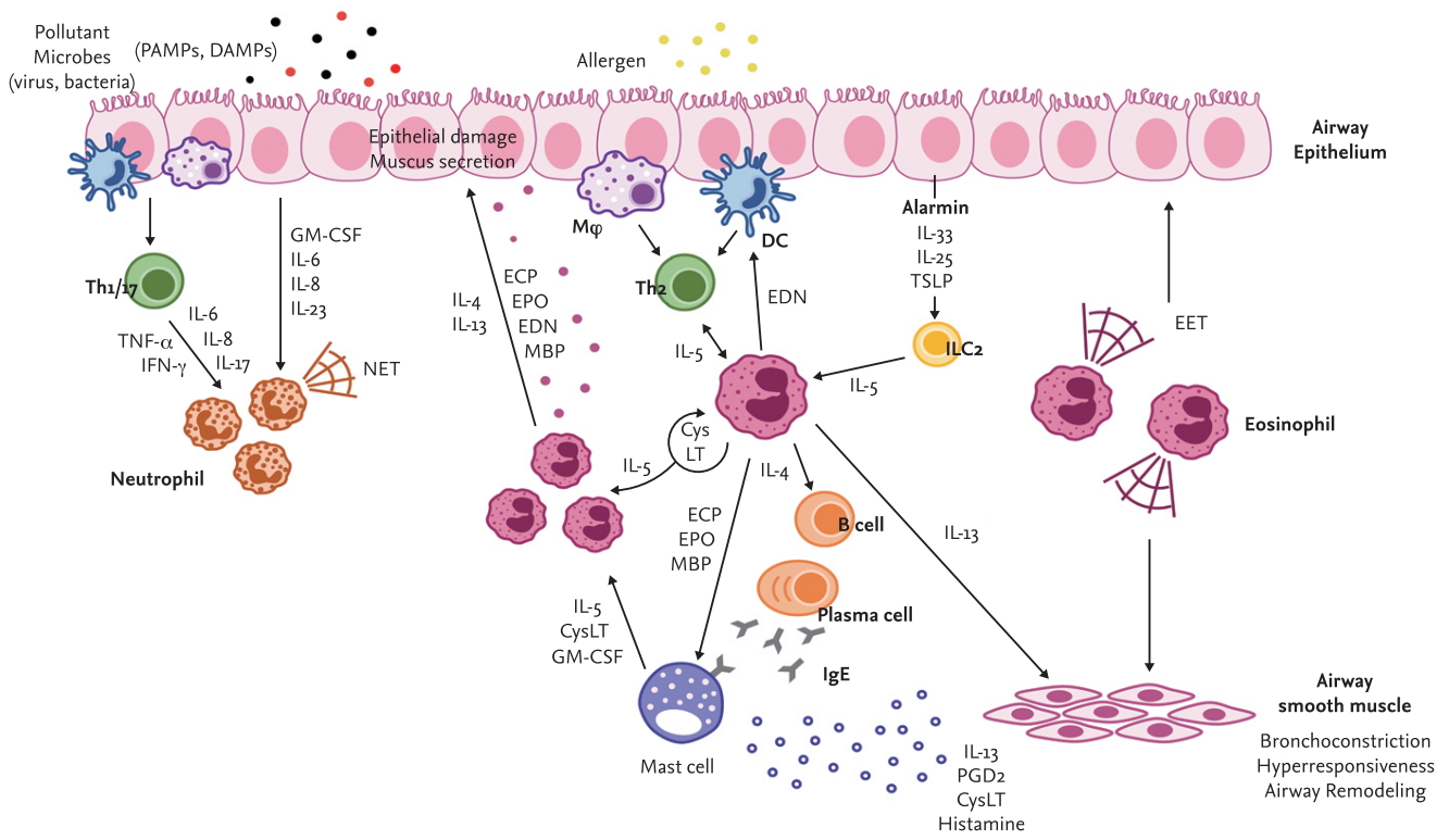
Eosinophils have been highlighted as the hallmark of severe eosinophilic asthma. They are major effector cells contributing to the pathogenesis of asthma by inducing type 2 inflammation and airway hyperresponsiveness (AHR) [26]. It has also been well demonstrated that eosinophils enhance type 2 immune responses by releasing several molecules such as cytokines, chemokines and granule proteins in response to parasitic helminth, bacterial, fungal and viral infection as well as allergens [27]. Persistent airway inflammation induced by eosinophils leads to constant tissue damage, resulting in smooth muscle thickening, goblet cell hyperplasia and extracellular matrix protein deposition called airway remodeling [28]. In inflammatory conditions, eosinophils produce cytokines (interleukin 2 [IL-2], IL-4, IL-5, IL-10, IL-12, IL-13, IL-16, IL-18, tumor necrosis factor-α [TNF-α], and transforming growth factor-α/β), chemokines (macrophage inflammatory protein 1 alpha [MIP-1α], regulated upon activation, normal T cell expressed and secreted [RANTES], and eotaxin-1) and other factors (vascular endothelial cell growth factor and metalloproteinases), and release granule proteins including major basic protein (MBP), eosinophil cationic protein (ECP), eosinophil peroxidase (EPO), and eosinophil-derived neurotoxin (EDN) which were proven to damage airway tissues in various ways [29]. Our recent study demonstrated significantly elevated levels of serum EDN in patients with severe asthma [30], suggesting that EDN, an indicator of eosinophils degranulation, is closely associated with asthma severity. In addition, emerging evidence has revealed that activated eosinophils produce novel molecules, such as extracellular traps or extracellular vesicles (EVs), which will be discussed in the following paragraphs.
Interactions between eosinophils and other immune cells exacerbate asthma symptoms (Fig. 1). Eosinophils certainly respond to IL-5 produced by T cells [31-33]. In the lungs, T cells are the main source of IL-5, which is critical for the recruitment, proliferation, survival and activation of eosinophils. Moreover, neutrophils are involved in eosinophil stimulation to induce airway inflammation by producing extracellular traps in severe asthma [34]. An important role of neutrophils interacting with eosinophils has also been suggested in occupational asthma [35]. Dendritic cell activation and migration could be promoted by EDN released from eosinophils [36]. Furthermore, eosinophils extensively communicate with tissue-resident mast cells [37]. Thus, activation, degranulation, interaction, survival and migration of eosinophils should be suppressed for the management of patients with severe asthma.

ROLE OF EOSINOPHIL EXTRACELLULAR TRAPS IN SEVERE ASTHMA

Immune function (innate and adaptive immunity) is classically defined as a host defense to recognize and eliminate pathogens. In innate immunity, neutrophils have been intensively studied as the first line of barrier against pathogen invasion. Neutrophil phagocytosis is a well characterized innate immune mechanism; however, an unexpected phenomenon has been also observed that neutrophil-forming extracellular DNA fibers bind to pathogens during host defense. These web-like chromatin structures were firstly found in neutrophils and termed neutrophil extracellular traps (NETs) [38]. NETs have been demonstrated to be implicated in multiple diseases along with several immune-modulatory functions [34,39,40]. Similar to neutrophils, extracellular traps have subsequently been observed from other cells including mast cells, monocytes, macrophages, and eosinophils [41-44].
Eosinophils release web-like chromosomes upon appropriate stimulation. DNA presented in extracellular traps was shown to be of mitochondrial origin, and multiple granule proteins were co-localized in DNA strands [45]. In addition, recent findings have suggested that most extracellular traps are composed of histone-bound DNA that is nuclear in its origins [46]. The formation of eosinophil extracellular traps (EETs) was once thought to be processed by the non-apoptotic cell death pathway [47,48] but these molecules were also found to be released from activated eosinophils independently of cellular cytoskeletal remodeling [49]. Moreover, EET formation was induced by nicotinamide adenine dinucleotide phosphate in an oxidase-dependent manner, which is distinct from apoptosis and necrosis [46].
Although EETs play an important role in innate immunity against extracellular pathogens, they have been related to host tissue damage, contributing to the pathogenesis of allergic diseases [50]. Previous studies have shown that EETs are often associated with blood and tissue eosinophilia [45,51]. Harmful effects of EETs on the airways of patients with asthma have also been demonstrated [44,47]. Furthermore, the proportion of eosinophils releasing extracellular traps was more elevated under the condition of severe airway inflammation [52,53]. Although the mechanisms by which EETs disturb immune responses have not been fully understood, our recent study demonstrated that peripheral EET-forming eosinophil and group 2 innate lymphoid cell (ILC2) counts are elevated in severe asthmatics compared to non-severe asthmatics, with a positive correlation between them and higher levels of peripheral/lung IL-33 and thymic stromal lymphopoietin (TSLP) [54,55]. An in vivo experiment showed that EETs could activate ILC2s in lung tissues through stimulation of airway epithelium to produce IL-33 and TSLP [54], which was attenuated by anti-IL-33 antibody treatment, suggesting that EETs play a crucial role in perpetuating type 2 airway inflammation in severe eosinophilic asthma. These findings suggest that biologics targeting epithelial cytokines may be beneficial in patients with severe eosinophilic asthma (with steroid resistance) via suppressive effects of the EET-ILC axis.

EOSINOPHIL EXTRACELLULAR VESICLES IN SEVERE ASTHMA

In the past, EVs were thought to be cell debris, but now it is certain that they are important mediators produced by cellular processes [56]. EVs are small membranous particles made up of lipid bilayers that contain biological information. Indeed, EVs are composed of a wide spectrum of molecules such as lipids, proteins, and nucleic acids. In terms of a heterogeneous collection of membrane-bound carriers, the function of EVs in cell-to-cell communication has been emphasized [57]. In addition, accumulating evidence supports that EVs are involved in pathophysiological processes of chronic inflammatory diseases such as cancer, metabolic disorders, and allergic disease [58-61]. EVs can promote airway inflammation through regulating recruitment, activation, and differentiation of immune cells and structural cells. Although every cell secretes EVs, especially eosinophils from patients with asthma have been shown to release larger amounts of EVs compared to those released from eosinophils of healthy subjects. The higher levels of EVs in asthmatic patients could lead to more serious symptoms when the EVs are stimulated to release their contents [62,63]. Furthermore, EV production was increased when eosinophils were stimulated with eotaxin-1 or TNF-α [64]. EVs derived from eosinophils contain the components of granule proteins such as MBP, ECP, and EPO; therefore, they similarly contribute to the pathogenesis of asthma. Moreover, EVs released from patients with asthma have been demonstrated to enhance eosinophil migration by up-regulating the expression of adhesion molecules [63]. A recent study has suggested that EVs drive the progression of severe asthma [65]. Diverse miRNAs in EVs have been proposed to be associated with asthma severity [66]. Despite growing interest, the exact mechanism of EVs in the pathogenesis of asthma or any applicable therapy has not yet been found. Further studies are needed to understand the role of eosinophil-derived EVs, which enables us to understand the complicated functions of eosinophils in asthmatic airways. It is suggested that EVs derived from eosinophils may be a potential biomarker for diagnosing asthma and classifying its phenotypes, especially severe eosinophilic asthma.

MANAGEMENT OF SEVERE ASTHMA

According to the Global Initiative for Asthma 2019 guidelines, severe asthma is defined as uncontrolled asthma despite proper adherence to optimized step 4/5 therapy and treatment of contributory factors, or asthma which worsens when doses of anti-asthmatic medications are decreased [67]. As severe asthma is associated with significant morbidity and mortality, several medications have been developed and used (Table 1). Conventionally, ICSs with long-acting beta-agonists (LABAs) are regarded as the first-line therapy for most patients with severe asthma [68]. In addition, systemic corticosteroids can often be administered as an add-on therapy to prevent asthma exacerbation [11,13]. ICSs, known as glucocorticoids, are known to directly or indirectly suppress various immune/structural cells and cytokines involved in airway inflammation [69]. At the gene expression level, they increase or decrease various transcription factors related to airway inflammation [70]. They could increase anti-inflammatory cytokines as well as decrease inflammatory cytokines, chemokines, inflammatory enzymes and adhesion molecules. At the cellular level, corticosteroids inhibit survival or recruitment of various inflammatory cells (such as eosinophils, T cells, and mast cells) and structural cells including epithelial cells in asthmatic airways [71]. Thus, ICS treatment could reduce the number of airway eosinophils and the recovery of epithelial cell injury, improving AHR/lung functions [72,73], and reducing asthma exacerbations [74]. Although an anti-inflammatory effect of corticosteroids is widely accepted, their use in clinical practice is still limited because adverse effects of corticosteroids (in a high-dose or long-term usage of systemic steroids) and decreased responsiveness to corticosteroids (insensitivity or steroid-dependence) have been found in some patients with severe eosinophilic asthma [12,75,76]. Also, since the dose-response curve of ICSs is flat, several addon therapies need to be included for the management of patients with severe asthma who are not effectively controlled with conventional anti-inflammatory medications such as medium-to-high doses of ICSs-LABAs and additional anti-leukotrienes (LTRAs) [77]. Several studies have shown that additional use of LABAs with ICSs is more effective than escalating the dose of ICSs in improving lung function and symptoms control [78] and in reducing the frequency of asthma exacerbations [79,80]. However, there is the possibility that regular use of LABAs could increase underlying inflammation in asthma, such as delays in eosinophil apoptosis [81] or blockade of apoptosis induced by corticosteroids [82]. LTRAs decrease eosinophil counts in blood and airways by blocking the cysteinyl leukotriene receptor 1 (cysLT1R) [83,84] and reduce exacerbations when combined with ICSs [84]. CysLTs are important pro-inflammatory mediators in asthma via increasing bronchoconstriction, AHR, vascular permeability and inflammatory cell recruitment [85]. Especially, CysLTs play important roles in the survival, maturation and differentiation of eosinophils as well as the release of IL-4, ECP, and EDN. Also, eosinophils are major sources of CysLT by autocrine or paracrine stimulation [86]. Recently, it has been reported that human ILC2s express CysLT receptors; thus, CysLTs are involved in ILC2 activation [87]. Corticosteroids do not effectively inhibit the CysLT synthesis pathway [71]. LTRAs do not completely suppress excessive CysLT release in severe eosinophilic asthma or AERD [88]. It is controversial whether LTRAs have an anti-inflammatory effect on eosinophils and ILC2s especially in relation to EETs, granule proteins, cytokines, and mediators. Therefore, there are unmet needs to develop alternative or additional medications for better control of eosinophils and severe asthma.
Many studies have been attempted to block the interaction between IL-5 and its receptor in eosinophilic inflammation because IL-5 is a key cytokine involved in eosinophil growth, maturation, activation and survival. Benralizumab is a monoclonal antibody against the alpha subunit of the IL-5 receptor. This antibody provides a potential benefit in decreasing exacerbation and improving lung function in uncontrolled severe asthma with elevated blood eosinophil counts by inducing antibody-dependent cell-mediated cytotoxicity [89-91]. Mepolizumab and reslizumab, humanized monoclonal antibodies against IL-5, neutralize circulating IL-5 and decrease the number of eosinophils in sputum and blood [92-95], leading to improvement in lung function in patients with severe eosinophilic asthma. IL-4 has also been suggested to play an important role in the differentiation and proliferation of type 2 helper T cells and B cells [96,97]. Dupilumab, a human monoclonal antibody against the alpha subunit of the IL-4 receptor (an overlapping receptor of IL-4 and IL-13), is regarded as a therapeutic agent of disease mediated by type 2 helper T cells. When this antibody was administered to patients with persistently elevated eosinophil levels, a significant decline in the frequency of severe exacerbation was observed [98,99]. Recently, our data has shown that anti-IL-33 antibody reduces AHR and decreases type 2 cytokine levels in an EET-induced inflammation model in vivo [54]. Considering the critical role of EETs in the pathogenesis of severe eosinophilic asthma, biologics targeting epithelial cytokines, especially IL-33, may provide a potential benefit.
Eosinophilic inflammation is strongly associated with type 2 cytokines, as well as allergen-specific IgE. In addition, alarmin-like cytokines such as IL-33 and TSLP, which are mainly released from airway epithelium, are involved in the development of severe asthma [55,100]. IL-33 activates myeloid and lymphoid innate cells to exacerbate airway inflammation but stimulates eosinophils as well [101]. TSLP is regarded as an IL-7-like cytokine known to be important for inducing type 2 cytokine production, leading to the activation of eosinophils [102]. In a previous study, the efficacy of human monoclonal anti-TSLP antibody in patients with allergic asthma was tested; a potential benefit in attenuating airway inflammation was suggested [103]. Moreover, the effect of anti-IL-33 or anti-TSLP antibody on reduction in AHR was demonstrated in vivo models [54]. Although antibodies against IL-33 and TSLP are under clinical trial, they can be a promising treatment for patients with severe type 2 asthma.
Although various pharmacotherapy and biologics have been approved for the management of severe asthma, there remain unresolved issues about selecting proper targets and patients for effective treatment, depending on its phenotypes/endotypes. Further studies are needed to find potential biomarkers for various phenotypes and endotypes to implement precision medicine.

CONCLUSIONS

Accumulating evidence strongly supports heterogeneity in severe asthma with distinct subtypes. Especially, an important role of eosinophils in type 2 severe asthma has been widely accepted as sputum/blood eosinophilia is associated with more severe symptoms, more frequent exacerbations and lower response to anti-inflammatory medications. Recent studies suggest that novel molecules, including extracellular traps and vesicles released from eosinophils, could enhance type 2 immune responses interacting with airway epithelium in the pathogenesis of severe eosinophilic asthma. However, current anti-asthmatic medications have limitations in completely controlling severe eosinophilic asthma; instead, new biologics targeting eosinophils or epithelial cells can provide potential benefits with some limitations. The development of effective biologics in terms of eosinophil function is essential for better management of severe asthma.

Conflict of Interest

No potential conflict of interest relevant to this article was reported.

Acknowledgments

This study was supported by a grant from the Korean Health Technology R & D Project, Ministry of Health and Welfare, Republic of Korea (HI16C0992).

Figure 1.
Interactions between eosinophils and various immune cells contributing to airway inflammation in severe asthma with a type 2 phenotype. PAMP, pathogen-associated molecular pattern; DAMP, damage-associated molecular pattern; Th1/2/17, type 1/2/17 T helper cells; ILC2, type 2 innate lymphoid cell; Mϕ, macrophage; DC, dendritic cell; GM-CSF, granulocyte-macrophage colony-stimulating factor; ECP, eosinophil cationic protein; EPO, eosinophil peroxidase; EDN, eosinophil-derived neurotoxin; TSLP, thymic stromal lymphopoietin; NET, neutrophil extracellular traps; EET, eosinophil extracellular traps; CysLT, cysteinyl leukotriene; PGD2, prostaglandin D2; TNF-α, tumor necrosis factor-α; IFN-γ, interferon-gamma; IL, interleukin.
kjim-2020-022f1.jpg
Table 1.
Various medications developed to manage asthma severity
Biologic Target Mechanism Patients population Effect
Mepolizumab Anti-IL-5 Prevents IL-5 from binding to its receptor Severe eosinophilic asthma (≥ 12 yr) Reduces exacerbations
Blood eosinophils ≥ 150–300/μL Improves lung function
Sputum eosinophils > 3% Reduces blood/sputum eosinophils
Adult-onset of asthma Decreases NPs; steroid-sparing effect
CRSwNPs
Reslizumab Anti-IL-5 Prevents IL-5 from binding to its receptor Severe eosinophilic asthma (≥ 18 yr) Reduces exacerbations
Blood eosinophils ≥ 400/μL Improves lung function
Sputum eosinophils > 3% Reduces blood and sputum eosinophils
CRSwNPs Decreases NPs; steroid-sparing effect
Benralizumab Anti-IL-5 receptor α Blocks IL-5Rα on eosinophils and basophils Severe eosinophilic asthma (≥ 12 yr) Reduces exacerbations
Blood eosinophils ≥ 300/μL Improves lung function
Adult-onset of asthma Reduces eosinophils counts
CRSwNPs Steroid-sparing effect
Dupilumab Anti-IL-4 receptorα Blocks IL-4Rα on T cells, B cells, macrophage, eosinophils and structural cells Severe eosinophilic/type 2 asthma (≥ 12 yr) Reduces exacerbations
Blood eosinophils ≥ 150/μL Improves lung function
FeNO ≥ 25 ppb Decreases NPs; steroid-sparing effect
Etokimab Anti-IL-33 Prevents IL-33 from binding to its receptor Severe eosinophilic asthma Improves lung function
Blood eosinophils ≥ 300/μL Reduces eosinophils counts
Pre-bronchodilator FEV1 < 80%
Tezepelumab Anti-TSLP Prevents TSLP from binding to its receptor Severe type 2 high or low asthma (> 2 exacerbations per year) Reduces exacerbations
Improves lung function
Reduces blood eosinophils
Reduces total serum IgE

IL, interleukin; CRSwNP, chronic rhinosinusitis with nasal polyp; NP, nasal polyposis; FeNO, fractional exhaled nitric oxide; FEV1, forced expiratory volume in 1 second; TSLP, thymic stromal lymphopoietin; IgE, immunoglobulin E.

REFERENCES

1. Kaur R, Chupp G. Phenotypes and endotypes of adult asthma: moving toward precision medicine. J Allergy Clin Immunol 2019;144:1–12.
crossref pmid
2. Corren J. Asthma phenotypes and endotypes: an evolving paradigm for classification. Discov Med 2013;15:243–249.
pmid
3. Kuruvilla ME, Lee FE, Lee GB. Understanding asthma phenotypes, endotypes, and mechanisms of disease. Clin Rev Allergy Immunol 2019;56:219–233.
crossref pmid pmc pdf
4. Wenzel SE, Schwartz LB, Langmack EL, et al. Evidence that severe asthma can be divided pathologically into two inflammatory subtypes with distinct physiologic and clinical characteristics. Am J Respir Crit Care Med 1999;160:1001–1008.
crossref pmid
5. Carr TF, Zeki AA, Kraft M. Eosinophilic and noneosinophilic asthma. Am J Respir Crit Care Med 2018;197:22–37.
crossref pmid pmc
6. Duvall MG, Krishnamoorthy N, Levy BD. Non-type 2 inflammation in severe asthma is propelled by neutrophil cytoplasts and maintained by defective resolution. Allergol Int 2019;68:143–149.
crossref pmid
7. Wenzel S. Severe asthma in adults. Am J Respir Crit Care Med 2005;172:149–160.
crossref pmid
8. Schleich FN, Chevremont A, Paulus V, et al. Importance of concomitant local and systemic eosinophilia in uncontrolled asthma. Eur Respir J 2014;44:97–108.
crossref pmid
9. Buhl R, Humbert M, Bjermer L, et al. Severe eosinophilic asthma: a roadmap to consensus. Eur Respir J 2017;49:1700634.
crossref pmid
10. Dolan CM, Fraher KE, Bleecker ER, et al. Design and baseline characteristics of the epidemiology and natural history of asthma: outcomes and treatment regimens (TENOR) study: a large cohort of patients with severe or difficult-to-treat asthma. Ann Allergy Asthma Immunol 2004;92:32–39.
crossref pmid
11. Moore WC, Bleecker ER, Curran-Everett D, et al. Characterization of the severe asthma phenotype by the National Heart, Lung, and Blood Institute’s Severe Asthma Research Program. J Allergy Clin Immunol 2007;119:405–413.
crossref pmid pmc
12. Hew M, Chung KF. Corticosteroid insensitivity in severe asthma: significance, mechanisms and aetiology. Intern Med J 2010;40:323–334.
crossref pmid
13. Chung KF, Wenzel SE, Brozek JL, et al. International ERS/ATS guidelines on definition, evaluation and treatment of severe asthma. Eur Respir J 2014;43:343–373.
crossref pmid
14. Song WJ, Lee JH, Kang Y, Joung WJ, Chung KF. Future risks in patients with severe asthma. Allergy Asthma Immunol Res 2019;11:763–778.
crossref pmid pmc
15. Wenzel S. Severe asthma: from characteristics to phenotypes to endotypes. Clin Exp Allergy 2012;42:650–658.
crossref pmid
16. Tran TN, Zeiger RS, Peters SP, et al. Overlap of atopic, eosinophilic, and TH2-high asthma phenotypes in a general population with current asthma. Ann Allergy Asthma Immunol 2016;116:37–42.
crossref pmid
17. Chapman KR, Albers FC, Chipps B, et al. The clinical benefit of mepolizumab replacing omalizumab in uncontrolled severe eosinophilic asthma. Allergy 2019;74:1716–1726.
crossref pmid pmc
18. Huang YC, Weng CM, Lee MJ, Lin SM, Wang CH, Kuo HP. Endotypes of severe allergic asthma patients who clinically benefit from anti-IgE therapy. Clin Exp Allergy 2019;49:44–53.
crossref pmid
19. Price DB, Rigazio A, Campbell JD, et al. Blood eosinophil count and prospective annual asthma disease burden: a UK cohort study. Lancet Respir Med 2015;3:849–858.
crossref pmid
20. De Groot JC, Storm H, Amelink M, et al. Clinical profile of patients with adult-onset eosinophilic asthma. ERJ Open Res 2016;2:00100–2015.
crossref pmid pmc
21. Steinke JW, Wilson JM. Aspirin-exacerbated respiratory disease: pathophysiological insights and clinical advances. J Asthma Allergy 2016;9:37–43.
crossref pmid pmc
22. Choi Y, Lee Y, Park HS. Which factors associated with activated eosinophils contribute to the pathogenesis of aspirin-exacerbated respiratory disease? Allergy Asthma Immunol Res 2019;11:320–329.
crossref pmid
23. Choi Y, Lee DH, Trinh HK, et al. Surfactant protein D alleviates eosinophil-mediated airway inflammation and remodeling in patients with aspirin-exacerbated respiratory disease. Allergy 2019;74:78–88.
crossref pmid
24. Choi Y, Lee DH, Lee JH, Shin YS, Kim SH, Park HS. Immunomodulatory function of surfactant protein D in eosinophilic asthma. Allergy 2019;74:192–195.
crossref pmid
25. Trinh HK, Pham DL, Choi Y, Kim HM, Kim SH, Park HS. Epithelial folliculin enhances airway inflammation in aspirin-exacerbated respiratory disease. Clin Exp Allergy 2018;48:1464–1473.
crossref pmid
26. Rosenberg HF, Dyer KD, Foster PS. Eosinophils: changing perspectives in health and disease. Nat Rev Immunol 2013;13:9–22.
crossref pmid pdf
27. Pagnoux C, Nair P, Xi Y, et al. Serum cytokine and chemokine levels in patients with eosinophilic granulomatosis with polyangiitis, hypereosinophilic syndrome, or eosinophilic asthma. Clin Exp Rheumatol 2019;37 Suppl 117:40–44.
pmid
28. Tagaya E, Tamaoki J. Mechanisms of airway remodeling in asthma. Allergol Int 2007;56:331–340.
crossref pmid
29. Acharya KR, Ackerman SJ. Eosinophil granule proteins: form and function. J Biol Chem 2014;289:17406–17415.
crossref pmid pmc
30. Lee Y, Lee JH, Yang EM, et al. Serum levels of eosinophil-derived neurotoxin: a biomarker for asthma severity in adult asthmatics. Allergy Asthma Immunol Res 2019;11:394–405.
crossref pmid pmc
31. MacKenzie JR, Mattes J, Dent LA, Foster PS. Eosinophils promote allergic disease of the lung by regulating CD4(+) Th2 lymphocyte function. J Immunol 2001;167:3146–3155.
crossref pmid
32. Mattes J, Yang M, Mahalingam S, et al. Intrinsic defect in T cell production of interleukin (IL)-13 in the absence of both IL-5 and eotaxin precludes the development of eosinophilia and airways hyperreactivity in experimental asthma. J Exp Med 2002;195:1433–1444.
crossref pmid pmc pdf
33. Emma R, Morjaria JB, Fuochi V, Polosa R, Caruso M. Mepolizumab in the management of severe eosinophilic asthma in adults: current evidence and practical experience. Ther Adv Respir Dis 2018;12:1753466618808490.
crossref pmid pmc
34. Choi Y, Pham LD, Lee DH, et al. Neutrophil extracellular DNA traps induce autoantigen production by airway epithelial cells. Mediators Inflamm 2017;2017:5675029.
crossref pmid pmc pdf
35. Choi Y, Lee Y, Park HS. Neutrophil activation in occupational asthma. Curr Opin Allergy Clin Immunol 2019;19:81–85.
crossref pmid
36. Yang D, Chen Q, Su SB, et al. Eosinophil-derived neurotoxin acts as an alarmin to activate the TLR2-MyD88 signal pathway in dendritic cells and enhances Th2 immune responses. J Exp Med 2008;205:79–90.
crossref pmid pmc pdf
37. Elishmereni M, Alenius HT, Bradding P, et al. Physical interactions between mast cells and eosinophils: a novel mechanism enhancing eosinophil survival in vitro. Allergy 2011;66:376–385.
crossref pmid
38. Brinkmann V, Reichard U, Goosmann C, et al. Neutrophil extracellular traps kill bacteria. Science 2004;303:1532–1535.
crossref pmid
39. Papayannopoulos V. Neutrophil extracellular traps in immunity and disease. Nat Rev Immunol 2018;18:134–147.
crossref pmid pdf
40. Castanheira FV, Kubes P. Neutrophils and NETs in modulating acute and chronic inflammation. Blood 2019;133:2178–2185.
crossref pmid pdf
41. Von Kockritz-Blickwede M, Goldmann O, Thulin P, et al. Phagocytosis-independent antimicrobial activity of mast cells by means of extracellular trap formation. Blood 2008;111:3070–3080.
crossref pmid pdf
42. Webster SJ, Daigneault M, Bewley MA, et al. Distinct cell death programs in monocytes regulate innate responses following challenge with common causes of invasive bacterial disease. J Immunol 2010;185:2968–2979.
crossref pmid pmc
43. Mohanan S, Horibata S, McElwee JL, Dannenberg AJ, Coonrod SA. Identification of macrophage extracellular trap-like structures in mammary gland adipose tissue: a preliminary study. Front Immunol 2013;4:67.
crossref pmid pmc
44. Ueki S, Konno Y, Takeda M, et al. Eosinophil extracellular trap cell death-derived DNA traps: their presence in secretions and functional attributes. J Allergy Clin Immunol 2016;137:258–267.
crossref pmid
45. Dworski R, Simon HU, Hoskins A, Yousefi S. Eosinophil and neutrophil extracellular DNA traps in human allergic asthmatic airways. J Allergy Clin Immunol 2011;127:1260–1266.
crossref pmid pmc
46. Ueki S, Melo RC, Ghiran I, Spencer LA, Dvorak AM, Weller PF. Eosinophil extracellular DNA trap cell death mediates lytic release of free secretion-competent eosinophil granules in humans. Blood 2013;121:2074–2083.
crossref pmid pmc pdf
47. Yousefi S, Simon D, Simon HU. Eosinophil extracellular DNA traps: molecular mechanisms and potential roles in disease. Curr Opin Immunol 2012;24:736–739.
crossref pmid
48. Ueki S, Tokunaga T, Fujieda S, et al. Eosinophil ETosis and DNA traps: a new look at eosinophilic inflammation. Curr Allergy Asthma Rep 2016;16:54.
crossref pmid pmc pdf
49. Yousefi S, Gold JA, Andina N, et al. Catapult-like release of mitochondrial DNA by eosinophils contributes to antibacterial defense. Nat Med 2008;14:949–953.
crossref pmid pdf
50. Mukherjee M, Lacy P, Ueki S. Eosinophil extracellular traps and inflammatory pathologies-untangling the Web! Front Immunol 2018;9:2763.
crossref pmid pmc
51. Simon D, Hoesli S, Roth N, Staedler S, Yousefi S, Simon HU. Eosinophil extracellular DNA traps in skin diseases. J Allergy Clin Immunol 2011;127:194–199.
crossref pmid
52. Gevaert E, Zhang N, Krysko O, et al. Extracellular eosinophilic traps in association with Staphylococcus aureus at the site of epithelial barrier defects in patients with severe airway inflammation. J Allergy Clin Immunol 2017;139:1849–1860.
crossref pmid
53. Choi Y, Le Pham D, Lee DH, Lee SH, Kim SH, Park HS. Biological function of eosinophil extracellular traps in patients with severe eosinophilic asthma. Exp Mol Med 2018;50:104.
crossref pmid pmc pdf
54. Choi Y, Kim YM, Lee HR, et al. Eosinophil extracellular traps activate type 2 innate lymphoid cells through stimulating airway epithelium in severe asthma. Allergy 2020;75:95–103.
crossref pmid
55. Li Y, Wang W, Lv Z, et al. Elevated expression of IL-33 and TSLP in the airways of human asthmatics in vivo: a potential biomarker of severe refractory disease. J Immunol 2018;200:2253–2262.
crossref pmid
56. Maas SLN, Breakefield XO, Weaver AM. Extracellular vesicles: unique intercellular delivery vehicles. Trends Cell Biol 2017;27:172–188.
crossref pmid
57. Raposo G, Stahl PD. Extracellular vesicles: a new communication paradigm? Nat Rev Mol Cell Biol 2019;20:509–510.
crossref pmid pdf
58. Becker A, Thakur BK, Weiss JM, Kim HS, Peinado H, Lyden D. Extracellular vesicles in cancer: cell-to-cell mediators of metastasis. Cancer Cell 2016;30:836–848.
crossref pmid pmc
59. Choi Y, Kwon Y, Kim DK, et al. Gut microbe-derived extracellular vesicles induce insulin resistance, thereby impairing glucose metabolism in skeletal muscle. Sci Rep 2015;5:15878.
crossref pmid pmc pdf
60. Chelakkot C, Choi Y, Kim DK, et al. Akkermansia muciniphila-derived extracellular vesicles influence gut permeability through the regulation of tight junctions. Exp Mol Med 2018;50:e450.
crossref pmid pmc pdf
61. Choi Y, Park H, Park HS, Kim YK. Extracellular vesicles, a key mediator to link environmental microbiota to airway immunity. Allergy Asthma Immunol Res 2017;9:101–106.
crossref pmid
62. Canas JA, Sastre B, Mazzeo C, et al. Exosomes from eosinophils autoregulate and promote eosinophil functions. J Leukoc Biol 2017;101:1191–1199.
crossref pmid
63. Mazzeo C, Canas JA, Zafra MP, et al. Exosome secretion by eosinophils: a possible role in asthma pathogenesis. J Allergy Clin Immunol 2015;135:1603–1613.
crossref pmid
64. Akuthota P, Carmo LA, Bonjour K, et al. Extracellular microvesicle production by human eosinophils activated by “inflammatory” stimuli. Front Cell Dev Biol 2016;4:117.
crossref pmid pmc
65. Mortaz E, Alipoor SD, Varahram M, et al. Exosomes in severe asthma: update in their roles and potential in therapy. Biomed Res Int 2018;2018:2862187.
crossref pmid pmc pdf
66. Sastre B, Canas JA, Rodrigo-Munoz JM, Del Pozo V. Novel modulators of asthma and allergy: exosomes and microRNAs. Front Immunol 2017;8:826.
crossref pmid pmc
67. Global Initiative for Asthma. Global Strategy for Asthma Management and Prevention, 2019. Fontana (WI): GINA, 2019.

68. Wegmann M. Targeting eosinophil biology in asthma therapy. Am J Respir Cell Mol Biol 2011;45:667–674.
crossref pmid
69. Adcock IM, Barnes PJ. Molecular mechanisms of corticosteroid resistance. Chest 2008;134:394–401.
crossref pmid
70. Rhen T, Cidlowski JA. Antiinflammatory action of glucocorticoids: new mechanisms for old drugs. N Engl J Med 2005;353:1711–1723.
crossref pmid
71. Barnes PJ. Inhaled corticosteroids. Pharmaceuticals (Basel) 2010;3:514–540.
crossref pmid pmc
72. Gibson PG, Saltos N, Fakes K. Acute anti-inflammatory effects of inhaled budesonide in asthma: a randomized controlled trial. Am J Respir Crit Care Med 2001;163:32–36.
crossref pmid
73. Erin EM, Zacharasiewicz AS, Nicholson GC, et al. Rapid effect of inhaled ciclesonide in asthma: a randomized, placebo-controlled study. Chest 2008;134:740–745.
crossref pmid
74. Pauwels RA, Pedersen S, Busse WW, et al. Early intervention with budesonide in mild persistent asthma: a randomised, double-blind trial. Lancet 2003;361:1071–1076.
crossref pmid
75. Sullivan PW, Ghushchyan VH, Globe G, Schatz M. Oral corticosteroid exposure and adverse effects in asthmatic patients. J Allergy Clin Immunol 2018;141:110–116.
crossref pmid
76. Liu S, Verma M, Michalec L, et al. Steroid resistance of airway type 2 innate lymphoid cells from patients with severe asthma: the role of thymic stromal lymphopoietin. J Allergy Clin Immunol 2018;141:257–268.
crossref pmid
77. Kankaanranta H, Lahdensuo A, Moilanen E, Barnes PJ. Add-on therapy options in asthma not adequately controlled by inhaled corticosteroids: a comprehensive review. Respir Res 2004;5:17.
crossref pmid pmc
78. Greening AP, Ind PW, Northfield M, Shaw G. Added salmeterol versus higher-dose corticosteroid in asthma patients with symptoms on existing inhaled corticosteroid. Allen & Hanburys Limited UK Study Group. Lancet 1994;344:219–224.
crossref pmid
79. Pauwels RA, Lofdahl CG, Postma DS, et al. Effect of inhaled formoterol and budesonide on exacerbations of asthma. Formoterol and Corticosteroids Establishing Therapy (FACET) International Study Group. N Engl J Med 1997;337:1405–1411.
crossref pmid
80. Gibson PG, Powell H, Ducharme FM. Differential effects of maintenance long-acting beta-agonist and inhaled corticosteroid on asthma control and asthma exacerbations. J Allergy Clin Immunol 2007;119:344–350.
crossref pmid
81. Kankaanranta H, Lindsay MA, Giembycz MA, Zhang X, Moilanen E, Barnes PJ. Delayed eosinophil apoptosis in asthma. J Allergy Clin Immunol 2000;106(1 Pt 1):77–83.
crossref pmid
82. Nielson CP, Hadjokas NE. Beta-adrenoceptor agonists block corticosteroid inhibition in eosinophils. Am J Respir Crit Care Med 1998;157:184–191.
crossref pmid
83. Calhoun WJ. Summary of clinical trials with zafirlukast. Am J Respir Crit Care Med 1998;157(6 Pt 2):S238–S248.
crossref
84. Knorr B, Matz J, Bernstein JA, et al. Montelukast for chronic asthma in 6- to 14-year-old children: a randomized, double-blind trial. Pediatric Montelukast Study Group. JAMA 1998;279:1181–1186.
crossref pmid
85. Dempsey OJ. Leukotriene receptor antagonist therapy. Postgrad Med J 2000;76:767–773.
crossref pmid pmc
86. Thompson-Souza GA, Gropillo I, Neves JS. Cysteinyl leukotrienes in eosinophil biology: functional roles and therapeutic perspectives in eosinophilic disorders. Front Med (Lausanne) 2017;4:106.
crossref pmid pmc
87. Helfrich S, Mindt BC, Fritz JH, Duerr CU. Group 2 innate lymphoid cells in respiratory allergic inflammation. Front Immunol 2019;10:930.
crossref pmid pmc
88. Ducharme FM, Lasserson TJ, Cates CJ. Long-acting beta2-agonists versus anti-leukotrienes as add-on therapy to inhaled corticosteroids for chronic asthma. Cochrane Database Syst Rev 2006;4:CD003137.
crossref
89. Kolbeck R, Kozhich A, Koike M, et al. MEDI-563, a humanized anti-IL-5 receptor alpha mAb with enhanced antibody-dependent cell-mediated cytotoxicity function. J Allergy Clin Immunol 2010;125:1344–1353.
crossref pmid
90. Bleecker ER, Wechsler ME, FitzGerald JM, et al. Baseline patient factors impact on the clinical efficacy of benralizumab for severe asthma. Eur Respir J 2018;52:1800936.
crossref pmid pmc
91. Park HS, Lee SH, Lee SY, et al. Efficacy and safety of benralizumab for Korean patients with severe, uncontrolled eosinophilic asthma. Allergy Asthma Immunol Res 2019;11:508–518.
crossref pmid pmc
92. Albers FC, Hozawa S, Bratton DJ, et al. Update: mepolizumab treatment in patients with severe eosinophilic asthma and prior omalizumab use. Allergy 2020;75:942–946.
crossref pmid
93. Nair P, Pizzichini MM, Kjarsgaard M, et al. Mepolizumab for prednisone-dependent asthma with sputum eosinophilia. N Engl J Med 2009;360:985–993.
crossref pmid
94. Ortega HG, Liu MC, Pavord ID, et al. Mepolizumab treatment in patients with severe eosinophilic asthma. N Engl J Med 2014;371:1198–1207.
crossref pmid
95. Castro M, Zangrilli J, Wechsler ME, et al. Reslizumab for inadequately controlled asthma with elevated blood eosinophil counts: results from two multicentre, parallel, double-blind, randomised, placebo-controlled, phase 3 trials. Lancet Respir Med 2015;3:355–366.
crossref pmid
96. Zhu J. T helper 2 (Th2) cell differentiation, type 2 innate lymphoid cell (ILC2) development and regulation of interleukin-4 (IL-4) and IL-13 production. Cytokine 2015;75:14–24.
crossref pmid pmc
97. Hofman FM, Brock M, Taylor CR, Lyons B. IL-4 regulates differentiation and proliferation of human precursor B cells. J Immunol 1988;141:1185–1190.
crossref pmid pdf
98. Wenzel S, Ford L, Pearlman D, et al. Dupilumab in persistent asthma with elevated eosinophil levels. N Engl J Med 2013;368:2455–2466.
crossref pmid
99. Castro M, Corren J, Pavord ID, et al. Dupilumab efficacy and safety in moderate-to-severe uncontrolled asthma. N Engl J Med 2018;378:2486–2496.
crossref pmid
100. Divekar R, Kita H. Recent advances in epithelium-derived cytokines (IL-33, IL-25, and thymic stromal lymphopoietin) and allergic inflammation. Curr Opin Allergy Clin Immunol 2015;15:98–103.
crossref pmid pmc
101. Stolarski B, Kurowska-Stolarska M, Kewin P, Xu D, Liew FY. IL-33 exacerbates eosinophil-mediated airway inflammation. J Immunol 2010;185:3472–3480.
crossref pmid
102. Wong CK, Hu S, Cheung PF, Lam CW. Thymic stromal lymphopoietin induces chemotactic and prosurvival effects in eosinophils: implications in allergic inflammation. Am J Respir Cell Mol Biol 2010;43:305–315.
crossref pmid
103. Gauvreau GM, O’Byrne PM, Boulet LP, et al. Effects of an anti-TSLP antibody on allergen-induced asthmatic responses. N Engl J Med 2014;370:2102–2110.
crossref pmid
TOOLS
METRICS Graph View
  • 24 Web of Science
  • 23 Crossref
  • 25 Scopus
  • 4,864 View
  • 234 Download
Related articles

Editorial Office
101-2501, Lotte Castle President, 109 Mapo-daero, Mapo-gu, Seoul 04146, Korea
Tel: +82-2-2271-6792    Fax: +82-2-790-0993    E-mail: kaim@kams.or.kr                

Copyright © 2024 by Korean Association of Internal Medicine.

Close layer
prev next