


INTRODUCTION
Appendiceal abscesses represent 2.3% of all the cases of acute appendicitis1) and usually present a mass in the right lower quadrant2). Occasionally, abscesses develop in the right subhepatic space, right subdiaphragmatic space and right pararenal space or liver3). However, an appendiceal abscess located in the epigastric space has never been reported. The management of appendiceal abscess is controversial4). Recently, the ultrasonic or CT guided percutaneous puncture technique has offered an alternative to conventional surgical treatment3,5). We experienced a patient with an epigastric appendiceal abscess, demonstrated by abdominal CT scan and barium enema, which resolved spontaneously.
CASE REPORT
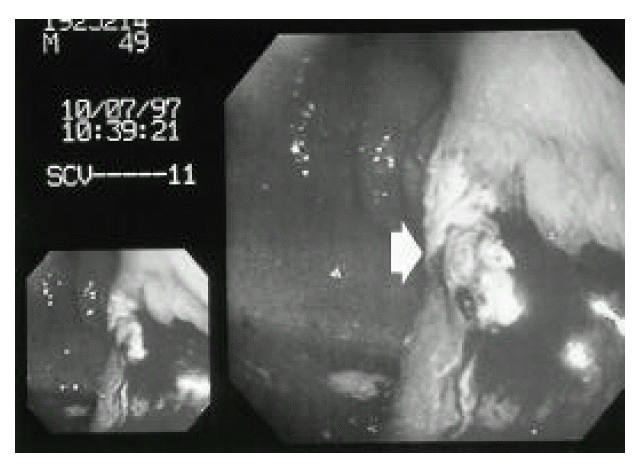
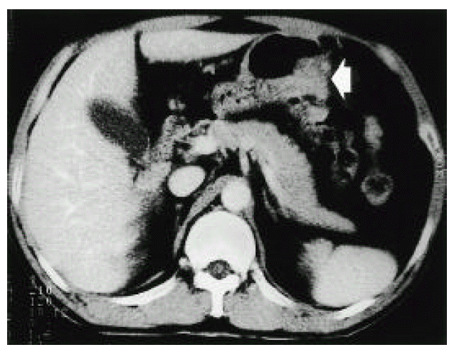
A 49-year-old man was referred to our institution for further evaluation and management of an intraabdominal abscess with the air-fluid level located outside of the greater curvature of the stomach, demonstrated on the abdominal CT scan checked 5 days before the referral (Fig 1). He experienced left upper quadrant abdominal pain with fever and chills for 15 days without any preceding trauma and was managed at a private clinic without improvement of symptoms. Upon abdominal examination, an adult fist-sized, ill-defined, round, nonmovable and tender mass was palpated at the left epigastrium. No abnormalities were observed in CBC, urinalysis, stool examination, serum amylase and liver chemistry. We treated the patient with intravenous antibiotics and bed rest. On the second day of hospitalization, an gastroscopy was performed which showed a bulging external mass effect with adherent purulent material and mucosal friability around a fistula-like lesion at the posterior wall of the mid-gastric body (Fig. 2). We took biopsy specimens which showed necrotic tissues along with an intact gastric mucosa.
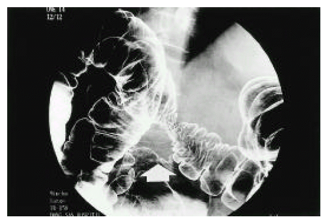
On the third day of hospitalization, a barium enema was performed and showed a hyperrotated cecum in the left upper quadrant and nonvisualization of the appendix with mucosal irregularity and focal luminal narrowing at the surrounding distal transverse colon on prone position film consistent with a hyperrotated cecum and appendiceal abscess(Fig. 3).
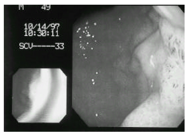
On the fifth day of hospitalization, after conservative management, the tender mass subsided. A follow-up gastroscopy, showed a focal regenerating edematous mucosa without the bulging effect noted in the initial examination (Fig. 4). A follow-up CT scan disclosed the abscess pocket had subsided with surrounding inflammatory change only(Fig. 5). The patient was discharged on the tenth day of hospitalization and has remained well for over 6 months.
DISCUSSION
The palpable mass associated with acute appendicitis may consist of phlegmon or abscesses of various sizes4). 2ŌĆō3% of the patients with appendicitis who are admitted to the hospital have abdominal masses4,6ŌĆō8) and appendiceal abscesses represent 50ŌĆō89% of the patients with an appendiceal mass4, 9ŌĆō12). Complicating abscesses have been reported in 2.3% of all cases of acute appendicitis1).
The main manifestations of acute appendicitis are fever, a palpable mass and leukocytosis4). Patients often have a palpable mass in the right lower quadrant (the ŌĆ£appendix massŌĆØ)2). Rarely does the abscess form in the right subhepatic space, right subdiaphragmatic space, liver or posterior pararenal space distant to the right lower quadrant. Therefore, the value of aggressive radiologic work-up and follow-up can not be overemphasized in suspected appendiceal abscesses, especially those present in locations remote from the appendix5). Jordan has reported that the distinction between an appendiceal mass and an appendiceal abscess could not be made by considering the patientŌĆÖs duration of symptoms, temperature and white blood cell count when the patient was admitted4). Careful follow-up, routine barium enema study, ultrasonography and abdominal CT scanning would prevent misdiagnosis and delayed treatment13). Ultrasonography has made it possible to distinguish an abscess from the phlegmon without operation14) and the abdominal CT scan has proven to be of considerable clinical value in characterizing periappendiceal inflammatory masses and in determining the relative size of the liquefied versus nonliquefied component15ŌĆō17).
Despite extensive clinical experiences, the surgical management of appendiceal abscesses remains controversial4,7,10).
However, most authors agree that the initial treatment must be conservative management, including bed rest, nasogastric suction, systemic antibiotics and drainage, rather than early surgery7,11,13). Initial conservative management with intravenous antibiotics, with or without percutaneous drainage of the abscess, is prudent, safe and effective17) and shows a high success rate (80ŌĆō90%) and low morbidity rate (15%)7,12,18) compared to the high complication rate of early surgery (15ŌĆō50%)2,4,7,9). Guided percutaneous drainage is an effective alternative to surgical drainage3,5) Nonoperative treatment and, if possible, ultrasonic percutaneous drainage of a verified abscess are safe procedures with few complications and late sequelae12). Recently, the ultrasonic percutaneous puncture technique has offered an almost atraumatic alternative to conventional surgical treatment14,19). Also, CT guided percutaneous abscess drainage is an effective alternative to surgery7,17).
The recurrence of appendicitis after conservative treatment is between 4ŌĆō80%2) or 0ŌĆō20%13). Sixty-six percent of the recurrent cases occurred within 2 years of the initial attack13). The abscess often spontaneously resolves or drains into the intestine14) with a low recurrence rate11).
Our case exhibited fever, chills and an epigastric abscess. At first, we suspected a pancreatic abscess because of its location, and managed initially with antibiotics and planned percutaneous drainage. However, the radiological studies, especially the CT scan and the barium enema, revealed an appendiceal abscess in the left upper quadrant of the abdomen due to a hyperrotated ceum.
The abscess resolved spontaneously, and we think that the abscess drained into the stomach through a small fistula between the stomach and abscess cavity. The patient has remained well for over 6 months.

 PDF Links
 PubReader
 ePub Link
 Full text via DOI
 Download Citation
 Print
Print


