


INTRODUCTION
Variceal bleeding is one of the most serious complications of portal hypertension. Despite many advances in the management of variceal bleeding, each episode of bleeding is associated with 30% to 50% mortality rate1ŌĆō3). Most deaths occur after early rebleeding, which occurs in 20% to 50% of patients1,4). This fact makes mandatory the application of therapeutic strategies to prevent recurrent bleeding. The surgical shunt has been considered one of the therapeutic approaches when pharmacologic and endoscopic treatments fail to control variceal bleeding. However, surgical shunt is associated with a high operative mortality and high incidence of encephalopathy and this has limited its application3,5ŌĆō7).
Transjugular intrahepatic portosystemic shunt (TIPS) is an interventional radiologic procedure that results in decompression of the portal venous system by means of a communication between the hepatic and the portal vein through the hepatic parenchyma with a percutaneous approach, avoiding the risk of major surgery8). To date, most studies have focused on the use of TIPS as a treatment for patients with recurrent variceal bleeding that has been refractory to pharmacologic and endoscopic treatments9,10).
The aim of this study was to assess the long-term effect and safety of TIPS in the treatment of variceal bleeding that is not controlled with pharmacological and endoscopic treatment.
PATIENTS and METHODS
1. Patients
Thirty-six patients (32 men, 4 women) who underwent transjugular intrahepatic portosystemic shunt (TIPS), due to refractory variceal bleeding, between March 1994 and September 1999 in the Chonnam National University Hospital were included in the study. The clinical characteristics of these patients are summarized in Table 1. They comprised 32 males and 4 females ranging in age from 25 to 73 years, with a mean age of 50.6 years. The etiology of liver cirrhosis was as follows: alcoholic (16 patients), chronic infection by hepatitis B virus (16 patients) chronic infection by hepatitis C virus (3 patients), and chronic infection by hepatitis B and C virus (1 patient). Two cases had hepatocellular cancer without portal invasion. The TIPS was performed for recurrent or uncontrolled variceal bleeding despite one or several sessions of endoscopic intervention and vasoactive therapy (octerotide or vasopression combined with nitroglycerine) during several days. The procedure was performed on an emergency basis for active bleeding in 14 patients (39%) and was elective in 22 (61%). Variceal bleeding was diagnosed by a history of hematemesis, hematochezia, or melena and endoscopic evidence of bleeding varices (spurting or oozing) or presence of varices with stigmata of bleeding and no other lesions that could account for the bleeding. Patients were excluded if they had the following: portal vein thrombosis; previous episodes of chronic recurrent hepatic encephalopathy; advanced hepatocellular carcinoma; severe hepatic failure. The sites of bleeding varices were esophagus in 15 (41.7%) patients, stomach in 19 (52.8%) patients, and duodenum in 2 (5.6%) patients. According to Child-Pugh classification at the time of TIPS procedure, 6 (16.7%) of the cases were A, 16 (44.4%) were B and 14 (38.9%) were C.
2. Endoscopic evaluation of varices and portal hypertensive gastropathy
Upper gastrointestinal endoscopy was performed to analyse the degree of varices and portal hypertensive gastropathy (PHG) before TIPS procedure and during 1 to 3 weeks after TIPS. The endoscopic findings of esophagogastric varices were classified according to the General Rules for Recording Endoscopic Findings set by the Japanese Research Society for Portal Hypertension11), in which the form of varices is graded as 0, none; 1, F1 (straight form); 2, F2 (enlarged tortuous form); and 3, F3 (nodular-beaded). PHG were classified according to the description of Mc-Cormack et al.12) and scored as follows: 0, none; 1, mild PHG (mosaic pattern, superficial reddening, scarlatina-type rash); 2, severe PHG (cherry red spots, diffuse hemorrhagic gastritis).
3. TIPS procedure
Studied patients were in stable hemodynamic condition after blood transfusion and fluid infusion. The TIPS procedure was performed using the standard technique of accessing the right internal jugular venous access8) and a free hepatic venogram or test injection of contrast medium was done. Free hepatic venous pressure (HVP) were measured before the puncture. We punctured blindly with Colapinto needle. The artificial shunt tract was created by bridging the hepatic vein with portal vein. Then insertion of guidewire was introduced deeply into the portal system followed by a catheter for portal pressure measurement and portal venogram. Portosystemic pressure gradient (PSG) was calculated as the difference between portal venous pressure (PVP) and HVP. After measurement of pressures, the tract was dilated with a balloon catheter and Wall stent was advanced to the tract and dilated to achieve an internal diameter of 10 mm. If filling of varices persisted on the portogram or the reduction in portal pressure was not significant, further dilatation, using a larger diameter balloon catheter or direct variceal embolization with metal coil, was done. Portal pressure measurement and venogram were obtained again before the end of procedure.
4. Follow-up
Doppler ultrasonography and endoscopy were carried out during 1 to 3 weeks after TIPS, every 3 months, thereafter and whenever stent dysfunction was suspected clinically. Doppler ultrasonographic measurements of peak flow velocity (Vmax) within stent of 50 cm/sec or less and reversal of intrahepatic portal venous flow direction from hepatofugal to hepatopetal are indicators of the stent dysfunction13) in this study.
Angiography was performed in surviving patients, if bleeding recurred, or if ultrasonography or endoscopy suggested stent dysfunction.
Patients were recalled to the hospital if they missed their outpatient appointment.
5. Statistical analysis
For analysis of the data, all results were expressed as mean┬▒SD. Quantitative variables were compared with the Student's t test. Survival was analyzed by the Kaplan-Meyer method. The statistical software program used was Statistical Package for the Social Sciences (SPSS/PC+7.0, Chicago, IL). A p-value of less than 0.05 was accepted as statistically significant.
RESULTS
TIPS were successfully placed in 36 of 38 patients (94.6%). Two technical failures occurred in patients with narrowing of intrahepatic portal vein. TIPS achieved hemostasis of variceal bleeding in 34 patients (94.4%).
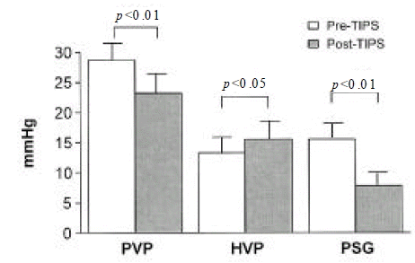
Portal venous pressure decreased from an initial average of 28.7┬▒7.9 to 23.2┬▒9.4 mmHg after TIPS (p<0.05). The portosystemic pressure gradient was significantly decreased from 15.5┬▒6.3 to 7.8┬▒4.1 mmHg (p<0.01) (Figure 1). Post-TIPS pressure gradient remained above 12 mmHg in 5 patients.
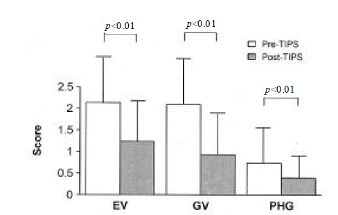
The degree of esophageal varices significantly improved after TIPS (mean score from 2.13┬▒ 1.14 to 1.23┬▒0.97 in all patients, p<0.0 1) (Figure 2). Variable sized esophageal varices were seen in 28 patients before TIPS. Those were reduced in 23 patients after the procedure.
The degree of gastric varices improved after TIPS (mean score from 2.09┬▒0.94 to 0.93┬▒0.87 in all patients, p<0.01) (Figure 2). Gastric varices were seen in 24 patients. Gastric varices disappeared in 6 patients and decreased in 15 patients, but the size of gastric varices did not change in the remaining 3 patients after TIPS.
PHG were seen in 15 patients before TIPS (mean score in all patients, 0.74┬▒0.86), and improved after TIPS (mean score 0.39┬▒0.49) (Figure 2). Eight patients with severe PHG improved markedly; severe PHG disappeared in 2 patients, and changed to mild PHG in 6 patients. In 2 of 5 patients with mild PHG, the PHG lesions disappeared.
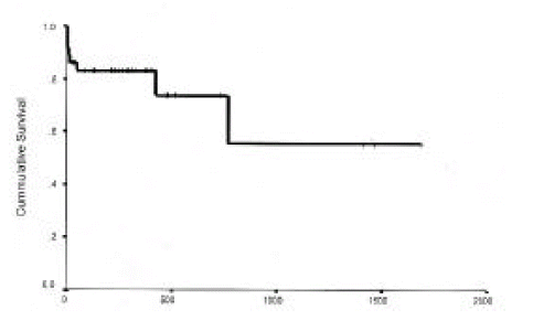
Eight patients had died between March 1994 and September 1999. Overall mortality was 22.2%. The causes of death were uncontrolled bleeding, respiratory failure, hepatic failure and recurrent variceal bleeding (Table 2). Uncontrolled bleeding led to death in two cases. One out of six cases of rebleeding died. Two cases died due to progressive hepatic failure. Two cases died of respiratory failure due to pneumonia. In the first two months after TIPS, six of eight deaths occurred. The actuarial probability of survival was 83% at one year and 74% at two years (Figure 3). The one who dropped out had undergone liver transplantation after TIPS.
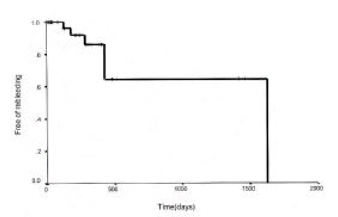
Variceal rebleeding occurred in six patients, five episodes of which were associated with shunt malfunction and responded to shunt revision. In the remaining one case, angiography was not possible because of septic shock. The cumulative probability of not bleeding at one and two years was 86% and 65% (Figure 4). Non-variceal hemorrhage occurred in two patients.
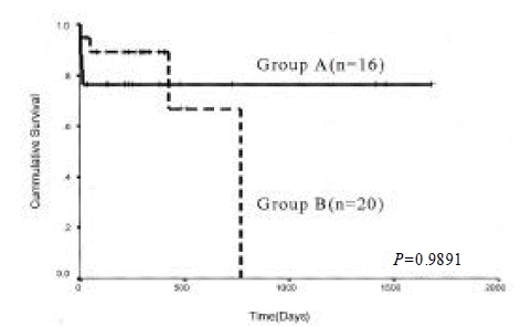
Six patients had hepatic encephalopathy before TIPS and it was aggravated in five patients after TIPS. Hepatic encephalopathy was newly developed in 15 patients after TIPS. In one patient, hepatic encephalopathy progressed and death ensued within two months. Most of them were controlled with medical treatments such as protein restriction and administration of lactulose. The presence of hepatic encephalopathy after TIPS, either aggravated status or newly developed encephalopathy, was not a significant predictive factor for survival (p=0.9891) (Figure 5).
Twenty-three episodes of angiographic evaluation were performed due to rebleeding or suggestion of stent malfunction in 16 patients. Overall 16 episodes of stent malfunction were diagnosed during follow-up (occlusion in 4, stent stenosis in 9, and hepatic vein stenosis in 2). Stent revision by means of angioplasty was successfully performed in 14 of these episodes while an additional stent was needed in 5.
Eleven patients (31%) had complications associated with the procedure. Accidental intrahepatic biliary duct puncture or hepatic capsule puncture was noted in eight patients (Table 3). There were two significant complications of the procedure. One patient developed retroperitoneal hematoma during the procedure but this complication was spontaneously resolved. The other patient developed hemobilia within 3 days after TIPS and died at the fifteenth day after the procedure.
DISCUSSION
Acute variceal bleeding refractory to endoscopic and pharmacologic treatment is a life-threatening complication of portal hypertension that produces a high mortality rate. Despite recent advances in endoscopic or pharmacologic treatment, a large number of episodes of variceal bleeding are refractory to conventional therapy1ŌĆō3). Theoretically, TIPS has advantages over shunt surgery in portal decompression without the risks associated with laparotomy in cirrhotic patients. It is, therefore, conceivable that TIPS would provide effective salvage therapy in patients at a high risk of dying after emergent surgery for active variceal bleeding despite conventional therapy14ŌĆō16). In this study, a selected group of patients, who continued to bleed despite endoscopic and pharmacologic treatments, were managed with TIPS.
Bleedings were arrested after TIPS creation in 34 of 36 patients with acute variceal hemorrhage. The TIPS procedures were associated with significant hemodynamic effects, consisting of a marked decrease in portosystemic pressure gradients and the grade of gastroesophageal varices, which were associated with a high hemostatic rate, similar to the rate obtained with shunt surgery5,17). Therefore, TIPS creation is a clearly effective means of controlling acute variceal hemorrhage.
PHG has been recognized as one of the sources of upper gastrointestinal hemorrhage. The direct relationship between values of portal pressure and severity of PHG has not been demonstrated18ŌĆō20). In only one report18), free PVP was significantly elevated in cirrhotic patients with severe PHG, whereas in other reports 19,20), there was no significant difference in free PVP with and without PHG. A few reports have described TIPS as an effective therapeutic means for PHG21,22). In this study, PHG improved in most cases after TIPS. Therefore, it may be possible that PHG is closely related to portal hypertension.
Gastric varices are the source of bleeding in 10ŌĆō36% of patients with variceal hemorrhage23ŌĆō27). Gastric varices have been reported to have a lower bleeding risk than esophageal varices; however, once bleeding occurs, patients with gastric variceal bleeding havea worse prognosis compared to patients with esophageal variceal bleeding24). It is well known that the hemostatic efficacy of endoscopic and pharmacologic therapy is lower in patient with gastric variceal bleeding than esophageal svariceal bleeding23ŌĆō26). Unfortunately, although surgery is highly successful in achieving hemostasis, it carries high rates of perioperative mortality5,28ŌĆō30). In the case of gastric variceal bleeding, we consider TIPS to be the treatment of choice, as indicated by the comparatively large proportion of patients in our series with gastric varices.
Newly developed or worsening spontaneous hepatic encephalopathy was seen in 55.6% of patients after TIPS, which was not a significant predictive factor for survival in this study. In all but one patient, the encephalopathy was transient and responded to simple medical treatments. Recent reports indicate that increasing age, liver failure and a previous history of encephalopathy are the main predictors of encephalopathy after TIPS31) and, perhaps, patients with these factors should have narrower stents placed.
Excluding hepatic encephalopathy and stenosis or occlusion of stent, the overall complication rate in several published series was less than 10%,9,14,16). Severe life-threatening complications occur in 1% to 2% of patients undergoing TIPS placement. These complications include hemoperitoneum, hemobilia, acute hepatic ischemia, cardiac puncture, pulmonary edema and septicemia9,14,16). In our series, serious procedurerelated complications occurred in only two patients (5.6%). Most complications were mild and spontaneously resolved. Therefore, TIPS is considered a safe procedure.
The incidence of rebleeding was reported to be between 6.7 and 38%32ŌĆō34). It developed in 14% of cases during a mean follow-up 12 months in our series. The proportion of patients who remained free of rebleeding one year after TIPS(86%) is close to the results in three previous studies14,15,33). Similar to other investigators33,34), we found shunt dysfunction in patients with variceal rebleeding. Shunt dysfunction is a major problem with the TIPS. The principle cause of rebleeding after TIPS is the recurrence of portal hypertension. This may result from TIPS thrombosis, stent retraction and kinking, or stent stenosis10). Angiography is the gold standard for the detection of stent malfuncton and is required for its treatment. However, angiographic procedure is expensive and invasive. In this study, therefore, regular examination by endoscopy and ultrasonography were performed for detection of stent dysfunction. Angiography was performed if bleeding recurred, or if ultrasonography or endoscopy suggested TIPS occlusion or stenosis, which was associated with a low frequency of radiologic procedures. Shunt patency can be easily recovered in most cases.
Our one and two year survival rates of 83% and 74%, respectively, are similar to the recent report by Merli et al.35) (84%, 73%, respectively). In other study, LaBerge et al.33) and Stanly et al.34) are reported lower than our results. But Garcia-Villarreal et al.36) recently reported a higher survival rate than our results. The difference of survival rate among the trials considered to be due to difference of study design; emergency or elective bases of the procedure, rate of shunt revision, and severity of the liver disease of patients.
In conclusion, TIPS is an effective and reliable nonoperative means of lowering portal pressure. This procedure has proved useful in the management of acute variceal bleeding refractory to endoscopic treatment. The major advantages of the procedure include comparatively low procedural morbidity and wide applicability. However, shunt dysfunction is common. Surveillance by ultrasonography or endoscopy and angiographic interventions are, therefore, necessary.
 PDF Links
 PubReader
 ePub Link
 Full text via DOI
 Download Citation
 Print
Print


