


Introduction
Ectopic gastric mucosa in the esophagus is considered to be the residue of columnar epithelium of the embryonic esophagus. Replacement of columnar epithelium by stratified squamous epithelium begins in the mid esophagus and extends proximally and distally. Therefore, rests of columnar epithelium occasionally may persist in the upper and distal end of the esophagus1). As shown in autopsy and endoscopic studies, ectopic gastric mucosa can occur as solitary or multiple mucosal patches, most frequently in the upper third of the esophagus, near the level of the cricopharyngeal muscle. It is frequently overlooked on routine endoscopic examination because of technical problems in observing the upper esophagus and its asymptomatic nature. However, certain complications have been reported, including dysphagia2), esophageal stricture3), tracheoesophageal fistula4), adenocarcinoma5) and ring6). It has been suggested that these complications develop from acid secretion from the heterotopic gastric mucosa. We report a case of heterotopic gastric mucosa in the upper esophagus with the secretion of acid demonstrated by continuous ambulatory pH monitoring.
CASE REPORT
A 33-year-old man visited the gastrointestinal department of the Gil Medical Center with throat discomfort for the previous month. He denied symptoms of dysphagia, but suffered from occasional hoarseness, coughing and heartburn. There was no known history of excessive alcohol intake and smoking. His past medical history and family history were unremarkable. Physical examination revealed that the vital signs were stable and the patientŌĆÖs pharynx and abdomen appeared unremarkable.
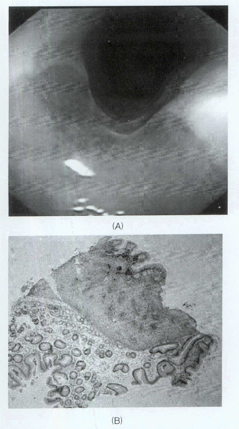
On gastroesophageal endoscopic findings, there was no definite abnormal findings on the lower esophagus and stomach. Upon withdrawal of the endoscope, a patch of salmon colored epithelium was clearly demarcated from the surrounding normal mucosa in the upper esophagus (Figure 1A). Its diameter was between one-quarter and one-half fully expanded, and it was located just below the upper esophageal sphincter. Endoscopic biopsy finding from the patches revealed gastric-type columnar epithelium with parietal cells and Helicobacter pylori was not found on the Giemsa stain (Figure 1B).
A 24-hour ambulatory pH monitoring was performed to demonstrate acid secretion from this patch. Esophageal manometry was performed to confirm the level of the lower and upper esophageal sphincters. The level of the lower esophageal sphincter was 39 to 42 cm and that of the upper sphincter was about 19 cm from the incisor.
A dual-probe antimony pH catheter with a diameter of 2.1 mm (Synectics Medical Inc., Irving, Tx.) was placed transnasally after an overnight fast. The distal probe was placed at 5 cm above the manometrically defined lower esophageal sphincter. The proximal electrode was placed at 19 cm from the incisor, which corresponded approximately with the endoscopic findings of the heterotopic gastric mucosa. The pH values from both intraesophageal electrodes were recorded continuously on an ambulatory Mark III Digitrapper (Synectics Medical Inc.), and the data were downloaded to an IBM-compatible computer for calculation and analysis by Gastrosoft (Synecitics Medical Inc.).
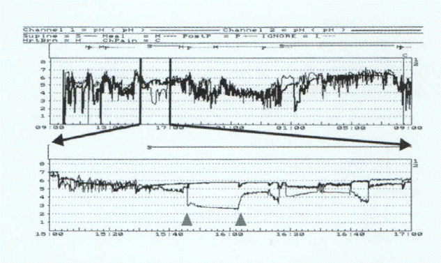
A 24-hour ambulatory pH monitoring showed that the pH in the proximal esophagus less than 4 (including supine and upright positions) were 3.8% of the time in total (normal value < 1), 3.3% (normal value < 1.3) in the upright position, and in 4.6% (normal value is 0) in the supine position, respectively. The pH less than 4 in distal esophagus was 9.2% (normal value < 4.5) of the time in total (including supine and upright positions), 13.1% (normal value < 6.3%) in the upright position and in 3.5% (normal value < 1.2) in the supine position, respectively. Despite the increased acid exposure in the proximal pH probe, there was no acid reflux in supine position from 15:45 to 16:10 (Figure 2). The patient was treated with 40 mg of omeprazole, once daily, which resulted in improvement of the throat discomfort, hoarseness and cough.
DISCUSSION
The occurrence of gastric fundic type epithelium in the esophagus has been noted since the 1800s. As opposed to BarrettŌĆÖs esophagus, gastric epithelium in the upper esophagus is thought to be a developmental anomaly. During embryonic development, the esophageal mucosa is initially lined with columnar epithelium, which is later replaced with a stratified squamous epithelium. The replacement begins in the middle of the esophagus and spreads gradually in both the caudal and cephalic directions. The occurrence of gastric fundic type epithelium in the upper esophagus is, therefore, thought to represent a failure of this spread to reach the cephalic end of the esophagus.
The prevalence of inlet patch on endoscopy varies considerably from 3.8%7) to 10%8). In most cases, the patients were thought to be asymptomatic, but in some cases the inlet patch causes pharyngeal and upper esophageal symptoms. The proposed mechanisms of the symptoms are cricopharyngeal spasm by acid secretion9) and increased tone of upper esophageal sphinicter in response to acid stimulation3). Since complications associated with heterotopic gastric mucosa have been reported2ŌĆō6), they are presumed to be related to acid secretion by this ectopic mucosa.
Some investigators have tried to demonstrate the secretary capability of this mucosa by different methods. Jabbari and Goresky7) measured the pH profile from the stomach to the esophageal inlet patch in five patients 30 minutes after administering 10 ╬╝g/kg of pentagastrin intravenously. The pH values decreased in the area of the heterotopic gastric mucosa in two patients with large patches. Hamilton and Thune10) used a sensitive stain and pentagastrin stimulation to study the production of acid by this mucosa. Four patients were infused with 1% congo red solution in the area of the inlet patch after pentagastrin infusion. A darkening of the stained area (consistent with a pH < 4.5) occurred in all patients. Nakajima and Munakata11) measured the pH profile of the esophagus in five patients with heterotopic gastric mucosa after tetragastrin stimulation, using a pH electrode attached to a standard fiberscope. Significant reduction in pH was observed at the heterotopic gastric mucosa or adjacent areas in three of five patients, and was confirmed with congo red staining. Galan et al12) reported a case of heterotopic gastric mucosa in the upper esophagus, where the acid secretion was demonstrated by continuous 24-hour pH monitoring, complicated with an upper esophageal stricture and dysphagia. These previous studies suggest that heterotopic gastric mucosa in the upper esophagus is capable of secreting acid. Furthermore, it was believed that this proximal acid exposure was not caused by acid reflux because only the upper esophageal pH dropped without falling in the lower esophageal pH. The normal pH finding in lower esophageal probe may be due to a relatively small volume of acid, which can be easily diluted and neutralized by saliva, and which is then rapidly cleared. In our case, the patient complained of pharyngeal symptoms and cough, and was diagnosed with a large inlet patch and showed pH discrepancy in the upper and lower esophagus. Despite the increased acid exposure in the distal esophagus, the proximal acid reflux could not be explained because there was no correlation between the proximal and distal acid exposure. The pharyngeal and laryngeal symptoms caused by inlet patch are relieved after administration of H2 antagonist or proton pump inhibitor. It leads to the speculation that the extraesophageal manifestations may also have been related to the acid secretion of the heterotopic gastric mucosa. Also the patientŌĆÖs cough and hoarseness resolved with proton pump inhibitor therapy dramatically in our case.
In summary, we report a case of heterotopic gastric mucosa in the upper esophagus, where the acid secretion was demonstrated by continuous 24-hour pH monitoring. In the supine position, the upper esophageal pH dropped below four without falling in the lower esophageal pH, and the patientŌĆÖs throat discomfort was improved after aggressive acid suppressive therapy.
 PDF Links
 PubReader
 ePub Link
 Full text via DOI
 Download Citation
 Print
Print


