


Introduction
Malignant proliferating trichilemmoma is a rare neoplasm derived from the outer sheath of the hair follicle. This condition preferentially arises in areas of dense hair follicle concentration; therefore 90% occurs on the scalp. It is usually a solitary lesion and most commonly seen in elderly women. The usual clinical presentation is a subcutaneous cystic nodule that has been present for years and slowly progresses to a large, nodular mass, often following a history of trauma or inflammation.
Histologically, the growth is made up of a massive proliferation of the outer sheath epithelium, foci of trichilemmal keratinization, palisading nuclei on the peripheral side with nuclear atypia and surrounding tissue invasion. The lesion may lead to a false impression of squamous cell carcinoma due to the presence of cellular atypia, dyskeratotic cells and mitotic figures. While the majority of these neoplasms are considered biologically benign, there are rare reports of local recurrences and distant metastases1–3). We experienced a patient displaying a scalp lesion with multiple distant metastases, who achieved a partial response following combination chemotherapy.
Case Report
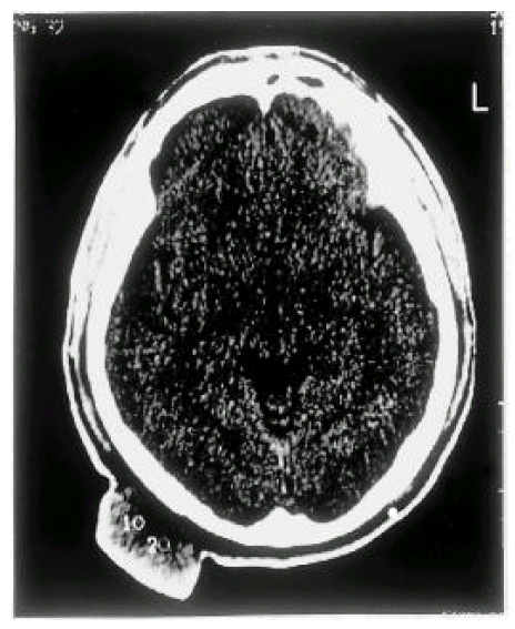
A 32-year-old Korean man had a large soft nodule on the right parietal scalp for 12 months. The lesion had been rapidly growing for the previous 3 months. It was covered with areas of superficial erosion and crusting. Upon examination, he appeared well overall but a round, subcutaneous, firm 8 cm-sized mass was palpated in the occipital scalp. No other regional lymphadenopathy was detected. Brain CT was performed (Figure 1). Additionally, under general anesthesia, a wide excision was performed with a 1cm margin. Frozen section examination of the margins confirmed adequate clearance. The biopsy specimen was diagnosed as a malignant proliferating trichilemmal tumor. The patient was hospitalized 4 months after wide excision due to a recurrence in the postauricular, suboc cipital area of a hard, fixed nodule measuring 2.5×1.0cm in maximal diameter. Aspiration cytology was accomplished, and the cytologic finding exhibited a metastatic malignant proliferative trichilemal tumor. Surgical extirpation was performed.
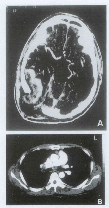
The patient revisited our clinic complaining of severe headache and nausea 6 months after surgical extirpation. Brain CT and chest CT were performed(Figure 2A, B). The brain CT imaging revealed a right superior cerebral, right inferior temporal-parietal parenchymal mass and a left superior frontoparietal scalp mass. Additionally, chest CT revealed multiple lymphatic metastases. The patient underwent partial resection of the tumor and a decompressive operation. He was treated with cisplatin (75mg/m2, day 1), etoposide(100/m2, days 1–3) combination chemotherapy, and achieved a partial response(Figure 3A, B). He has been followed up for 6 months without progression.
Histopathology
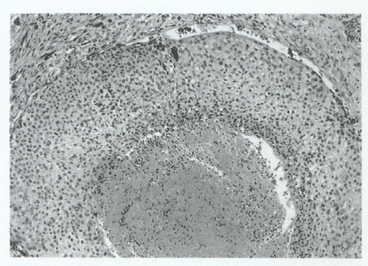
The excised specimen of the tumor consisted of an ovoid mass-like soft tissue, covered by an elipse of skin, measuring 8×6.5×4cm. Upon section, the cut surface of the mass is yellow-white and relatively homogeneous. The tumor is composed of a mainly lobular arrangement of large eosinophilic, sometimes clear squamoid cells and scattered small nests of small hyperchromatic round or somewhat spindle cells in a cellular and fibrotic stroma. The lobule of squamoid cells shows abrupt trichilemmal keratinization and eosinophilic necrosis in the center(Figure 4). The squamoid cells show marked nuclear pleomorphism and frequent mitoses(Figure 5). At the periphery of the lobule, there are transitions from large squamoid cells to small atypical hyperchromatic cells, or plump spindle cells. Immunochemistry was done in order to identify the origin of tumor cells. It disclosed positive staining for cytokeratin and epithelial membrane antigen(EMA) and negative staining for neuron-specific enolase (NSE) and chromogranin. The findings were consistent with a light microscopic diagnosis of malignant proliferating trichilemmal tumor.
Discussion
The proliferative trichilemmal tumor is an uncommon neoplasm, derived from the outer root sheath of the hair follicle4). Brownstein and Arluk5) reported 50 patients, 84% of whom were women. In about 90% of all reported cases, the lesion is located on the scalp, with the remainder on the back, forehead, wrist and chest6). It usually consists of a solitary lesion and the usual clinical presentation is a subcutaneous cystic nodule that has been present for years and slowly progresses to a large, nodular mass, as a consequence of a history of trauma or chronic inflammation. Proliferating trichilemmal tumors can become large if neglected; a lesion as large as 25cm has been reported7).
Histologically, the growth is made up of massive proliferation of pilar sheath epithelium showing multiple central areas of trichilemmal keratinization and formation of homogeneous keratin cysts8). The tumor masses are surrounded by a layer of basaloid cells in which the tumor cell nuclei palisade and the cell membranes rest over an outer layer of PAS-reactive basement membrane9). Trichilemmal keratinization, as demonstrated by keratin foci without keratohyaline granules, usually is observed at the center of the cyst, and pale-staining, glycogen-rich clear cells also are a frequent finding. This is a peculiar form of cell maturation normally observed in the stratified epithelium of the outer root sheath of hair follicle5,6,10–12). Occasional cells with hyperchromatic nuclei, mitotic figures and scattered dyskeratotic cells may be present, but these are not indicative of malignant transformation11). Saida and colleagues3) classified the oncological development of the trichilemmal type of tumor into three stages: 1)trichilemmal cyst, adenomatous stage: 2)proliferating trichilemmal cyst, epitheliomatous stage; and 3)malignant proliferating trichilemmal cyst, carcinomatous stage. Mehregan and Lee9) suggested that the malignant transformation of a proliferating trichilemmal tumor should be considered in cases of longstanding cystic and nodular lesions that demonstrate rapid growth or exophytic enlargement.
A truly malignant proliferating trichilemmal tumor is a rare incidence3). Amaral et al2) have reviewed seven cases of malignant proliferating trichilemmal tumors reported in the literature and added a case of their own with a lesion originating in the inguinal area and ending with pulmonary and liver metastasis.
In Korea, two cases13,14) have been reported. Both cases received surgical excision and one case14) received palliative radiotherapy. In our case, the tumor grew rapidly and metastasized to multiple distant sites following wide excision. The efficacy of chemotherapy in metastatic disease cannot be evaluated because of the small number of published cases. Weiss et al15) reported that one case responded to chemotherapy with cisplatin and fluorouracil. Our patient was treated with cisplatin and etoposide combination chemotherapy, and achieved a partial response. Malignant proliferating trichilemmal tumor is a rare disease and its pathogenesis has not been completely elucidated. We experienced a case of malignant proliferating trichilemmal tumor that showed a partial response to chemotherapy, suggesting that some of these tumors may respond to aggressive treatment with chemotherapy.


 PDF Links
 PubReader
 ePub Link
 Full text via DOI
 Download Citation
 Print
Print


