Idiopathic Granulomatous Hepatitis Manifested with Fever of Unknown Origin:
— Case Report —
Article information
Abstract
Idiopathic granulomatous hepatitis is a rare disease of unknown cause that is characterized by recurrent fevers and granuloma in the liver. Attempts to define an exact etiology of the fever of granulomatous hepatitis frequently do not yield a precise diagnosis. Idiopathic granulomatous hepatitis was confirmed after a thorough work up and negative cultures and serologies were obtained, and in the absence of another condition that could lead to granulomas in the liver.
We have experienced a 67-year-old female patient who presented with prolonged fever for 2 months and revealed granuloma in liver biopsy. She was treated with glucocorticosteroid and defervescence resulted.
INTRODUCTION
The syndrome of idiopathic granulomatous hepatitis as a cause of fever of unknown origin is now well established1–3) but only a few follow-up reports have established the clinical outcome.
Idiopathic granulomatous hepatitis is a rare disease of unknown cause that is characterized by recurrent fevers and granuloma in the liver. Establishing the diagnosis may be laborious because other diseases that cause hepatic granulomas (such as lymphoma, sarcoidosis, drug allergy and fungal or mycobacterium infections) must be excluded. The finding of granulomas in the liver frequently correlates with a systemic disease previously diagnosed in a patient. However, liver granulomas may reflect an occult systemic disease or may be associated with no detectable underlying process. When no cause is obvious, these granulomas are often manifested by nonspecific symptoms, such as fever, malaise and anorexia, or by asymptomatic liver function abnormalities. Previous reports have underscored the diversity of etiologic factors4). In these studies, no primary cause could be found after extensive workup in 5 to 36%. Other groups reported that idiopathic granulomatous hepatitis was the most common (50%) cause in their study5).
We report a case of idiopathic granulomatous hepatitis manifested with fever of unknown origin.
CASE REPORT
A 67-year-old Korean female was admitted to our hospital because of intermittent fever for 2 months.
Four years before admission, she had visited a hospital because of fever and abdominal pain, and cholecystectomy was done due to acute calculous cholecystitis.
The fever was persistent and she had taken oriental herb medicine, thereafter, she had been in excellent health until 2 months earlier, when diabetes mellitus developed which was treated by oral hypoglycemic agent. In spite of enthusiastic management in the private clinic, fever developed again and was persistent.
The patient was a housewife, non-smoker and did not drink at all. There was no history of pulmonary tuberculosis. She complained of general weakness, fatigue, fever, weight loss (2.5 kilograms over 2 months) but denied cough, dyspnea, abdominal pain, nausea, vomiting, hemoptysis, frequent voiding and bladder or bowei dysfunction. The body temperature was 37.5°C, the pulse rate was 100/min, the respiration rate was 18/min and the blood pressure was 110/60 mmHg. On physical examination, she was pale and chronically ill-looking with alert mental state. She had pale conjunctiva and dehydrated tongue, and lymph nodes were impalpable. The lung, heart and abdomen were normal except for post-operative abdominal scar on the right upper quadrant.
The hemoglobin was 10.5 g/dl and the hematocrit 31.4%. The white cell count was 6,700/mm3, with 77.9% neutrophils, 0.9% lymphocyte, 6.9% monocytes and 2.3% eosinophils. The platelet count was 206,000/mm3. The erythrocyte sedimentation rate was 49 mm/hr. The aspartate aminotransferase (AST) and alanine aminotransferase (ALT) were 13.6 and 9.0 IU/L, the alkaline phosphatase 71.2 IU/L, the total protein and albumin 6.1 and 3.3 g/dl, the direct bilirubin 0.2 mg/dl, the blood urea nitrogen and creatinine 5.9 and 0.5 mg/dl, the total cholesterol 134.1 mg/dl and total calcium was 7.6 mg/dl. The urine was normal. The alphafeto protein was below 5 ng/ml and the carcinoembryonic antigen was 1.44 ng/ml.
The thyroid function test was normal. The angiotensin converting enzyme was 23.1 U/ml (normal: 8–52U/ml). The brucella antibody was negative. The anti-mitochondrial antibody was negative. The sputum smear and culture for mycobacteria and fungus were negative. The chest X-ray showed a fibrosteak density in the right upper lobe and increased interstitial marking in both lungs. This finding was suggestive of inactive pulmonary tuberculosis (Fig. 1). The abdominal CT (computerized tomography) showed mildly dilated both intrahepatic ducts without internal calcific density and the common bile duct was not dilated (Fig. 2). The bone marrow biopsy showed normocelluarity (about 30% cellularity) with polymorphous cellular population and adequate megakaryocytes. The culture of bone marrow grew neither bacterium nor fungus. The needle biopsy of the liver showed relatively intact lobular architecture and hepatic cord arrangement and the portal area was some-what widened by infiltrated inflammatory cells and fibrosis, but the limiting plate was intact. There were a focal hepatocytic necrosis, perivenular cholestasis and a few ill-defined aggregates of epitheloid cells. No acid-fast bacillus was found (Fig. 3).
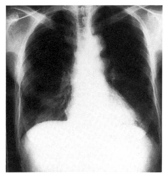
Chest X-ray showed fibrostreak density in right upper lung and increased interstitial marking in both lungs.

Abdominal CT scan with contrast enhancement showed mild dilated both intrahepatic ducts without internal calcific densities.
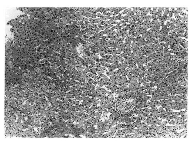
Low power view of the liver showed a focus of granuloma in the lobule (H-E, ×100)
The antituberculosis medication was prescribed for 2 weeks as a therapeutic trial. Even after that, the temperature was 38.7°C. Thereafter, we prescribed prednisolone 1 mg/kg under the diagnosis of idiopathic granulomatous hepatitis.
DISCUSSION
We and other authors6) have defined idiopathic granulomatous hepatitis as hepatic granulomas with no cause or comorbid condition discernible on extensive evaluation. Because of the diverse etiology, the clinical features of chronic granulomatous hepatitis are sufficiently indistinct as to make establishment of the exact cause in a given case very difficult.
Many researchers have reported various results, and they found granulomas in 2–35% of liver biopsies and up to 30% of these cases remain idiopathic7–9). The microscopic appearance of hepatic granulomas from differing causes is not specific enough to permit etiologic classification by histologic criteria alone. Moreover, only infrequently does one find acid-fast bacilli or fungi in special stains; when a specific etiology is found on a liver biopsy, this information is very useful. The culture of liver biopsied tissue grew mycobacteria in < 10% of submitted specimens from patients with tuberculosis.
As a result of the release of interleukin-1 and other endogenous pyrogens from activated mononuclear phagocytes in the granulomas10). These agents act upon the hypothalamus to alter the thermoregulatory set point. The fever associated with granulomatous hepatitis is often relapsing in character, but continuous and remittent fever usually developed11). The immunopathogenesis of hepatic granuloma formation has been the subject of several reviews. In brief, many different antigens can act as stimuli to initiate the granulomatous inflammatory response. These antigens can be infectious agents of viral, bacterial, fungal or parasitic origin. Immune complexes, medications, foreign bodies and malignancies can also initiate granuloma formation. Continued exposure to the antigen results in accelerated and idiopathic granulomatous hepatitis.
Classic sarcoidosis was effectively ruled out in the idiopathic groups by the clinical absence of adenopathy, iritis, rash, arthropathy, pulmonary infiltrates, eosinophilia and hypercalcemia6). Serum angiotensin-converting enzyme (ACE) levels were not measured, since ACD levels do not have sufficient positive or negative predictive values to be useful in establishing or ruling out the diagnosis of sarcoidosis. The relatively low frequency of abnormal findings on chest roentgenograms and increased levels of angiotensin converting enzyme among patients with sarcoidosis may be attributed to the quiescent nature of pulmonary involvement in thest patients6).
The diagnosis of primary biliary cirrhosis was based primarily on an increased antimitochondrial antibody. Rarely, the histologic features of primary biliary cirrhosis may be partly or predominantly granulomatous. Some drugs are known to cause granulomatous hepatitis and fever of unknown origin. Among them, methydopa is a well-known cause of both granulomatous hepatitis and fever, and there are reports of granulomatous hepatitis secondary to hydrochlorothiazide, benzodiazepines and allopurinol, as well13).
On the basis of several studies, authors recommend that an evaluation for granulomatous hepatitis include at least chest roentgenography, appropriate cultures for bacteria (including Brucella), mycobacteria and fungi; serologies for Q fever, Brucellosis, syphilis, hepatitis B and hepatitis C; a tuberculin skin test with controls, and an assay for antimitochondrial antibody.
When no etiology for granulomatous hepatitis is found, the clinician must make up his mind about therapy for the febrile patient. Our patient was managed with glucocorticosteroid. Prednisolone is safe and highly effective in patients with idiopathic granulomatous hepatitis and FUO if an infectious etiology has been excluded.
In our case, we recommend oral prednisolone in a dose of 0.75–1.0 mg/kg/day. This generally results in a prompt defervescence and symptomatic improvement. Careful follow-up is critical to avoid an advancing occult infection. After 3–4 weeks, the dosage can be slowly tapered until the glucocorticosteroid has been discontinued or fever returns. If that happens, and there is no suggestion of infection, the dosage of glucocorticosteroid can be increased, and maintenance dose prescribed along with regular follow-up for sign of glucocorticosteroid toxicity. After several months of maintenance therapy, the dosage of glucocorticosteroids usually can be tapered and discontinued while the clinical response is observed. In a few patients, continuous glucocorticosteroids are required. Indomethacin has not been used frequently. However, on theoretical grounds, it offers several advantages over glucocorticosteroids, since indomethacin has none of the immunosuppressive effects of prednisolone.
The meticulous and regular examination of the patient would prevent the complication of the disseminated mycobacterial or fungal infection14,15).
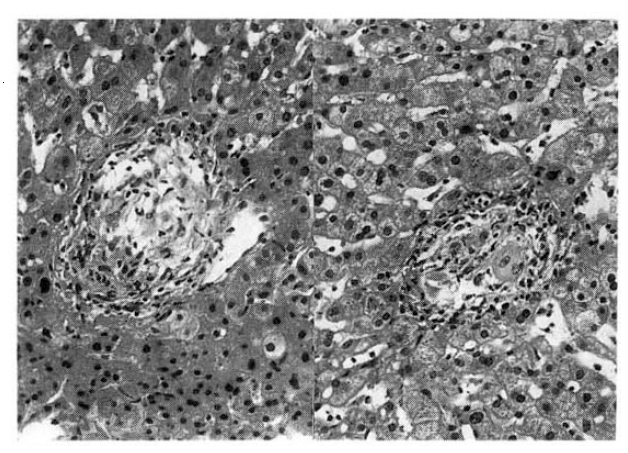
The granuloma consisted of epithelioid cells, multinucleated giant cells, and surrounding lymphocytic infiltration and fibrosis (H-E, ×400)