An Adult Case of Pulmonary Sling with Complete Tracheal Ring
Article information
Abstract
Pulmonary sling is a rare congenital condition in which the left pulmonary artery arises from the right pulmonary artery forming a sling around the trachea causing tracheal compression. Many cases are associated with cardiovascular and tracheo-bronchial anomalies. When combined with complete tracheal ring, it is called “ring-sling complex”. The average onset of symptoms is 2 months and about half have symptoms from birth. The anomaly, if not corrected, can be fatal within the first year of life. In an adult, it is most often asymptomatic and usually found incidentally. We report an adult case who was diagnosed as pulmonary sling with complete tracheal ring by CT and bronchoscopy along with a literature review.
INTRODUCTION
Pulmonary sling is a vascular abnormality wherein the left pulmonary artery arises from the right pulmonary artery and then traverses between the esophagus and the trachea toward the hilum of the left lung. This produces a sling around the distal trachea and the proximal main bronchi. It was first described by Glaevecke and Doehle in 1897. Since then, about 100 cases have been reported in the world1). The average age at the onset of symptoms, such as respiratory distress and cyanosis, is 2 months and about half have symptoms from birth. Many patients die within the first year of life. So, adult cases are very rare, are most often asymptomatic and usually found incidentally2). Many cases are associated with cardiovascular and tracheobronchial anomalies. When combined with complete cartilaginous tracheal rings in the absence of posterior membranous portion, it is called “ring-sling complex”. In that case, respiratory distress is severe and prognosis is poor1).
We here report an adult case who was diagnosed as pulmonary sling with complete tracheal ring by CT and bronchoscopy along with a literature review.
CASE REPORT
A 21 years old female was admitted for the evaluation of mild dyspnea. She had had symptoms of airway obstruction, such as wheezing, hoarseness and mild dyspnea since a young infant. She was premature at delivery with body weight of less than 2 kg. There was no family history of congenital anomalies.
On admission, blood pressure was 140/90 mmHg, pulse rate was 80/min, body temperature was 36.5°C and respiration rate was 16/min.
On physical examination, she was alert, but had a slight dyspneic appearance. There were no cyanosis, chest deformities and cardiac murmurs.
Laboratory findings on admission were within normal ranges. There were no abnormalities on EKG. FEV1 was 1.52L (66.2% of predicted value) and FVC was 1.90L (54.5% of predicted value) on pulmonary function test.
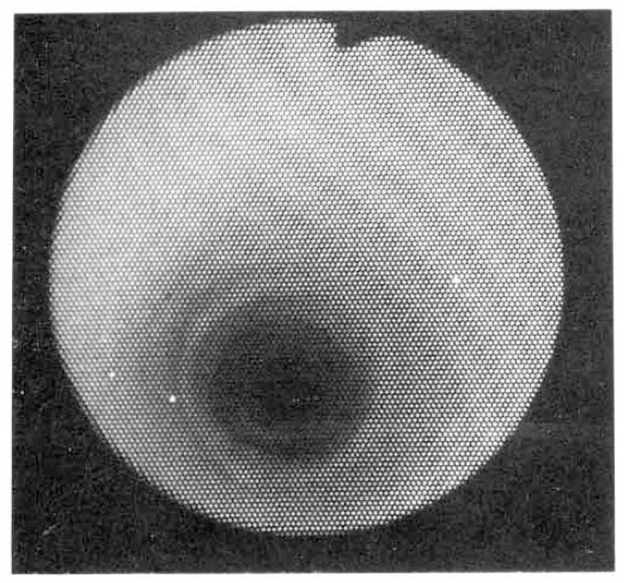
On bronchoscopy, the trachea was too narrow from the initiation to pass through. Complete cartilaginous ring without posterior membranous portion was found on the entire trachea (Fig. 1).
The plain chest X-ray showed a right mediastinal mass-like density and a long-segment tracheal narrowing (Fig. 2). The Chest CT revealed the anomalous left pulmonary artery originating from the right pulmonary artery and its posterior course between the trachea and the esophagus (Fig. 3).

Chest CT findings Anomalous left pulmonary artery originating from the right pulmonary artery and crossing between the trachea and the esophagus toward the hilum of the left lung.
She did not have any other cardiovascular and gastrointestinal anomalies. She was discharged without specific treatment because of mild symptoms which are mainly caused by complete tracheal ring and not by tracheal compression due to vascular anomaly.
DISSCUSSION
The term “Vascular Ring” was introduced to describe the mediastinal vascular anomalies causing tracheobronchial compression by Dr. Robert Gross who reported the first successful operation of a vascular ring. The pulmonary sling is a kind of vascular ring and the term was first used by Contro et al. in 1958 to distinguish it from other vascular anomalies4). Carl et al. reported that when vascular rings were classified into four groups, as follows, 1) complete vascular rings with double aortic arch or right aortic arch 2) pulmonary sling 3) innominate artery compression and 4) miscellaneous, pulmonary sling was about 4.5%5).
Embryologically, it has been suggested that it is caused by a maldevelpment of the left ventral pulmonary artery bud due to growth failure or reabsorption. The left lung may capture its arterial supply by connection of the left postbronchial plexus with the right sixth arch through capillaries caudal to the lung bud. Then, as the lung bud grows caudally, the course of the left pulmonary artery is behind the developing trachea-bronchial tree6). About half have cardiac or tracheo-bronchial anomalies which determine the prognosis. According to Christoph Dohleman’s review, the associated cardiac anomalies were 49% and among those, patent ductus arteriosus, ventricular septal defect and atrial septal defect were most common. Tracheo-bronchial anomalies were associated in 56%. Tracheal stenosis with complete tracheal rings was the most common anomaly, followed by stenosis of main stem bronchus, tracheal bronchus and tracheo-esophageal fistula7).
Wells et al. proposed a classification system assigning pulmonary sling without complete tracheal rings to type 1 and pulmonary sling with complete tracheal ring to type 2. corresponding to the ring-sling complex. Ring-sling complex can be classified into a lethal form and a form in which there is enough pars membranacea left to allow sufficient tracheal growth. The two forms can be distinguished only by their clinical course, i.e., either severe respiratory symptoms with progression or mild symptoms without progression8).
Pulmonary sling can be diagnosed by angiography, CT or MRI. Bronchoscopy and echocardiography should be performed to evaluate the combined cardiac or tracheo-bronchial anomalies. The plain radiographic features include unequal aeration due to compression of the right main bronchus by the anomalous vessel, low carina, horizontal equal-length right and left mainstem bronchi and long-segment tracheal stenosis. Pulsating mass compressing esophagus can be visualized on fluoroscopy with barium esophagography9). Most common finding in an asymptomatic adult is a right mediastinal mass. Diagnostic considerations such as dilated azygous vein, lymphadenopathy, foregut cyst or esophageal neoplasm should be excluded10). We found a right mediastinal mass in our case. Most cases can be diagnosed by CT or MRI, but misinterpretation can occur because of a bifurcation-like structure of the pulmonary artery. The left pulmonary artery can be missed because of its different level of crossing the carina or main bronchus. Slice thickness should be as little as 3 mm11,12).
Potts et al. was the first to propose the now most common corrective surgical procedure: division and reimplantation of the anomalous left pulmonary artery into the pulmonary trunk anterior to the trachea with division of the ligamentum arteriosum13,14). Because of high operation mortality and loss of function of the left lung due to obstruction of the reconstructed vessel, a conservative approach with symptomatic management was recommended initially15,16). But recent development of operation technique has resulted in low operation mortality and good patencty of the reconstructed vessel. So, in view of the seriousness of this condition and the fact that reports of surgical management have been encouraging, current policy is to offer an operation as soon as the diagnosis is made17).

