Multiple Myeloma Presenting as a Testicular Mass
Article information
Abstract
Primary testicular plasmacytoma is extremly rare and only a few cases have been reported1–5). Testicular involvement in the course of multiple myeloma is just as rare and it implies a poor prognosis, expressing an acceleration of the malignant process6–9).
We present the case of a patient with multiple myeloma presenting as a testicular mass. The literature is reviewed and testicular involvement in plasma cell neoplasms is discussed.
INTRODUCTION
Most plasma cell neoplasms form multiple tumors in the bone and cause a generalized marrow plasmacytosis. These neoplasms rarely present as identified most frequently in the upper respiratory tract and have also been recorded in other organs, including lymph nodes and spleen, skin, gastrointestinal tract, and thyroid3,10,11).
On the other hand, extramedullary involvement in the course of multiple myeloma, in the form of macroscopic and or microscopic plasma cell infiltrates, is observed in approximately 2/3 of the patients with myeloma and who are subject to autopsy8). The localization can be limited to the paraskeletal areas or involve distant organs and tissue-the spleen, liver, lymph nodes and kidney, in particular. However localization in the testis is relatively rare. In addition, most of them were detected at autopsy and symptomatic plasma cell neoplasms of the testis were only a few. Kapadia detected the presence of microscopic infiltration of extraosseous tissues in 67% of patients with multiple myeloma at autopsy and testicular involvement appeared in only 6 cases6).
Primary testicular plasmacytoma is even more rare; Corwin reviewed 288 cases with solitary plasmacytoma involving various organs. None of them involved the testis.
The prognosis of plasma cell neoplasm invading extramedullary soft tissues, solitary or otherwise, is poor. The poor prognosis is certainly characteristic of cases with testicular plasmacytoma7–9).
We therefore report a patient with multiple myeloma, presenting as a testicular mass, who expired only 6 months after diagnosis in spite of chemotherapy.
CASE
A 49-year old man was admitted to our hospital because of painless swelling of both testes and multiple bone pain. Five months before admission, he noticed a slight enlargement of the right testis. The enlargement of the right testis progressed slowly and, a few weeks later, the left testis was enlarged also.
On admission, he showed an acutely-ill looking appearance and was slightly thin. The vital signs were within normal range. There were no abnormal findings in skin and hair. He had slightly pale conjunctivae and anicteric sclerae. Peripheral lymph nodes were not palpable. His breath sound was clear and no adventitious sound was heard. His heart beat was regular and no murmur was heard. On abdominal palpation, liver, spleen or kidney were not palpable.
Both testes were nearly the same in size and were palpable as nontender, hard, movable masses. The size of the right and left testes were about 10 cm×15 cm and 10 cm×12 cm in diameter, respectively. Bony tenderness was prominent especially on both thighs.
Laboratory findings were as follows; Complete blood counts were hemoglobin 9.5 gm/dl, hematocrit 28%, leukocyte 14,000/mm3 and platelet 140,000/mm3. Blood chemistry tests showed Ca 11.7 mg/dl, P 6.4 mg/dl, total protein 9.5 g/dl, albumin 2.9 g/dl, LDH 509 IU/I, BUN 55 mg/dl, creatinine 7.1 mg/dl, IgG 3,460 mg/dl, IgM 64 mg/dl, IgA 43 mg/dl, IgD 1.1 mg/dl. He had proteinuria (5,905 mg/24 hours) and creatinine clearance was measured as 9 ml/min. Serum protein electrophoresis showed M-peak in γ-region and M-component was measured as 43.4%. Serum Immunoelectrophoresis showed monoclonal gammopathy in IgG, k band. Urine PEP and IEP showed kappa type light chain (M-peak). Radiologic examination revealed multiple punched-out osteolytic lesions in the skull. Bone marrow aspiration showed 65% plasma cell infiltration. Bone marrow biopsy revealed hypercellular marrow packed with immature plasma cells. Testis ultrasonography showed heterogeneous hypoechogenicity. Testicular needle biopsy showed diffuse infiltration of atypical large lymphoid cells with feature of poorly differentiated plasma cell nature between seminiferous tubules and inside of them as well (Fig. 1). Immunohistochemical studies revealed positivity for L26 (CD20) and Ig k predominance on infiltrating neoplastic plasmacytoid cells (Fig. 2).
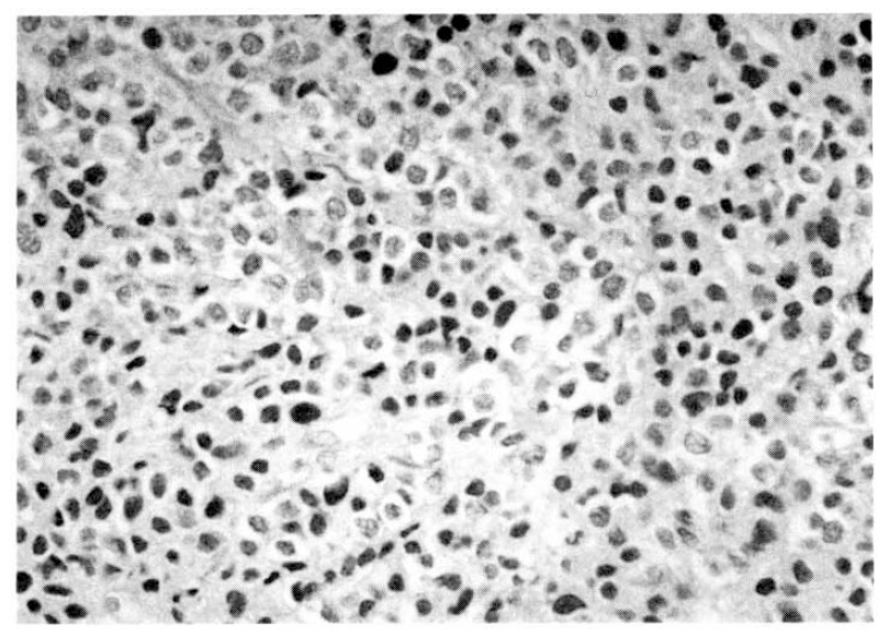
Microphotograph of H&E section shows diffuse infiltration of atypical large lymphoid cells suggestive of poorly differentiated plasmacytic nature. Reminiscence of seminiferous tubular structure is also noted in the left lower part of the figure.

Microphotograph of immunohistochemical stain for L26(CD20) shows positive findings on scattered neoplastic plasmacytoid cells.
The patient was initially treated with combination chemotherapy of vincristine, melphalan and prednisolon. After the 2nd cycle of treatment, the testicular enlargement was not decreased and multiple bone pain was aggravated. Palliative radiotherapy was given on both testes with some improvement. After that, he was treated with combination chemotherapy of vincristine, adriamycin and dexamethasone. One month later, however, numerous soft tissue nodules newly appeared on the left forearm, both lower legs, and chest wall. Somewhat later, follow-up chest x-ray showed multiple, variable sized round opacity in both whole lung fields with pleural effusion. The patient had a rapidly progressive downhill course and died 6 months after initial diagnosis.
DISCUSSION
The reported incidence of extramedullary lesions in multiple myeloma ranges from 11 to 73%, with the liver, spleen, lymph nodes, kidney and lung being the most frequently involved organs. The incidence of testicular involvement in patients with multiple myeloma is rare and it has been reported to between 1 and 3 percent12). However, Levin and Mostofi found only 7 cases of surgically resected plasmacytomas in approximately 6000 cases (0.1%) of testicular and peritesticular primary and secondary tumors on file in the American Testicular Tumor Registry3). These figures attest to the rarity with which the lesion is clinically encountered.
The diagnosis of testicular tumor must be made taking into consideration the classic morphological and immunohistochemical aspects; these are first obtained by not very aggressive techniques, such as fine needle aspiration, which will reveal the plasmocytic and malignant nature of the cells. This can be later confirmed by biopsy if necessary.
The appearance of extraosseous localization in the course of multiple myeloma usually means poor prognosis, expressing an accelleration of the malignant process. This is the case of testicular plasmacytoma, characterized by an average survival of 3–4 months. Typically, an insensitivity to chemotherapy is observed8). The extreme is the “terminal aggressive phase” of myeloma and it is associated with high fever of unknown cause, appearance of rapidly spreading extramedullary tumor masses, reduction of the monoclonal component, no response to chemotherapy and a rapidly fatal course, ultimately7–9). Because testicular involvement in multiple myeloma is rare, there is no uniformity in the choice of treatment yet. Although the combined treatment of radiotherapy and chemotherapy has produced acceptable results in one report13, it may not well be established yet and further study will be required.