Pelvic Bone Fractures Mimicking Bone Metastases in a Patient with Hepatitis B Virus-Associated Liver Cirrhosis and Hepatocellular Carcinoma
Article information
To the Editor,
Metabolic bone disease has been reported in patients with chronic liver disease, especially those with cholestatic type and those awaiting liver transplantation. Osteoporotic fractures occur in 3% to 6.7% of patients with cirrhosis [1]. Within the general population, common fracture sites induced by osteoporosis are the spine, distal radius, and proximal femur. Among these, vertebral fractures are almost always observed in cirrhotic patients because other fractures commonly occur about a decade later than vertebral fractures, and the life expectancy of patients with cirrhosis is shorter than a decade [2]. We herein present a case of multiple nonvertebral fractures mimicking bone metastases in a patient with hepatitis B virus (HBV)-related liver cirrhosis and hepatocellular carcinoma (HCC).
A 65-year-old Korean woman visited the emergency department complaining of sudden-onset right hip pain without trauma. She was previously diagnosed with HBV-related liver cirrhosis and had begun taking entecavir 7 months previously. The patient was neither a heavy drinker nor a smoker. She had been postmenopausal for the last 15 years. Her height, weight, and body mass index at the time of presentation were 149.0 cm, 37.9 kg, and 17.07 kg/m2, respectively. Four months previously, she underwent transarterial chemoembolization chemoembolization for treatment of a 15 mm single nodular HCC in segment six of the liver. Three months prior to presentation, a compression fracture at the third lumbar vertebra was found on simple X-ray films during an evaluation of lower back pain. She was diagnosed with osteoporosis and was started on calcium, vitamin D, and ibandronate at another hospital. Physical examination showed tenderness of the right hip area and limitation of motion due to pain. Initial laboratory findings were as follows: white blood cell count, 7,700/µL; hemoglobin, 8.5 g/dL; platelet count, 63,000/µL; total protein, 6.3 g/dL; albumin, 3.3 g/dL; total bilirubin, 2.0 mg/dL; alkaline phosphatase, 230 IU/L; aspartate aminotransferase, 32 IU/L; alanine aminotransferase, 10 IU/L; prothrombin time, 16.8 seconds (INR, 1.42); and alpha-fetoprotein, 6.1 IU/mL. She had Child-Pugh class B liver disease. The latest hepatitis B e antigen and serum HBV-DNA level detected by polymerase chain reaction were negative. Simple pelvic X-ray (Fig. 1) and hip computed tomography (CT) (Fig. 2A) showed a sclerotic lesion with a focal bulging contour at the left ischial tuberosity and multiple fractures of the pelvic bone. The patient underwent several treatments of immobilization with pain control for these minor pelvic fractures after consultation with an orthopedic surgeon.
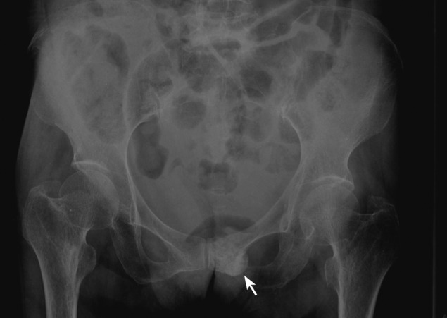
Anteroposterior radiograph of the pelvis demonstrated a focal sclerotic bulging contour lesion at the left inferior pubic ramus (arrow).

(A) Hip computed tomography with 3D reconstruction revealed a sclerotic lesion with a fracture line at the left pubic ramus (arrow). (B) Pelvic bone magnetic resonance imaging showed a bone marrow signal change and fracture line at the left pubic ramus without evidence of a tumor (arrow).
To evaluate the possibility of bone metastases, pelvic magnetic resonance imaging (MRI) (Fig. 2B), bone scintigraphy (Fig. 3A and 3B), and 18F-fluorodeoxyglucose positron emission tomography (18F-FDG-PET)/CT (Fig. 3C and 3D) were performed. They demonstrated multiple fractures in both sets of ribs, sternum, T-L spine, and both pelvic bones. However, there were no abnormal findings or hypermetabolic lesions suggesting malignancy. Moreover, there was no definite mass or osteolytic bony lesion on pelvic MRI. Dual energy X-ray absorptiometry showed a femur neck and total hip joint T score of -4.3, which is within the osteoporosis category. Blood tests revealed secondary hyperparathyroidism and low vitamin D levels as follows: corrected calcium, 8.5 mg/dL; serum parathyroid hormone (PTH), 150 pg/mL (normal range, 8 to 76); and total vitamin D level, 4.2 ng/mL (recommended range, > 20). After obtaining these values, treatment with a calcium/vitamin D combination and bisphosphonate therapy was continued. Two months after the initial emergency department visit, analgesic therapy was stopped.

(A, B) Bone scintigraphy demonstrated multiple fractures in both sets of ribs, sternum, T-L spine, and both pelvic bones. (C, D) 18F-fluorodeoxyglucose positron emission tomography/computed tomography revealed a fracture with hypermetabolism at the left pubic ramus (standardized uptake value 3.4; arrows).
Hepatic osteodystrophy, which was first reported in 1956, is a generic definition of a metabolic bone process that has a multifactorial origin and is associated with chronic liver disease. The biological mechanism of hepatic osteodystrophy is not clear. However, both decreased 25-hydroxylation of vitamin D in the liver and a defect of Kupffer cell-mediated cleavage of PTH from hepatic dysfunction can contribute to the higher prevalence of metabolic bone diseases in patients with chronic liver disease compared with those without liver disease [3]. Despite its high incidence, however, the clinical importance of hepatic osteodystrophy is undervalued in practical fields. Almost all fractures originating from hepatic osteodystrophy are limited to the vertebra [1]. Our report might be the first case report of multiple pelvic bone fractures induced by hepatic osteodystrophy and mimicking bone metastases from HCC.
Bone was once considered to be a rare site of metastasis from HCC. However, improving long-term survival rates combined with technical advances in diagnostic modalities have led to an increased incidence of bone metastases. Although 18F-FDG-PET/CT is very a sensitive modality with which to detect metastatic HCC, Ho et al. [4] reported that 38% of bone metastasis reveals no uptake on FDG-PET scans. Thus, we used various imaging modalities (simple X-ray, hip CT, and pelvic MRI) to evaluate the possibility of bone metastasis in the present case, and the results revealed no osteolytic lesions suggesting typical bone metastasis from HCC. Because osteoblastic bone metastasis from HCC has never been reported, these imaging findings suggest that the pelvic fractures in this case were induced by hepatic osteodystrophy rather than bone metastasis.
In this case, the patient was elderly, underweight, and postmenopausal and had a previous history of compression fractures. Moreover, she had been closely monitored because of liver cirrhosis of Child-Pugh class B. Because of these risk factors, fractures of multiple sites occurred despite treatment with bisphophonates. Only a few small, randomized controlled studies of interventions for preventing osteoporosis and fractures in patients with chronic liver disease have been performed. Most were conducted on patients with primary biliary cirrhosis, and none showed significant differences in intervention and reduction in the risk of osteoporotic fractures [5]. Collier et al. [5] suggested that diagnostic workup and treatment of osteoporosis are necessary in patients with T scores of less than -2.5 or previous fragility fractures. According to their guidelines, hypogonadal female patients are recommended to undergo treatment with hormonal replacement therapy (HRT). However, multiple fractures arose during treatment with bisphosphonates in the present case; therefore, we continued the calcium/vitamin D combination therapy and bisphosphonates instead of HRT.
In conclusion, although multiple pelvic bone fractures associated with hepatic osteodystrophy is a very rare complication, we should consider this possibility in patients with severe chronic liver disease, even HCC. After fragility fractures have occurred, a diagnostic workup and active intervention for metabolic bone disease are mandatory.
Notes
No potential conflict of interest relevant to this article is reported.