Homonymous hemianopia in a patient with Behcet's disease
Article information
A 49-year-old male with a 10-year history of Behcet's disease (BD) presented with sudden visual field defects in both eyes the first occurred when walking. Laboratory testing revealed an elevated serum C-reactive protein (1.3 mg/dL) and erythrocyte sedimentation rate (47 mm/hr). The Humphrey visual field test showed right homonymous hemianopia (Fig. 1), while no abnormal findings were identified on fundal examination. Suspecting homonymous hemianopia, magnetic resonance imaging (MRI) was performed; T2 fluid-attenuated inversion recovery MRI showed circumferential high-intensity lesions of the left lateral geniculate body (Fig. 2A). Consequently, he was diagnosed with acute neuro-BD and given steroid pulse therapy (1 g/day) for 3 days, followed by highdose prednisolone (1 mg/kg/day) and a cyclophosphamide pulse (750 mg/day). The lesions in the lateral geniculate body were reduced on MRI taken 2 weeks after treatment (Fig. 2B), but the homonymous hemianopia remained nearly unchanged. We plan to administer two cyclophosphamide pulse treatments per month. Ocular manifestations are one of the main features of BD, especially uveitis and retinal vasculitis that progressively damage the vision. Homonymous hemianopia with hemianopic visual field loss on the same side in both eyes is extremely rare in BD. The pathogenesis of the lesions in the geniculate bodies in these cases is believed to be related to underlying vascular involvement of the inflammation. Early immunosuppressant use is necessary; intravenous cyclophosphamide combined with corticosteroids can improve the prognosis. When patients with BD have visual defects, clinicians should consider brain MRI to detect cerebral lesions, as well as a fundal examination.
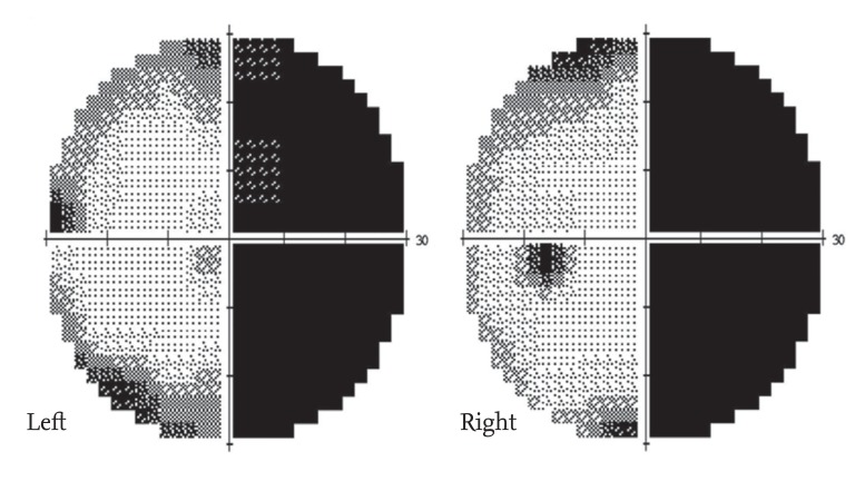
Humphrey visual field testing showed the loss of half of the field of view on the right side in both eyes; i.e., right homonymous hemianopia.
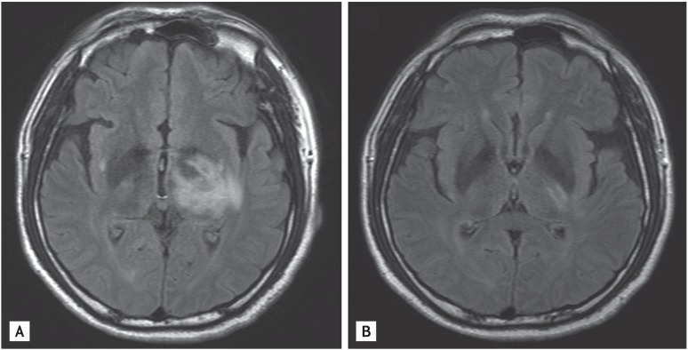
(A) T2 fluid-attenuated inversion recovery magnetic resonance imaging (MRI) showed circumferential high-intensity lesions in the left lateral geniculate body. (B) These lesions were reduced in MRI images taken 2 weeks after treatment.
Notes
Conflict of interest: No potential conflict of interest relevant to this article was reported.