Complete recovery of pyoderma gangrenosum after successful treatment of underlying hairy cell leukemia with cladribine
Article information
To the Editor,
Hairy cell leukemia (HCL) is a chronic B-cell disorder characterized by the presence of typical hairy cells in the peripheral blood and marrow, pancytopenia, and splenomegaly. Cutaneous lesions referable to thrombocytopenia, such as ecchymoses and petechiae, infection, and vasculitis, are common during the disease course, but lesions caused by infiltration of the skin by hairy cells are unusual. Pyoderma gangrenosum (PG) is a rare idiopathic ulcerative neutrophilic inflammatory skin disease characterized by a variable clinical presentation. PG is often a cutaneous manifestation of a systemic disease [1]. Here, we report a patient who was diagnosed with HCL presenting with a PG lesion and recovered after treating the underlying disease with cladribine.
A 43-year-old male was admitted to our hospital with a painful ulcer on his left tibial surface. The lesion had first appeared 4 weeks ago accompanied by 38ºC to 39ºC fever. A physical examination revealed blood pressure, 120/85 mmHg; heart rate, 92 beats per minute; respiration rate, 22 breaths per min; and body temperature, 36.5ºC. Two painful ulcers were found on his left tibial surface; one was 5 × 12 cm and the other was 4 × 8 cm in size (Fig. 1). The liver was palpable under the costal margin, and the spleen was 20 cm in length. Laboratory tests revealed pancytopenia (leukocytes, 1,230/mm3; neutrophils, 180 /mm3; hemoglobin, 6.6 g/dL; platelets, 51,000 /mm3). Cells with abundant agranular cytoplasm and multiple cytoplasmic projections were seen on the peripheral smear. A bone marrow examination showed diffuse infiltration of hairy cells and immunophenotyping using flow cytometry showed that these cells were CD103 (+), CD19 (+), CD20 (+), CD25 (+), CD3 (−), CD5 (−), and CD10 (−). HCL was diagnosed based on these findings.

Pyoderma gangrenosum before treatment of hairy cell leukemia on the left tibial surface of the patient.
Cultures and biopsies of the ulcerated skin lesions on the tibial surface were taken. Local and parenteral antibiotics were applied and dressings and debridement were performed regularly. No bacterial growth was seen on the cultures. The histopathological examination of this skin lesion proved the diagnosis of PG (Fig. 2).
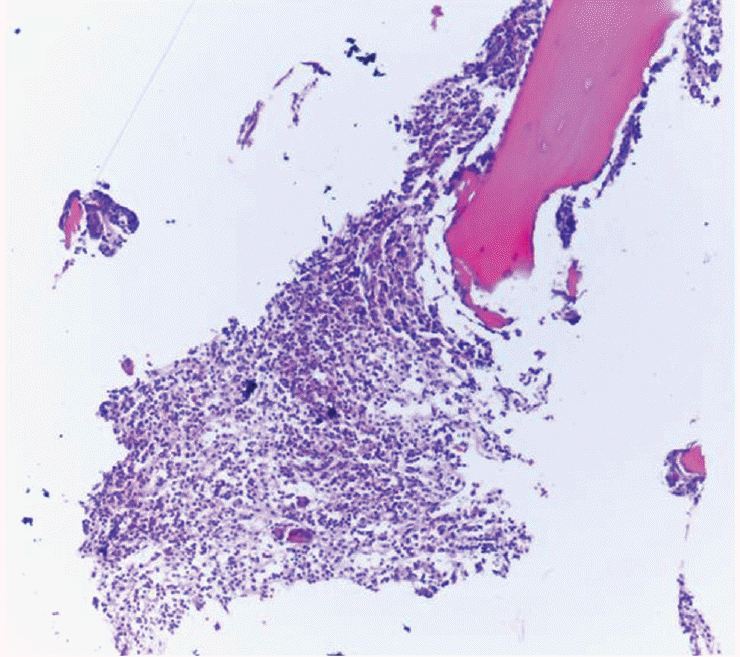
Diffuse necrosis and inflammatory cell infiltration were seen in the histopathological examination of the skin lesion, and this was compatible with pyoderma gangrenosum (H&E, ×40).
Cladribine was administered at a rate of 0.1 mg/kg/day for 7 days for the HCL. The neutropenia resolved after 20 days of cladribine monotherapy. The pancytopenia had resolved completely, and spleen size was normal at the follow-up. The PG resolved completely after the third month of cladribine treatment by treating the primary disease and changing the dressings regularly (Fig. 3).
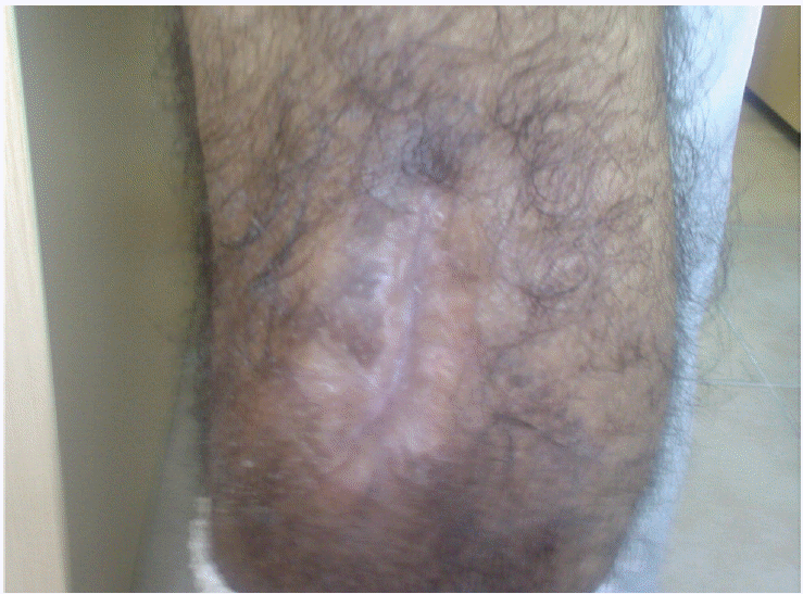
Pyoderma gangrenosum completely resolved after the cladribine treatment.
PG is a disease with unclear etiology. It is probably a hyperergic reaction, connected with a systemic disease or with an immunological compound. Approximately 50% to 70% of patients with PG have an underlying systemic disease, and the most commonly associated conditions are inflammatory bowel disease, polyarthritis, hematological disease (acute myelogenous leukemia and HCL), monoclonal gammopathies, hepatitis, and collagen vascular diseases. PG can begin at any age, but is most common in 30- to 50-year-old patients of either sex [1]. The incidence of PG is approximately 3 per million people per year in the United States. The frequency of malignant neoplasms in cases of PG is not exactly known, but it has been assessed to be 7% [2]. These cases are most often associated with acute or chronic leukemia.
PG skin lesions are painful, erythematous papules, sterile pustules, or fluctuant nodules that may progress to expanding ulcers. The lesions can develop individually at any cutaneous site but are typically found on the lower extremities and trunk [1]. The diagnosis of PG is based primarily on the clinical presentation, as immunohistopathological findings in patients with PG are nonspecific [1]. Biopsies may demonstrate edema, mixed inflammatory infiltrates (predominantly neutrophilic infiltrate), lymphocytic vasculitis, necrosis, and hemorrhage.
A few reported cases of HCL have the presenting symptoms of PG [3-5]. Patients presenting with PG should be carefully examined for an underlying hematological malignancy with detailed anamnesis, a physical examination, and laboratory testing. HCL can be easily diagnosed in patients with pancytopenia, splenomegaly, and typical hairy cells, and skin ulcers can be related to PG, as in our patient. Although the histopathological changes are not specific to PG, biopsies of the lesions must be performed, as these lesions can also be related to infiltration of leukemic cells. Once HCL and PG are diagnosed, the main treatment target should be HCL, as PG will completely recover in a few months with the successful treatment of HCL using cladribine, as in our patient.
Notes
No potential conflict of interest relevant to this article was reported.