Widespread intracranial calcifications in a patient with hypoparathyroidism
Article information
A 52-year-old woman presented with facial rigidity and jerky movements of all four limbs. These movements lasted for 12 hours. She had circumoral paresthesia and spasm of the carpal and pedal muscles. She was diagnosed with anxiety disorder 2 months ago, and had previously undergone cataract operations on both eyes separately (left eye, 2 days prior to admission; right eye, 4 years ago). She had no history of surgery or irradiation around the neck or central nervous system disturbances such as seizure. She seemed anxious but fully conscious and well oriented. The physical examination revealed an ipsilateral contraction of the facial muscles when tapping the facial nerve (Chvostek sign). Spasm of the hand muscles was induced by inflating a blood pressure cuff above the systolic blood pressure (Trousseau sign). Laboratory tests showed the following: serum calcium, 4.8 mg/dL (normal range, 8.2 to 10.2); ionized calcium, 0.68 mmol/L (normal range, 1.13 to 1.32); phosphorus, 4.6 mg/dL (normal range, 2.5 to 4.5); albumin, 4.4 g/dL (normal range, 3.8 to 5.3); magnesium, 1.9 mg/dL (normal range, 1.9 to 4.1); intact parathyroid hormone, 1.20 pg/mL (normal range, 8 to 76); and 25(OH) vitamin D, 20.95 ng/mL (normal range, 30 to 100). Brain computed tomography showed extensive bilateral symmetrical calcifications in the cerebellar dentate nuclei (Fig. 1A), basal ganglia (Fig. 1B), and periventricular white matter (Fig. 1C). Because no specific cause of hypoparathyroidism was identified, the patient was diagnosed with idiopathic hypoparathyroidism. With the use of intravenous calcium, oral calcium supplements, and a vitamin D analog, the patient’s symptoms improved gradually. Intracranial calcification is physiological in 0.3% to 1.5% of subjects. The most common pathologic causes are hypoparathyroidism and pseudohypoparathyroidism. Other causes are tuberous sclerosis, mitochondrial disease, Fahr syndrome, familial idiopathic basal ganglia calcification, and post-infection. The pathogenic mechanism of basal ganglia calcification in hypoparathyroidism is not clear. Its occurrence is believed to be high serum calcium-phosphorous products and poor calcium control.
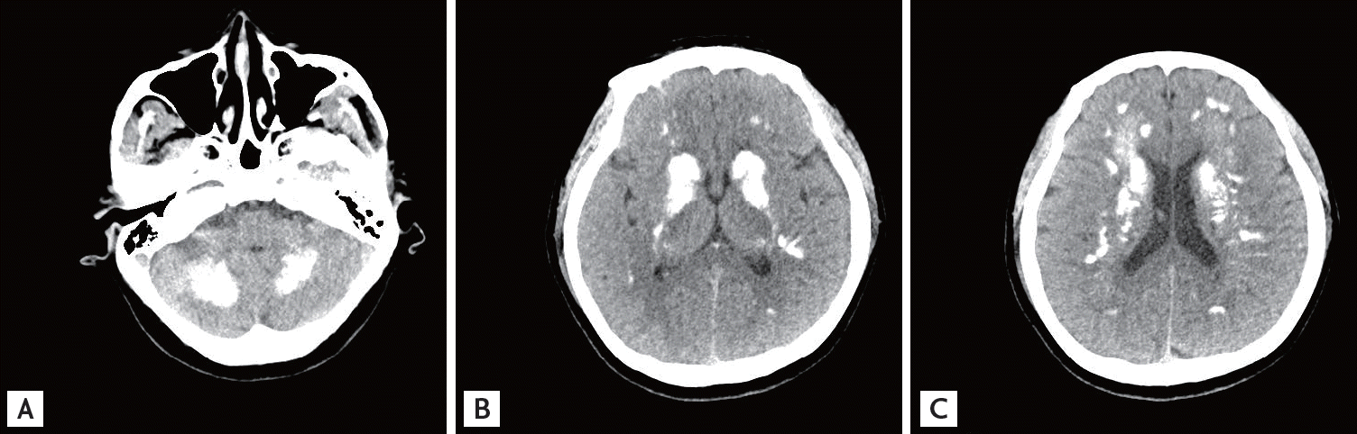
Brain computed tomography shows diffuse symmetric parenchymal calcifications involving the cerebellar dentate nuclei (A), basal ganglia (B), and periventricular white matter (C).
Notes
No potential conflict of interest relevant to this article was reported.