Synchronous parathyroid carcinoma and papillary thyroid carcinoma in a patient with long-standing schizophrenia
Article information
To the Editor,
Parathyroid carcinoma is one of the rarest malignancies, which accounts for 0.1% to 5% of primary hyperparathyroidism (PHPT) cases [1]. Parathyroid carcinoma is usually associated with higher serum calcium, parathyroid hormone (PTH) levels and related complications compared with adenomas [2]. Concomitant thyroid disease often occurs in patients with PHPT [3]; however, the simultaneous occurrence of parathyroid and thyroid carcinomas is extremely rare, and, as far as we are aware, only eight case reports have been published previously, and no published reports describe this in a Korean patient. Here, we describe a patient with a history of schizophrenia that spanned approximately 20 years who was found to have concurrent parathyroid carcinoma with PHPT and papillary thyroid carcinoma.
A 68-year-old female patient was referred to our endocrinology clinic for the evaluation of elevated serum calcium levels, alkaline phosphatase levels, and osteoporosis that had been incidentally found during a routine checkup. Her history of schizophrenia spanned about 20 years, and she had received medications for dyslipidemia and osteoporosis for 1 year. At presentation, she was receiving chlorpromazine 200 mg, flunitrazepam 1 mg, magnesium hydroxide 1,000 mg, pravastatin 40 mg, fenofibrate 160 mg, calcium citrate 750 mg, cholecalciferol 400 IU in a day, and risedronate 35 mg once weekly. The patient did not report that she had undergone head or neck irradiation or that a neck mass was present. The patient looked lethargic and complained of pain in the right thigh area that restricted her ambulation. Her daughter mentioned that she had been more lethargic and speechless during the preceding year. Her clinical examination was unremarkable. The initial laboratory test results showed a serum calcium concentration of 12.8 mg/dL (reference range, 8.4 to 10.2), ionized calcium of 6.65 mg/dL (reference range, 4.6 to 5.3), phosphorus of 1.9 mg/dL (reference range, 2.5 to 5.5), intact PTH of 1,247 pg/mL (reference range, 8 to 51), and a serum alkaline phosphatase of 6,240 IU/L (reference range, 104 to 338), findings that were suggestive of PHPT. The patient had an elevated 24-hour urinary calcium excretion of 700 mg/day (reference range, 100 to 300) and bone turnover markers; C-terminal telopeptide of 2.88 ng/mL (reference range, 0.104 to 1.008) and osteocalcin of 452 ng/mL (reference range, 25 to 46). Her serum creatinine, 25-hydroxyvitamin D, thyroid stimulating hormone, free thyroxine, and calcitonin levels were within the reference ranges. The patient’s bone mineral density was within the osteoporotic range, with T-scores of −4.3 for the lumbar spine and −2.7 for the femoral neck which was measured by dual-energy X-ray absorptiometry. Radiographs of the femurs were suggestive of osteolytic lesions in the right proximal femur, and computed tomography scanning showed a well-defined osteolytic lesion with a sclerotic rim in the right subtrochanteric femur (Fig. 1A). Additional 3-phase bone scans revealed focal increases in activity within the right subtrochanteric area and femoral neck, and within the mandible, right scapula, both iliac bones, and the ischium (Fig. 1B). Ultrasound scanning of the neck demonstrated a heterogeneous hypoechoic mass that measured 4 × 3 cm, which was believed to be an enlarged parathyroid gland that was inferior to the left thyroid lobe. The margins of this mass were poorly circumscribed (Fig. 2A). In addition, the patient had a homogenous hypoechoic nodule without microcalcification in the left thyroid lobe that measured 0.5 × 0.5 cm (Fig. 2B). Technetium-99m sestamibi scintigraphy revealed an area of increased radioactivity uptake at the inferior pole of the left thyroid lobe, which suggested a parathyroid adenoma corresponding to the mass seen during ultrasound scanning (Fig. 2C). A fine-needle aspiration biopsy of the nodule in the left thyroid lobe demonstrated an atypia of undetermined significance or follicular lesion of undetermined significance (AUS/FLUS). The patient underwent parathyroidectomy of her left inferior parathyroid gland and simultaneous left hemithyroidectomy. No suspicious cervical lymph nodes were found during the intraoperative exploration. The intact PTH levels were 1,247 pg/mL before surgery and 15.8 pg/mL 3 hours after surgery. The gross examination of the parathyroid mass that measured 4.2 × 3.3 × 3.1 cm revealed a slightly firm, white-grayish mass with a partial hemorrhage and cystic changes that adhered to the adjacent fat tissue (Fig. 3A). In addition, there was a well circumscribed, grayish-white firm nodule that measured approximately 0.5 × 0.5 cm in the left thyroid lobe. A histopathological examination showed that the parathyroid mass was a parathyroid carcinoma with evidence of invasion of the adjacent fat (Fig. 3B), and a 0.5 cm papillary carcinoma with a follicular variant was found in the left thyroid lobe (Fig. 3C and 3D). Postoperatively, the patient was prescribed orally administered calcium and vitamin D3, and she had normocalcemia in conjunction with intact PTH levels that ranged from 15.8 to 30.2 pg/mL. About 6 months after the operation, the patient looked more alert and active compared with the first time she visited our clinic.
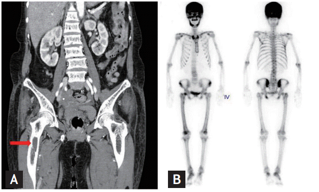
(A) Computed tomography scanning demonstrating a well-defined osteolytic lesion with a sclerotic rim in the right subtrochanteric femur (arrow). (B) The 3-phase bone scans revealed focal increases in activity within the right subtrochanteric area and femoral neck, and within the mandible, right scapula, both iliac bones, and the ischium.

(A) Ultrasonogram (sagittal view) demonstrating a 4 × 3 cm sized heterogenous hypoechoic mass with ill-defined margin, which was believed to be an enlarged parathyroid gland that was inferior to the left thyroid lobe. (B) Ultrasonogram (transverse view) demonstrating a 0.5 × 0.5 cm sized homogenous hypoechoic nodule without microcalcification in the left thyroid lobe. (C) Technetium-99m sestamibi scintigraphy 10 minutes (left panel) and 2 hours (right panel) after injection of the isotope revealing an area of increased radioactivity uptake at the inferior pole of the left thyroid lobe, which suggested a parathyroid adenoma corresponding to the mass seen during ultrasound scanning.
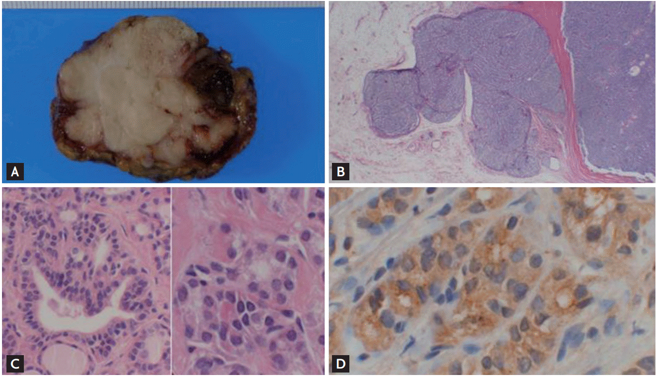
(A) Through-cut section of the surgical specimen demonstrating the parathyroid tumor, measuring 4.2 × 3.3 × 3.1 cm and weighing 5 g. It showing a slightly firm, white-grayish mass with a partial hemorrhage and cystic changes that adhered to the adjacent fat tissue. (B) Histopathologic section of parathyroid carcinoma demonstrating adjacent fat tissue invasion of the tumor and a trabecular pattern with fibrous septations (H&E, ×100). (C) Histopathologic section of the papillary thyroid carcinoma removed from the left thyroid lobe, which demonstrating typical features of a papillary carcinoma with a follicular variant. (H&E, left ×200, right ×400). (D) Histopathologic section of the papillary thyroid carcinoma with a follicular variant showing positive of the immunohistochemical staining for galectin 3 (×400).
Parathyroid carcinoma is an extremely rare cause of PHPT, and it commonly has an indolent growth with a tendency for local invasion [1]. Most parathyroid carcinomas are hormonally functional, and the initial manifestation usually occurs precipitously. The serum calcium and PTH levels are often much higher than in those cases with adenomas [2]. Parathyroid carcinoma is characterized by higher incidence rates of renal dysfunction, metabolic bone disease, and gastrointestinal symptoms [2]. The current patient presented with a large parathyroid mass, a very high PTH level, which showed an 18-fold increase compared with the normal upper value, and metabolic bone disease, which suggested a parathyroid carcinoma. Although the clinical manifestations of hyperparathyroidism are more severe in patients with parathyroid carcinomas compared with those who have parathyroid adenomas, it remains difficult to differentiate between carcinomas and adenomas without pathologic confirmation, but parathyroid carcinoma should be considered in patients with PHPT.
Among the various signs and symptoms associated with PHPT, the psychiatric vary from mild personality changes or nervousness to severe depression, obsessive-compulsive behavior, and paranoia. These symptoms can last for several months to several years [4]. Our patient looked lethargic, and her daughter advised that she had been more lethargic and speechless over the preceding year. It seemed that the assumption underlying this patient’s management had been that these symptoms comprised an aggravation of her schizophrenia or part of the aging process. In fact, it was quite difficult to clearly distinguish whether her mental change was a consequence of PHPT or an aggravation of her underlying disease. Given this patient’s mild recovery in relation to her mental status and wordlessness during the 6 months that followed the operation, it should be emphasized that any changes in the mental statuses of patients with schizophrenia should not be attributed to “functional” psychosis too readily. Some psychiatric patients, like our case, may present with organic mental disorders requiring medical or surgical interventions.
Concomitant thyroid disease is not unusual in patients with PHPT [3]. It has recently been reported that of 360 patients who underwent cervical explorations for PHPT, 206 patients (57.2%) underwent simultaneous thyroid surgery for diseases of the thyroid gland [3]. A pathological association between thyroid disease and parathyroid disease has been reported in 15% to 70% of patients with PHPT, and, in particular, thyroid cancer accompanied by PHPT has been encountered in 3.1% to 17% of cases [5]. However, synchronous parathyroid and thyroid carcinomas are extremely rare. Indeed, to the best of our knowledge, only eight documented cases have been described in published reports, and the current case is the first to be reported from Korea. With respect to the reasons for this association, some researchers have described these concomitant diseases as coincidental, while other authors have described the increases in endogenous calcium levels or growth factors, including epithelial growth factors and insulin-like growth factors, as goitrogenic factors [5]. In our case, a fine-needle aspirate of the nodule in the left thyroid lobe showed an AUS/FLUS and it revealed the presence of a papillary carcinoma with a follicular variant after operation.
The present case report demonstrates several important points that deserve emphasis. First, although parathyroid carcinoma is an uncommon cause of PTH-dependent hypercalcemia, it should be given consideration. Second, hyperparathyroidism that presents in association with psychiatric symptoms, which may be considered mild or atypical. Thus, the physician must be alert to the possibility of a new pathology that includes hyperparathyroidism for any changes in the mental statuses of patients with schizophrenia and the patient must undergo a careful examination for signs of organic disturbance. Finally, the possibility of a thyroid malignancy must also be considered in the context of PHPT with thyroid nodules. Hence, physicians should pay particular attention to patients who are undergoing surgery for diseases of the parathyroid gland or thyroid gland to ensure they are carefully evaluated both preoperatively and intraoperatively for abnormalities in both glands to ensure that any coexistent disease is recognized and treated in a single procedure. As a patient’s prognosis depends largely on the successful resection of all malignant tumors at the time of the initial operation, any thyroid or parathyroid lesions detected during imaging and/or pathologic investigations must be taken seriously.
Notes
No potential conflict of interest relevant to this article was reported.