Pheochromocytoma Complicated With Cardiomyopathy After Delivery:
– A Case Report and Literature Review –
Article information
Abstract
Pheochromocytoma in pregnancy is very rare but it is associated with very high maternal and fetal mortality. Therefore, it is important to include pheochromocytoma in the differential diagnosis of hypertension associated with pregnancy. It is difficult to make a diagnosis of pheochromocytoma in pregnancy before delivery. The characteristic symptoms of pheochromocytoma could be initiated during delivery because the process of delivery, general anesthesia, fetal movement, induce acute surge of catecholamine release, which could also induce cardiomyopathy. Early diagnosis and intensive care can affect the prognosis of cardiomyopathy induced by pheochromocytoma. Proper management with alpha-blockade, beta-blockade and angiotensin converting enzyme inhibitor could acutely reverse the course of cardiomyopathy.
INTRODUCTION
Pheochromocytoma is an unusual cause of hypertension. It accounts for only 0.1% of the hypertension found in adults1). Pheochromocytoma in pregnancy is extremely rare, and only about 200 cases have been reported in literature reviews. This disorder carries a high risk of maternal and fetal mortality if undiagnosed before delivery2). The various clinical manifestations of hypertension could resemble those of toxemia in pregnancy, preeclampsia, a ruptured uterus and shock immediately after delivery2). Therefore, in the case of fluctuating hypertension with characteristic symptoms during pregnancy, the physician should suspect pheochromocytoma. However, antepartum diagnosis is missed in about two-thirds of the cases. In many patients, the symptoms of pheochromocytoma are first-manifested during delivery.
Catecholamine induced cardiomyopathy has been reported in some of the literature3,4). It is very important to manage such a patient with aggressive and appropriate medical therapy because the initial management is closely related to the prognosis4). In world literature, only about five cases of pheochromocytoma induced cardiomyopathy in pregnancy have been reported. In Korea, no such case has been reported. Here, we report the case of a 34-year-old pregnant woman with pheochromocytoma who developed catecholamine induced cardiomyopathy after delivery and made a dramatic recovery through proper medical management.
CASE REPORT
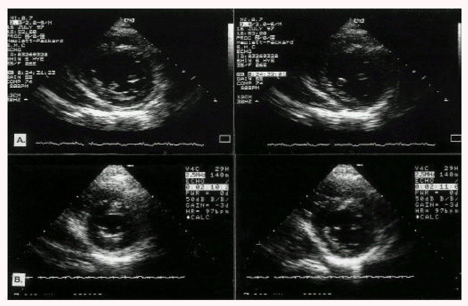
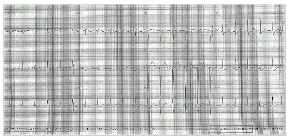
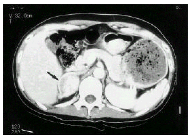
A 34-year-old pregnant woman underwent a normal vaginal delivery. She had no previous history of hypertension and there was no evidence of toxemia in pregnancy or preeclampsia in the prenatal examination. At the terminal period of pregnancy, the patient showed mild hypertension, which was well controlled by nifedipine. Immediately after delivery, her blood pressure rose rapidly and showed marked fluctuation (140/90~210/90 mmHg). The pulse rate increased from 70/min to 150/min. The patient complained of a severe headache, palpitations, chest discomfort, dyspnea and postural dizziness. The blood pressure and pulse rate could not be controlled by nifedipine and her symptoms worsened. Her breathing sounds were normal and the heart sound was regular; there was no murmur. The abdominal mass was not palpated. No periumbilical bruit was heard. Blood tests and simple chest X-ray showed no abnormal findings. Serum CK/CK-MB/LDH levels were 268/23/607 U/L. There was no interval change in serum CK/CK-MB/LDH levels in serial follow-up. The electrocardiograph showed sinus tachycardia, left ventricular hypertrophy and a non-specific ST change in the precordial leads (Fig. 1). In the transthoracic echocardiograph, left ventricular contraction was depressed (ejection fraction = 25%) and akinesia of the septal, anterior and inferior walls was noted (Fig. 2). In a 24-hour urine collection, the total metanephrine level was 15.4 mg/day (normal value; 0~1.2 mg/day), the vanilmandelic acid (VMA) level was 26.6 mg/day (normal value; 2.0~10 mg/day), the epinephrine level was 1682 ug/day (normal value; 0~40 ug/day) and the norepinephrine was 3111 ug/day (normal value; 0~80 ug/day). The serum norepinephrine and epinephrine levels were 4791 pg/ml (normal value; 100~410 pg/ml) and 1657 pg/ml (normal value; ä120 pg/ml), respectively. The abdominal computerized tomography (CT) showed a well-enhanced oval-shaped mass in the right adrenal gland (4.5cm × 3.6cm) (Fig. 3). The patient was diagnosed with pheochromocytoma on the 2 day after delivery. Cardiomyopathy induced by increased catecholamine was also diagnosed because of the abnormal echocardiographic findings.

Transthoracic eohocardiographic findings (left panel: end-systole; right panel: end-diastole). A. Depressed left ventricular contraction and regional wall motion abnormalities. B. Increased left ventricular ejection fraction and recovered wall motion abnormalities after therapy.
Phenoxybenzamine was used to control the hypertension and metoprolol was added after the phenoxybenzamine use. Enalapril was prescribed to treat the myocardial damage. After medication, the patient’s blood pressure decreased to the normal range and her symptoms disappeared. Serial transthoracic echocardiography was performed on the second day after the medical management. It revealed a dramatic increase in the left ventricular ejection fraction (to 49%) and complete resolution of the regional wall motion abnormalities (Fig. 2). In the early treatment period, the patient complained of postural dizziness and there was evidence of postural hypotension. The dosage of phenoxybenzamine was reduced in order to prevent the development of such symptoms. A right adrenalectomy was performed after 20 days of medical management. The mass was 5×5 cm in size and there was no evidence of malignancy. After the surgery, the patient’s blood pressure was well controlled by metoprolol and enalapril without phenoxybenzamine. In the 24-hour urine collection after the right adrenalectomy, the total metanephrine level decreased to 0.5 mg/day and the VMA level was 4.5 mg/day. The baby was healthy without neonatal growth retardation, and the patient discharged without complication.
DISCUSSION
Prevalence
Pheochromocytoma in pregnancy was first reported in 1911, but an antenatal diagnosis was not made until 19555). Pheochromocytoma is extremely rare in pregnancy and it is estimated to occur once in approximately 50,000 term pregnancies6). In Korea, there have been 5 case of pheochromocytoma in pregnancy since 19827). At the Royal Womens Hospital, Melbourne, with over 7,000 deliveries per year, there were three cases of pheochromocytoma in pregnancy in the 20 years since 19768). There were 35 patients with pheochromocytoma in pregnancy in the Japanese literature from 1958 to 19932). Among these 35 patients, the diagnosis of pheochromocytoma was confirmed before delivery in 31%, after delivery in 63%, and at autopsy in 3%2). In our case, diagnosis could be made on the 2 day after delivery. The overall maternal mortality rate was about 40%, but this rate has decreased because of antepartum diagnosis, proper management of pheochromocytoma and advances in surgical techniques9). For example, in Japan, the maternal mortality before 1969 was 48%, in the period 1969–1979 the rate was 26% and between 1980 and 1989 the rate was 17%2). The fetal mortality has dropped from 50% to 19%2,10).
Cardiomyopathy induced by pheochromocytoma has been reported in many cases, but its occurrence in pregnancy is very rare. Consequently, only about four cases have been reported in world literature4,5,11,12).
Clinical symptoms and pathophysiology
Symptoms of pheochromocytoma in pregnancy are not so different from those in non-pregnant women12). Fetal hypoxia, death and neonatal growth retardation can also occur in patients with pheochromocytoma. In over 60% of patients, the symptoms appear in the third trimester of pregnancy2). But the antenatal diagnosis is thought to be missed in about two-thirds of the cases. In our case, the patient showed mild hypertension without characteristic symptoms of pheochromocytoma before delivery. However, she developed full-blown symptoms immediately after delivery, which enabled us to make the diagnosis. It was suggested that the increased intra-abdominal pressure and uterine contraction, even vigorous fetal movement during vaginal delivery, might play a major role of initiating symptoms of pheochromocytoma13). Cardiomyopathy and/or myocarditis can be rarely associated with pheochromocytoma.
Chest pain, dyspnea, hypotension, pulmonary edema and shock may occur in the case of cardiomyopathy. Histologic findings of cardiomyopathy in pheochromocytoma are focal myofiber necrosis, myofibrillar degeneration and mononuclear cell infiltration. These findings are similar to those seen with catecholamine excess14). Recent studies have shown that cardiomyopathy with pheochromocytoma is mediated by alpha-adrenergic receptor stimulation in conjunction with a high concentration of norepinephrine14,15).
At autopsy of one case of pheochromocytoma-induced cardiomyopathy, the patient had normal-sized heart and there was evidence of catecholamine induced heart disease in the form of focal myocardial necrosis in histologic examination.16) Usually, the recovery of the left ventricular dysfunction was apparent after surgical removal of the tumor14,17). In one report, the improvement of the left ventricular dysfunction after medical therapy with alpha adrenergic blockade occurred from 15 to 60% over 14 days18). Therefore, our patients acute resolution of left ventricular dysfunction from 25 to 49% only over 3 days of aggressive medical management is uncommon. We suggest that aggressive medical management, including angiotensin converting enzyme inhibitor and short duration of typical symptoms of pheochromocytoma, i.e. 3 days in our case, could lead to rapid reverse of ventricular function.
Diagnosis
It is very difficult to detect pheochromocytoma in pregnancy because there are many diseases that mimic its symptoms. Hence, the most important diagnostic tool is to suspect the condition. The diagnosis of pheochromocytoma in pregnancy is not so different from that in non-pregnant women except in a few points.
After biochemical test such as 24 hour urine total metanephrine and VMA level, ultrasonography is the next step for the localization of the tumor because of its convenience and the absence of the risk of radiation exposure. However, this test has some limitations, such as the inability to detect small extra-adrenal tumors. Magnetic resonance imaging (MRI) has some advantage over CT because there is no risk of radiation.
For the diagnosis of cardiomyopathy, serial electrocardiographs and cardiac enzyme follow-up are essential. As in our case, if the cardiomyopathy is properly managed, serial echocardiography could show progressively increased left ventricular ejection fraction and improved regional wall motion abnormalities3).
Management
Immediately after diagnosis, it is essential to treat with alpha-blockades for the control of hypertension and preoperative management. Even though phenoxybenzamine, an irreversible alpha-adrenergic receptor antagonist, is the first-line agent for use in non-pregnant women, because of its teratogenic effect, labetolol and prazocin are commonly used during pregnancy. However, the safety of labetolol and prazocin has not currently been confirmed. Beta-blockade may be used to control the tachycardia and arrhythmia, but should be delayed until there has been sufficient alpha-blockade treatment to avoid generalized unintended vasoconstriction in response to alpha-receptor stimulation.
The initial management of cardiomyopathy induced by pheochromocytoma is very important because these manifestations are associated with a very high level of fatalities. For example, there was one mortality case among four patients with cardiomyopathy induced by pheochromocytoma in pregnancy16). Beta-blockade and an angiotensin converting enzyme inhibitor may be used for the management of cardiomyopathy induced by pheochromocytoma19). Angiotensin converting enzyme inhibitors have been used only in non-pregnant women due to its teratogenic effect, but the resistance usually ensues rapidly20). For the treatment of congestive heart failure, conservative management includes bed rest, oxygen, diuretics and vasodilators such as hydralazine. In our case, because the pheochromocytoma was diagnosed after delivery, we did not have to limit our medication. However, breast feeding was prohibited.
The definitive treatment of pheochromocytoma is surgical removal of the tumor. The timing of tumor resection remains controversial. If the diagnosis is made before 23 weeks of gestation, it is appropriate to remove the tumor after alpha-blockade medication2,9). After 24 weeks of gestation, it is difficult to remove the tumor because of the enlarged uterus. Therefore, alpha-blockade is continued until the fetus is mature and then an elective Cesarean section should be performed, followed immediately by exploration for the tumor18). Cesarean section is the safest method of delivery for both mother and infant because vaginal delivery has a higher mortality (31%) than has Cesarean section (19%)9).
SUMMARY
The prognosis for pheochromocytoma in pregnancy is greatly influenced by antenatal diagnosis. The diagnosis of pheochromocytoma in pregnancy is not different from that in non-pregnant women. Clinical suspicion is the most important factor for diagnosis. Consequently, the physician should include pheochromocytoma in the differential diagnosis of pregnancy-associated hypertension.
Our case was complicated with cardiomyopathy. Cardiomyopathy induced by pheochromocytoma in pregnancy may worsen the prognosis. Severe left ventricular dysfunction in this patient showed a marked improvement after the medical management.

