A Case of Sternal Insufficiency Fracture
Article information
Abstract
We report a case of insufficiency fracture of the sternum in a 70-year-old female patient with a review of the literature. She complained of sudden onset chest pain and aggravating dyspnea. She has been managed with corticosteroid due to chronic obstructive pulmonary disease for 15 years. Diagnosis of sternal insufficiency fracture presented with thoracic kyphosis was made on the basis of absence of trauma history, radiologic findings of lateral chest radiograph, bone scintigraphy and chest computed tomography. Thoracic kyphosis and osteoporosis secondary to menopause, corticosteroid therapy and limited mobility due to chronic obstructive pulmonary disease were considered as predisposing factors of the sternal insufficiency fracture in this patient.
INTRODUCTION
Insufficiency fractures occur in weakened bones unable to withstand even the stresses of normal daily activities. Spine, pelvis and long bones of lower extremities are common sites of insufficiency fracture1). Cases of sternal insufficiency fracture have been rarely reported in an elderly patient with osteoporosis or chronic obstructive pulmonary disease (COPD) taking corticosteroid2) Osteoporosis often leads to thoracic kyphosis secondary to thoracic compression fracture. Thoracic kyphosis may cause a bending stress on the sternum, which may result in sternal insufficiency fracture3). To make the diagnosis of sternal insufficiency fracture, spontaneous fracture of sternum secondary to secondary neoplasm, lymphomatous infiltration and myelomatosis must be excluded4–6). It has been emphasized that sternal insufficiency fracture should be considered in the differential diagnosis of acute chest pain in the elderly7–8). Bone scintigraph contributes to the early diagnosis and chest computed tomography can confirm the diagnosis and rule out the possibilities of metastatic spread9). Therapy for underlying causes, as well as pain control, must be included for the treatment of sternal insufficiency fracture.
We describe here a case of sternal insufficiency fracture in a patient with long-standing COPD who had osteoporosis and thoracic kyphosis.
CASE REPORT
A 72-year-old woman, a smoker with a 15 years history of COPD, was admitted to our hospital because of anterior chest pain for 20 days. She denied any trauma history. She has been managed with corticosteroids after she was diagnosed as having COPD. Recently, her dyspnea was aggravated with the onset of acute chest pain. On examination, she appeared acutely ill. Body temperature was 37°C, respiration rate 30/min and blood pressure 120/70 mmHg. Auscultation of the chest showed coarse breathing sound without rale and heart sound was normal. There was a tenderness on the sternum. Her initial laboratory findings were as follows: hematocrit 42.1%, leukocyte count 11,600 × 109/mm3 (neutrophil 71.8%, lymphocyte 20%, monocyte 7.4%, eosinophil 0.3%), platelet count 235,000 × 109/mm3. The values for glucose, liver function, renal function, muscle enzyme and electrolytes were normal. Urinalysis was normal. Arterial blood gas on room air revealed pH 7.443, PO2 of 64.7 mmHg, PCO2 of 38.9 mmHg, bicarbonate 26 mmol/L and O2 saturation of 93.7%. The alveolar-arterial oxygen difference was 29.2 mmHg. Electrocardiogram revealed no definite abnormality. Lateral radiograph of the sternum showed buckling fracture of the upper body of the sternum (Fig. 1), but her previous lateral radiograph of the sternum obtained 3 months ago revealed normal appearance. Lateral chest radiograph demonstrated compression fracture of the 5th and 8th thoracic spine and osteoporosis through the spine (Fig. 2). Bone scintigraph showed increased uptake at the upper body of the sternum, costovertebral junction of both 2nd and 4th ribs, and compression fracture with hot uptake at 2nd, 3rd lumbar vertebrae (Fig. 3). Chest CT revealed cortical breakage at the posterior aspect of the sternum with soft tissue swelling (Fig. 4). Lumbar spine bone density measured by dual energy x-ray absorptiometry was reduced to 0.735 g/cm2 (T score;−3.21) at the level of L2. Her chest pain and dyspnea were declined by the administration of calcitonin and analgesics.
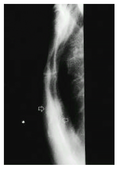
Lateral view of sternum reveals buckling fracture. The upper portion of sternal body was located posterior to the lower portion (arrow).
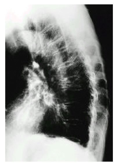
Lateral chest radiograph shows compression fractures of T5 and T8. Note osteoporosis and kyphosis throughout the thoracic spine.
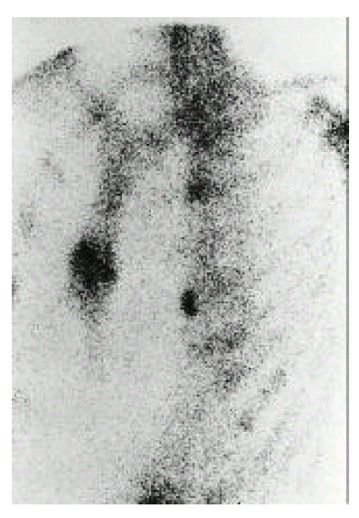
Bone scintigraph reveals increased radioisotope uptake at body of sternum around 3rd costosternal area. Upper (2nd) and lower (4th) costochondral junction of both ribs show increased uptakes.
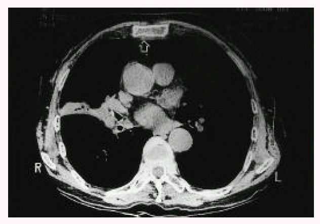
Chest CT with bone setting image reveals cortical disruption (arrow) and adjacent soft tissue swelling at the posterior aspect of the sternum.
DISCUSSION
Stress fractures are classified as fatigue and insufficiency fracture depending on the amount of stress applied to bone and on the elastic properties of the bone. Insufficiency fracture occurs when the elastic resistance of bone is inadequate to withstand the stresses of normal activity. The spine, pelvis and lower extremities are commonly affected by these fractures1). Spontaneous sternal insufficiency fractures have been rarely reported in elderly patients2,3,7–10). The sternum is an integral part of the thoracic cage, and slight movement at the manubirosternal joints aids expansion of the chest during inspiration. The stresses of sudden forward angulation of the thoracic spine in violent flexion are transmitted to the sternum by the ribs and clavicles. Bone loss with age and loss of elasticity associated with ossification of the costal cartilage are major predisposing factors for sternal insufficiency fracture. Bones weakened by osteopenia have insufficient elastic resistance to withstand even the minimal stress of normal daily activity. Sternal insufficiency fracture, which has been attributed to chronically applied flexion compression stress to the sternum caused by accentuated thoracic kyphosis, results from acute flexion-compression stress applied to the weakened sternum, particularly during forward bending9). The degree of sternal displacement may vary with the extent of the kyphosis. But Chen et al. reported that sternal insufficiency fracture may occur with or without thoracic kyphosis10). They classified sternal insufficiency fracture into two types- nonbuckling or buckling- and reported that all buckling fractures were associated with thoracic kyphosis, whereas nonbuckling fracture may be presented with or without thoracic kyphosis. Our patient had buckling type fractures with thoracic kyphosis. Hameed et al. reported spontaneous sternal fractures in four patients with chronic obstructive pulmonary disease taking corticosteroid and suggested that corticosteroids treatment and limited mobility secondary to their lung disease caused accelerated trabecular bone loss, which, in turn, lead to progressive thoracic kyphosis and deforming stress to the sternum2). They also suggested that coughing may also cause sternal fracture. Our patient has suffered from COPD and has been taking corticosteroid for 15 years. In most cases, sternal insufficiency fractures occur at the junction between the superior third and the two inferior thirds of the sternal body7). If sternal fractures were accompanied by osteolysis and adjacent soft-tissue swelling, pathologic fracture must be considered4). Spontaneous sternal fractures have been also reported in patients with myeloma, metastatic cancer, lymphomatous infiltration, during labour or athletic activities. In our patient, pathologic fractures could be excluded on the basis of clinical findings and absence of osteolysis. Patients with sternal insufficiency fracture usually have symptoms of chest pain4,6,11,12). However, patients with buckling sternal insufficiency fracture may be asymptomatic. The chest pain may be similar to that of myocardial infarction, acute pericarditis, pulmonary embolism and reflux esophagitis. Cardiopulmonary disorders as underlying causes of chest pain could be ruled out by the electrocardiograph, laboratory findings and clinical course in our patient. Dyspnea in COPD patients with sternal insufficiency fracture can be aggravated by paradoxical movement of the upper fractured fragment. If sternal insufficiency fracture is clinically unsuspected, radiological investigation and accurate diagnosis might be delayed2). Sternal insufficiency fracture can be confirmed by lateral chest radiographs which provide information about the location of the fracture, the stage in the process of callus formation, additional thoracic vertebral fractures and thoracic kyphosis10). Bone scintigraphy is a very useful diagnostic tool in the diagnosis of insufficiency fracture in various sites, such as the sternum13). Bone scintigraphy, which shows an intense horizontal or oblique uptake, contributes to an early diagnosis. In most cases, increased uptake on bone scan is observed within 72 hours at the fracture sites, but the bone scan may detect insufficiency fracture as early as 1 day after occurrence. It is also useful in monitoring the healing process through gradual decrease in uptake intensity. The minimum time for return to normal is 5 months and, in 90% of patients, the abnormal uptake disappears within 2 years14). Computed tomography provides information on the location, extent and stage of repair and helps exclude the possibility of infection and malignancy15). Bone biopsy must be considered only in cases which are suspicious of malignancy and infection. Abundant granulation tissue with reactive bone and hyaline cartilage are the main histological features in bone biopsy specimens from the insufficiency fracture14). Analgesics or NSAID treatment can relieve the pain. Calcitonin therapy seems to be effective through both its analgesic and anti-osteoclastic effects16). In addition, the underlying causes for osteoporosis should be recognized and specifically treated.
In summary, sternal insufficiency fracture should be included in the differential diagnosis of acute chest pain or aggravating dyspnea in COPD patients, and radiologic studies, such as lateral chest radiography, bone scintigraph or CT, must be considered.