An unusual cause of a huge abscess in the thigh
-
Dae Gon Ryu, Su Bum Park
 , Cheol Woong Choi, Su Jin Kim, Hyeong Seok Nam
- Received July 20, 2023; Revised August 18, 2023; Accepted September 18, 2023;
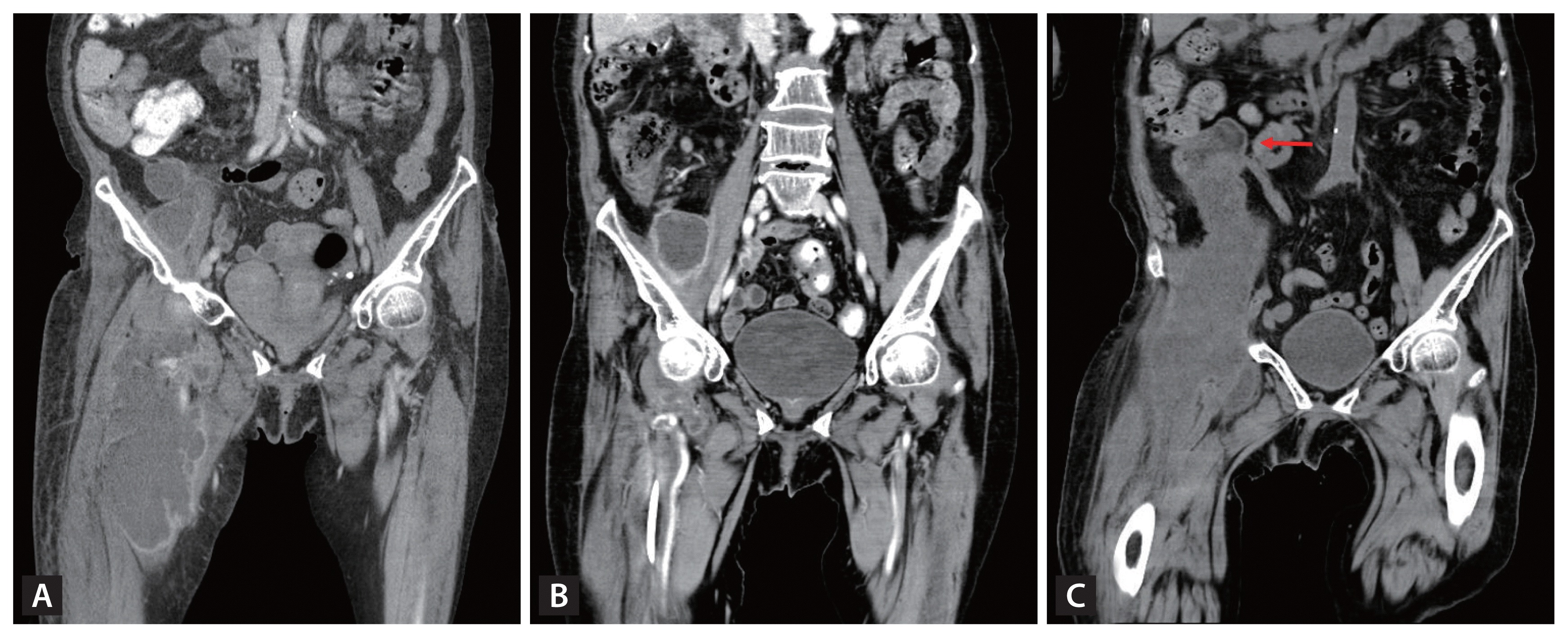
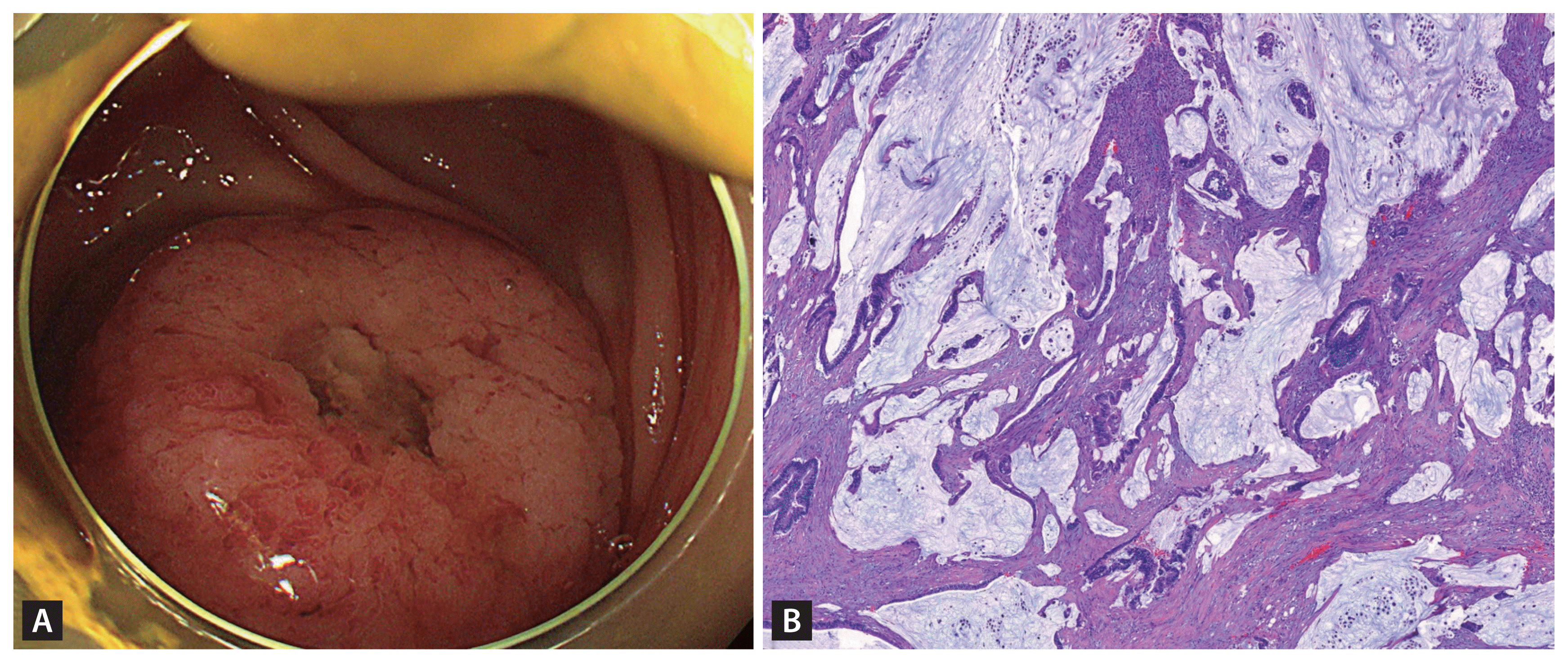
A 91-year-old woman was transferred for the management of a huge abscess of the right thigh. The abscess was of approximately 30 cm craniocaudal sized in computed tomography (Fig. 1A). Since the patient was very old, percutaneous catheter drainage (PCD) was performed without further evaluation. After PCD, the abscess improved significantly (Fig. 1B), however, after 4 months, the patient was transferred again due to abscess aggravation. In computed tomography, a mass in the cecum was adjacent to the abscess (Fig 1C). Colonoscopy was performed without bowel preparation. A 3 cm sized mass with central opening in the cecum was observed (Fig. 2A). Because cecal cancer was considered the cause of abscess, right hemicolectomy with lymph node dissection was performed. Histological findings of the resected specimen showed adenocarcinoma with extracellular mucin production of approximately 40% (Fig. 2B). Thus, the patient was diagnosed with mucinous adenocarcinoma without lymph node metastasis (pT4bN0). Although she had surgery, cancer recurred and died two months after the surgery.
An abscess caused by colon cancer is mostly caused by perforation. Mucinous adenocarcinoma has high enzymatic activity, which can break the intestinal wall and infiltrate, resulting in the formation of an abscess. There have previously been cases of intraperitoneal or psoas muscle abscess caused by mucinous adenocarcinoma of the colon. We present a case of mucinous adenocarcinoma of the cecum manifested as a huge leg abscess with images.
- Notes
- Notes
-
CRedit authorship contributions Dae Gon Ryu: conceptualization, writing - original draft, visualization; Su Bum Park: writing - review & editing, supervision; Cheol Woong Choi: data curation, formal analysis; Su Jin Kim: resources, investigation; Hyeong Seok Nam: investigation, visualization
- Conflicts of Interest
- Conflicts of Interest
-
Conflicts of interest The authors disclose no conflicts.
- Notes
- Notes
-
Funding This study was supported by a 2023 research grant from Pusan National University Yangsan Hospital.
Figure 1
(A) On computed tomography (CT), intramuscular abscess was noted at right ilioposas, adductor, and vastus muscles. (B) After percutaneous catheter drainage, the abscess improved significantly. (C) Four months later, a huge abscess recurred and a mass (arrow) in the cecum was observed adjacent to the abscess on abdominal CT.