A systematic approach for the diagnosis and treatment of idiopathic peptic ulcers
Article information
Abstract
An idiopathic peptic ulcer is defined as an ulcer with unknown cause or an ulcer that appears to arise spontaneously. The first step in treatment is to exclude common possible causes, including Helicobacter pylori infection, infection with other pathogens, ulcerogenic drugs, and uncommon diseases with upper gastrointestinal manifestations. When all known causes are excluded, a diagnosis of idiopathic peptic ulcer can be made. A patient whose peptic ulcer is idiopathic may have a higher risk for complicated ulcer disease, a poorer response to gastric acid suppressants, and a higher recurrence rate after treatment. Risk factors associated with this disease may include genetic predisposition, older age, chronic mesenteric ischemia, smoking, concomitant diseases, a higher American Society of Anesthesiologists score, and higher stress. Therefore, the diagnosis and management of emerging disease should systematically explore all known causes and treat underlying disease, while including regular endoscopic surveillance to confirm ulcer healing and the use of proton-pump inhibitors on a case-by-case basis.
INTRODUCTION
Helicobacter pylori infection and non-steroidal anti-inflammatory drugs (NSAIDs) are common causes of peptic ulcer disease (PUD). However, in recent years, the global occurrence of non-H. pylori, non-NSAID PUD has increased. Management of this emerging disease is increasingly important. Although clinicians tend to describe non-H. pylori, non-NSAID PUD as idiopathic, the term “idiopathic” refers to a disease whose cause is not known or appears to arise spontaneously. Therefore, in addition to a missed diagnosis of H. pylori and the unrecognized use of NSAIDs/aspirin, a diagnosis of idiopathic peptic ulcer disease (IPUD) should also exclude all other recognized etiologies of PUD, which requires a systematic approach that considers all possible causes.
IS THE PROPORTION OR ABSOLUTE NUMBER OF IDIOPATHIC PEPTIC ULCERS INCREASING?
The management of PUD changed dramatically after the discovery of H. pylori as a major cause of chronic gastritis, peptic ulcer, and gastric cancer. Over half of the world’s population is infected with H. pylori, especially in developing countries [1,2]. Historically, H. pylori infection was found in 90% to 100% of patients with duodenal ulcer (DU) and 60% to 100% of patients with gastric ulcer (GU) [3]. A test-and-treat strategy for H. pylori infection has been adopted as first-line management for patients with PUD, while a screen-and-treat strategy for H. pylori infection in the asymptomatic population has been considered as an effective approach to decrease future risk of gastric cancer [4,5]. These strategies have led to a continuous decline in the incidence of H. pylori-related peptic ulcers. Studies in Asia have also found that the incidence of H. pylori infection in younger patients has decreased [6-10], probably due to improved sanitation and hygiene. Conversely, the global use of NSAIDs/aspirin is increasingly prevalent in an aging population and medical comorbidities are frequent. Thus, non-H. pylori PUD is relatively more common because of increased use of ulcerogenic drugs [11].
Graham [12] proposed a model to illustrate the changing proportion of H. pylori-positive and -negative ulcer disease. If the H. pylori prevalence were to decline from 80% to 40%, and the risk of PUD from causes other than H. pylori were to remain stable at about 250 per 100,000 persons, the total number of ulcers would decrease from 1,050 to 650 per 100,000 persons, but the proportion of H. pylori-negative PUD would increase from 24% to 38% [12]. Epidemiological studies have consistently reported an increasing proportion of H. pylori-negative PUD, especially in Asian countries. Studies in the 1990s in the United States found that only about 6% of DUs were not associated with H. pylori infection, particularly in Caucasians [13]. However, later studies contradicted these results. A study in Orlando, Florida, found that only 32% of DU patients were H. pylori-positive, and only 25% of bleeding ulcers were associated with H. pylori [14]. A larger scale study including 305 cases showed that ~35% of PUD was not associated with H. pylori or NSAIDs [15], while a multicenter French study found that about 21.6% of patients with PUD had neither H. pylori infection nor a history of using ulcerogenic drugs [16]. Thus, it appears that the incidence of idiopathic ulcer remains stable in Western countries, in contrast to the increasing trend in Asian countries in recent years (Fig. 1) [15-43].
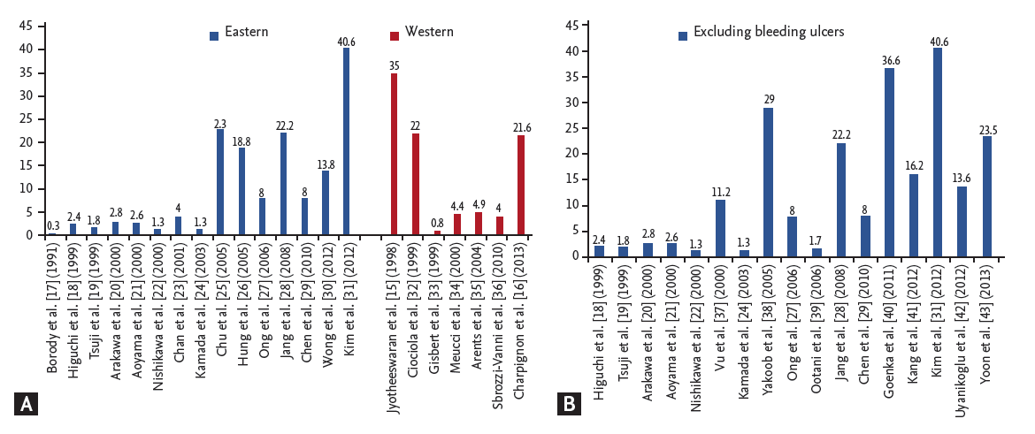
(A) Global incidence of clinical idiopathic peptic ulcer disease from 1991 to 2013 reported in large-scale studies with a sample size > 300 patients. (B) Reports of idiopathic peptic ulcer disease in Asian countries, excluding studies on bleeding peptic ulcers.
In Asian countries, a study published in 1991 revealed that only 6% of DUs were H. pylori negative, and when ulcerogenic agents were excluded the incidence was as low as 0.3% [17]. In a 2006 Japanese study, DU was idiopathic in one third of cases [38], and the proportion of idiopathic ulcers was found to be as high as 40.6% in a Korean study conducted in 2007 [31]. The trend of increasing idiopathic ulcers appears to follow an exponential curve if we focus on large-scale studies with a study population > 300) (Fig. 1A) and exclude studies that included bleeding PUD (Fig. 1B). As further evidence for the changing etiology, research conducted in Hong Kong found that only the absolute number of H. pylori-related bleeding ulcer was declining, while the number of H. pylori-negative bleeding ulcers remained stable [33,44-46]. Thus, the accumulated evidence supports the concept proposed by Graham [12]: non-H. pylori- and non-NSAIDs-related ulcer diseases are more likely to be found in older, sicker patients, are more resistant to acid suppression therapy, and are associated with a greater risk of bleeding and recurrence and a greater overall mortality rate as compared with traditional H. pylori-positive PUD [44,45].
IDIOPATHIC PEPTIC ULCER DISEASE: A DIAGNOSIS OF EXCLUSION
Because IPUD is defined as PUD that arises without an identifiable cause, it is a diagnosis of exclusion. Possible etiologies to be excluded are shown in the Table 1 (upper panel), which may include missed H. pylori infection, unidentified use of ulcerogenic medications, rare systemic diseases with upper gastrointestinal tract manifestations, hyperacidity of the stomach, and other rare infections involving the upper gastrointestinal tract.

Etiologies to be excluded for the diagnosis of idiopathic peptic ulcer and associated risk factors
Incorrect diagnosis of H. pylori
There are several methods available to detect H. pylori infection. These methods are characterized according to whether a mucosal specimen is needed for analysis. Biopsy-based tests include histological evaluation, culture, polymerase chain reaction, and the rapid urease test (RUT). Alternatively, non-invasive methods may include the urea breath test (UBT), serology, and the stool antigen test (SAT). A meta-analysis has revealed the following pooled sensitivity and specificity for different methods: RUT 0.67 and 0.93; histology 0.70 and 0.90; culture 0.45 and 0.98; UBT 0.93 and 0.92; SAT 0.87 and 0.70; and serology 0.88 and 0.69 [46], respectively, which clearly demonstrates that each of the different tests has its own limitations, and a single negative test does not exclude H. pylori infection.
Current or recent use of antibiotics or proton pump inhibitors (PPIs) is known to influence the accuracy of H. pylori tests. Bacterial concentrations are decreased by these medications, leading to false-negative results. A number of studies have demonstrated a lower yield in testing for H. pylori infection in patients receiving antibiotic therapy. Borody et al. [17] showed that ~22% of H. pylori-negative DU patients were reported to have used antibiotics recently. Gisbert et al. [33] also found a lower prevalence of H. pylori in DU patients who had prior antibiotic therapy compared with those who did not (78% vs. 96%). In another study, the prevalence of H. pylori-negative DU was shown to be much lower when patients taking antibiotics within 4 weeks of testing were excluded [47]. In addition, the use of PPIs and histamine-2 receptor antagonists (H2RAs) can also cause false-negative results by reducing gastric acid secretion and inhibiting the growth of H. pylori. Approximately one-third of patients who remained positive for H. pylori infection had a negative UBT result while receiving PPIs [48]. The study even showed that the proportion of patients whose UBT results turned out to be positive after completion of PPIs therapy were 91% at 3 days, 97% at 7 days, and 100% at 14 days [48]. Chey et al. [49] also demonstrated that both PPIs and H2RAs could affect the sensitivity of UBT, with an equivocal or false-negative result of 61% and 18%, respectively.
RUT can also yield a false-negative result during an actively bleeding stage of PUD. Lee et al. [50] have found that the prevalence of H. pylori infection in patients presenting with bleeding DU was 72.7%, which was lower than the prevalence of 92.8% in those with non-bleeding peptic ulcers (p < 0.05). The false-negative rate of RUT for bleeding ulcer has been reported to be up to 25% [50]. In another study, 55.1% of patients with bleeding ulcers with an initially negative RUT result were discovered to have a positive result at follow-up endoscopy [23]. Lee et al. [51] have demonstrated that the low sensitivity of RUT (61%) for diagnosis of H. pylori infection during bleeding peptic ulcer can be overcome by increasing the number of biopsies from the gastric antrum to 74% or from the gastric body to 73%. Some investigators speculate that these results reflect a buffering effect of serum albumin on the pH indicator of the RUT [52].
The location and number of biopsies taken are also important for the diagnostic accuracy of both RUTs and histological results [33,45,53,54]. The distribution of H. pylori can be sporadic and variable in the stomach when intestinal metaplasia or atrophic gastritis develops. By histological examination, < 3% of antral biopsy specimens yielded a false-negative interpretation, compared with 6% to 9% of those from the corpus (p = 0.02) [54]. Another study based on 1,000 biopsy specimens has shown that the area of H. pylori colonization was larger than the area of active chronic gastritis, suggesting that H. pylori colonization may precede the development of active chronic gastritis [53]. In the presence of atrophic gastritis or intestinal metaplasia, H. pylori is prone to disappear from the gastric mucosa with such a histological change [55]. Therefore, it is postulated that H. pylori may migrate proximally from the antrum to the corpus because of lack of acid in the atrophic antrum. A meta-analysis has suggested that the sensitivities of RUT and histology could be improved to 78% and 83%, respectively, if biopsies were taken from both the antrum and the corpus of the stomach [46].
Unrecognized use of ulcerogenic drugs
Surreptitious use of medications or lack of medication history with regard to ulcerogenic drugs may be of paramount importance when encountering H. pylori-negative PUD. By testing the blood for drugs, some studies have shown that a substantial number of users of ulcerogenic drugs cannot provide a correct description of their medication history [56,57]. Using the platelet cyclo-oxygenase activity test, 12.7% more users of aspirin could be identified than the number obtained by clinical history alone. Sixty-six percent of NSAID users were actually taking aspirin, alone or in combination with other NSAIDS, and 59.3% of patients who claimed no (non-aspirin) NSAID use were actually using NSAIDs [57]. Blood salicylic acid concentrations revealed that about half of patients with intractable PUD who denied using aspirin were actually aspirin users [56]. Ong et al. [27] also found that when serum thromboxane B2 levels were checked, more than 30% of patients who were thought to have IPUD were found to have taken NSAIDs. Other possible non-NSAID, non-aspirin, ulcer-related drugs include steroids, potassium chloride, nitrogen-containing bisphosphonates, and some immunosuppressive medications [58,59]. Visible gastric mucosal damage was found in 38% of patients who received alendronate as compared to 13% in the placebo group [59]. The results demonstrate that a valid review of medication history is mandatory before making the diagnosis of IPUD.
Excluding malignancies
Follow-up endoscopy is necessary in IPUD, especially in those patients who have alarming symptoms or recurrent ulcers, to determine whether there are secondary causes or undiscovered malignancies (Fig. 2). In fact, ~5% of endoscopically benign-appearing GUs are malignant [60,61]. Endoscopic surveillance of the gastric pre-malignant condition has been useful in identifying early-stage malignancy at 3- to 6-month follow-up visits scheduled to confirm that the GU is healed [62,63]. Although the optimal interval for endoscopic surveillance remains to be determined, repeat endoscopic biopsies, obtaining sufficient tissue involving submucosa, or targeted optical biopsy by image-enhanced endoscopy is a reasonable approach for IPUD [64].
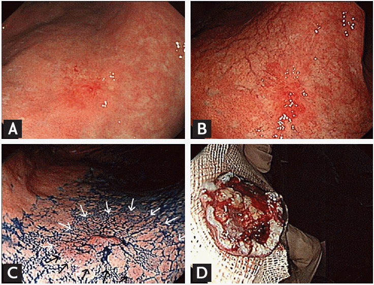
A case of gastric ulcer with a missed diagnosis of gastric cancer. (A) One healing gastric ulcer was seen at the lesser curvature of the lower body. A biopsy showed only chronic inf lammation. (B) The ulcer remained present on follow-up endoscopy at 3 months despite continuous proton-pump inhibitor treatment. A repeated biopsy showed a well-differentiated adenocarcinoma. (C) An image of the tumor margin delineated by arrows under chromoendoscopy with indigo carmine spraying before endoscopic resection. (D) An image of the en bloc resected specimen.
Excluding rare etiologies
Rare etiologies for PUD may include unusual systemic diseases and unusual infectious pathogens. Uncommon systemic diseases, such as Crohn’s disease (Fig. 3), mastocytosis, amyloidosis, sarcoidosis, vasculitis, eosinophilic gastroenteritis, and Zollinger-Ellison syndrome (Fig. 4), are possible sources of the upper gastrointestinal manifestations of PUD. Screening of the lower gastrointestinal tract for Crohn’s disease and specific laboratory and radiological tests (such as repeated measurement of serum fasting gastrin, secretin stimulation test, quantification of gastric acid secretion, measurement of serum chromogranin A, radiological studies, and endoscopic ultrasound for Zollinger-Ellison syndrome) are needed to exclude these uncommon diseases. Rare infectious causes of PUD may include Helicobacter heilmanii, tuberculosis, syphilis, cytomegalovirus (Fig. 5), herpes simplex virus, and fungal infection (Fig. 6).
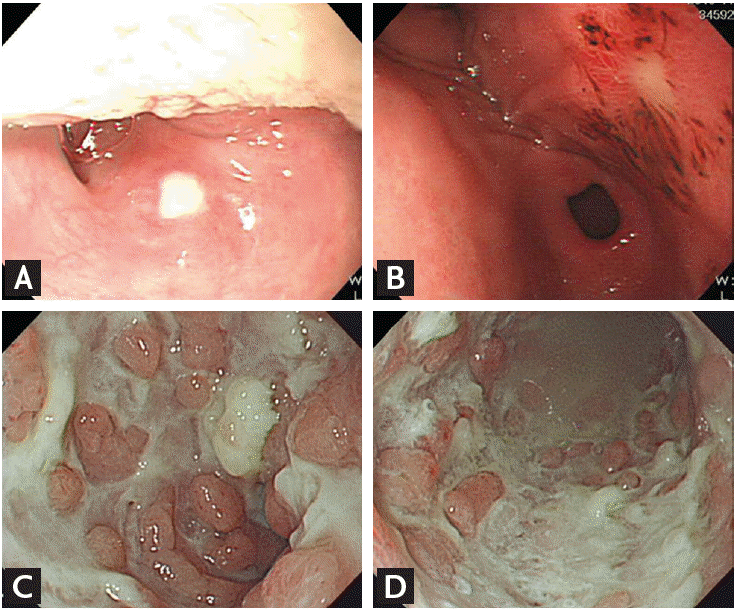
A case of Crohn’s disease with upper gastrointestinal manifestations. (A) One oral ulcer was found on the soft palate. (B) One gastric ulcer was noted on the posterior wall of the antrum. (C) Upon colonoscopic examination, multiple colonic ulcers were seen with a cobblestone appearance over the ascending colon and (D) descending colon.
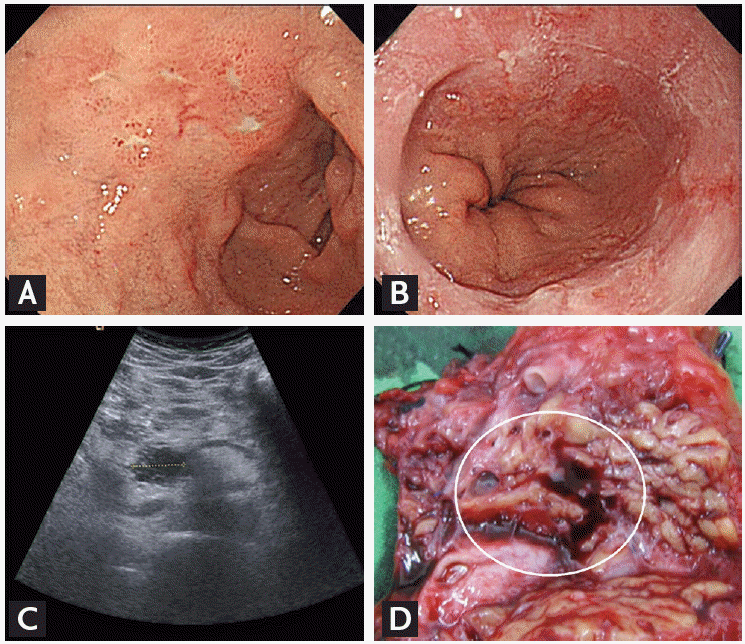
A case of hyperacidity of the stomach due to Zollinger-Ellison syndrome. (A) Repeat upper endoscopy showed poorly healed duodenal ulcers and (B) reflux erosive esophagitis, suggestive of gastric acid over-production. (C) Upon abdominal sonography, a pancreatic tumor originating from the uncinate process of the pancreas was noted. (D) The pancreatic tumor specimen was confirmed as a neuroendocrine tumor (circle).
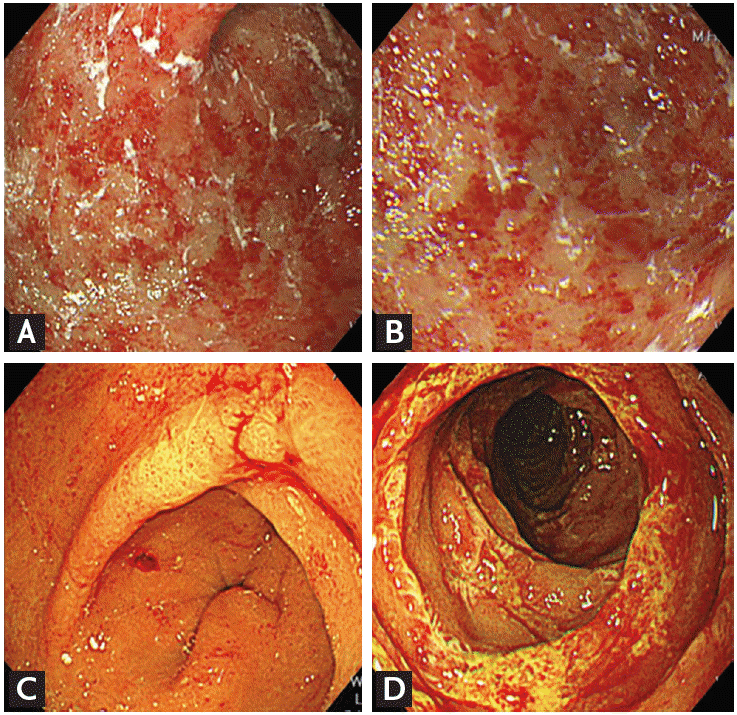
A case of gastric and duodenal ulcers due to cytomegalovirus infection. Multiple areas of erythema were found over the lower body (A) and antrum of the stomach (B) with a negative Helicobacter pylori test. (C) A bleeding ulcer was noted on the posterior wall of the gastric antrum. (D) Diffuse ulcerations in the jejunum found by the balloon-assisted enteroscopy. A biopsy was positive for cytomegalovirus inclusion bodies.
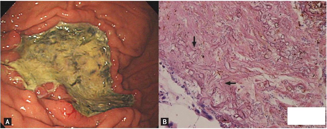
A case of a mucormycosis-related gastric ulcer. (A) Upon upper endoscopy, a gastric ulcer with greenish coating was noted in the greater curvature of the middle body. (B) Pathology showed numerous right-angled, pauci-septated, and ribbon-like hyphae (arrows), indicating a fungal infection (H&E, ×100).
In summary, accurate diagnosis of classic IPUD requires a systematic approach as outlined in Table 2. Confirmation of H. pylori infection status of the stomach and duodenum by at least three parallel tests is recommended to decrease the risk of a false-negative result. Thorough review of medication history is important to identify the use of potential ulcerogenic drugs. Moreover, a complete histological study of a benign-appearing ulcerative lesion is crucial to rule out the possibility of malignancy. Repeat endoscopic biopsies, optical targeted biopsy, or obtaining tissues deeper than the mucosal layer may improve diagnostic accuracy. Zollinger-Ellison syndrome should always be kept in mind when encountering poorly-healed peptic ulcers. Finally, rare infectious agents should be considered in the differential diagnosis, especially in immunocompromised subjects.

Considerations to review prior to making a diagnosis of idiopathic peptic ulcer
RISK FACTORS AND THE CLINICAL COURSE OF CLASSICAL IPUD
Risk factors for classical IPUD may include demographic risk factors, use of psychoactive substances, genetic risk factors, comorbid diseases, chronic mesenteric ischemia, and higher psychological stress (Table 1, lower panel).
Demographic factors, such as being Caucasian and older age, are associated with IPUD. A greater prevalence of H. pylori-negative ulcers was found in whites than in non-whites in previous studies [13,15]. In multi-racial studies, a greater prevalence of H. pylori-negative DUs was found in Malays and Chinese than in Indians [7,65]. Molecular changes in gastric protective factors, such as mucin, are also hypothesized to be associated with risk of ulcers. The gastric protective layer, which prevents enzymatic attachment by gastric acid, pepsin, and other aggressive factors, is created by secreted mucin. Structural changes of membrane-bound mucin and glycan side-chain sialic acids are associated with protective efficiency against offending factors. Based on an immunochemical study of ulcer tissues, cytoplasmic MUC17 staining intensity was significantly lower in patients with IPUD than in those with H. pylori-related peptic ulcer [66]. In addition to genetic or epigenetic changes in the gastric mucin molecule, the HLA-DQA1*0102 allele was associated with increased risk of IPUD [67,68]. Ethnic differences in the prevalence of H. pylori-negative PUD may correlate with different lifestyles or unknown genetic factors. Several studies in different areas have demonstrated that patients with IPUD are significantly older than those with PUD associated with H. pylori or NSAID use [23,26,27,34,69].
Older age was associated with a significantly lower level of prostaglandin concentration in the fundus, antrum, and post-bulbar duodenum [70]. Because prostaglandins protect the gastric mucosa from damage, diminished synthesis in elderly subjects makes their mucosa more susceptible to aggressive factors, followed by the ulcerogenic process. Moreover, older patients have higher risk for vascular disease, especially chronic mesenteric ischemia [71]. A combination of risk factors, including reduced blood flow, decreased production of prostaglandins by the stomach, a higher prevalence of concomitant diseases, and a higher American Society of Anesthesiologists score in the elderly render the mucosa more fragile and, thus, PUD is more likely to develop [16,72,73]. Cigarette smoking also increases xanthine oxidase activity, production of leukotrienes and nitric oxide, and neutrophil infiltration into the gastric mucosa [72]. An association between psychological stress and PUD has long been suggested. Two recent studies of Japanese victims of the Great East Japan Earthquake and Tsunami that occurred in 2011 have found that the incidence of PUD increased significantly after the psychological trauma [74,75]. In the first 3 months after the earthquake, the incidence of PUD was increased 1.5 fold, and the incidence of bleeding PUD was increased 2.2 fold [74,75]. Furthermore, after the earthquake, the proportion of IPUD doubled from 13% in 2010 to 24% in 2011 (p < 0.05) [75]. A population-based study of 3,379 Danish adults in 1982 to 1983 revealed a greater incidence of ulcers among subjects in the highest tertile of stress scores (3.5%) than in those in the lowest tertile (1.6%; p < 0.01) and high psychological stress has been identified as an independent risk factor for PUD on multivariate analysis (odds ratio, 1.12 per point increment of stress index; 95% confidence interval [CI], 1.01 to 1.23; p = 0.03) [76].
The clinical course of IPUD remains poorly understood because of the heterogeneity of diagnostic methods and populations recruited for studies. Nevertheless, idiopathic ulcer disease tends to cause longer-lasting ulceration, a greater recurrence rate and mortality rate, and greater symptoms of dyspepsia. In addition, it is more refractory to treatment than H. pylori-positive ulcer disease [26,77-84]. Furthermore, IPUD is also more likely to present with hemorrhage, and multiple and larger ulcers [84]. After a bleeding episode, the risk of recurrent ulcer complications within 12 months was higher in patients with H. pylori-negative PUD than in those with H. pylori-related PUD after eradication therapy (13.4% vs. 2.5%, p < 0.001) [26]. In Hong Kong, a 7-year cohort study has shown that the cumulative incidence of recurrent bleeding from ulcers was higher in the H. pylori-negative ulcer cohort than in the positive cohort (42.3% vs. 11.2%, p < 0.0001), and more patients died in the H. pylori-negative ulcer cohort than in the H. pylori-positive ulcer cohort (87.6% vs. 37.3%, p < 0.0001) [84]. A randomized trial with a 2-year follow-up period consistently showed that the prognosis of H. pylori-negative ulcers was poorer [81]. The rates of recurrent ulcer or ulcer that has not healed (35% vs. 26%), and relapse of dyspepsia symptoms (16% vs. 7%) were higher in those with H. pylori-negative ulcers than in those with H. pylori-positive ulcers [81].
MANAGEMENT OF IDIOPATHIC PEPTIC ULCER DISEASE
As IPUD is increasingly encountered, whether we should modify the standard treatment of PUD, including short-term PPI therapy and once-off H. pylori eradication, is of clinical significance. Based on present knowledge regarding a poorer response to acid-suppressing therapy in IPUD than in H. pylori-positive ulcers, anti-secretory medication remains the mainstay of treatment [77]. In a Danish study, outcomes in PPI-treated patients with DU did not differ according to H. pylori status, which suggested that PPI therapy was effective in the prevention of recurrence of IPUD [81]. In contrast, another study from Hong Kong demonstrated that the risk of re-bleeding (2.9 per 100 person-years vs. 1.1 per 100 person-years, p < 0.001) and mortality (hazard ratio, 1.1; 95% CI, 0.6 to 1.7) were not reduced by gastro-protective agents in patients with bleeding IPUD [30]. Gillen et al. [85] found that H. pylori infection may potentiate the anti-secretory effect of PPIs. During PPI administration, median basal intragastric pH was higher in the H. pylori-positive (7.95) versus -negative (3.75) subjects (p < 0.002) [85]. The poorer response to the anti-secretory therapy and altered gastric physiology of IPUD could indicate the necessity of longer duration and higher dose of acid-suppressing agents. Quan and Talley [45] have proposed a flow chart for the management of IPUD (Fig. 7). After excluding gastric H. pylori infection (by multiple testing with a parallel interpretation of results) and the use of common ulcerogenic medications, the diagnosis of clinical IPUD is made. PPIs administration for 4 to 8 weeks is recommended, and a longer duration of therapy may be needed for complicated ulcer (e.g., bleeding or perforated). Otherwise, patients with uncomplicated ulcer disease may wean off therapy with a wait-and-see strategy, or receive on-demand/maintenance PPI therapy if symptoms recur. Re-evaluation of IPUD by endoscopy should be considered. If healing is slow or absent, items listed in Table 2 should be re-evaluated to confirm whether classic IPUD is the correct diagnosis.
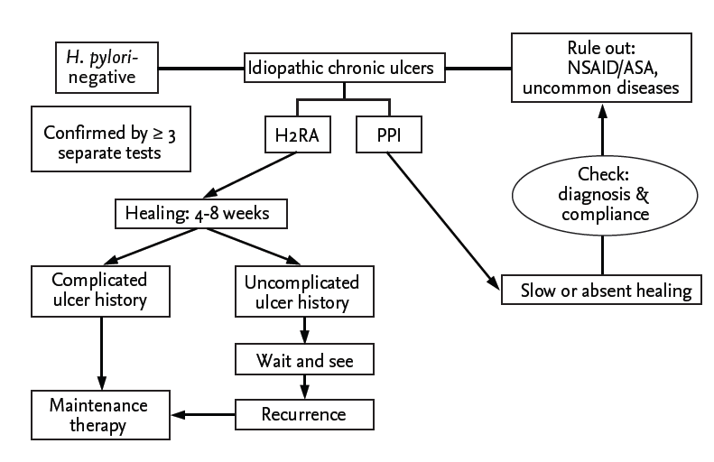
A f low chart for the diagnosis and management of an idiopathic peptic ulcer disease. H. pylori, Helicobacter pylori; NSAID, nonsteroidal anti-inflammatory drug; ASA, acetylsalicylic acid (aspirin); H2RA, histamine-2 receptor antagonists; PPI, proton pump inhibitor. Adapted from Quan et al. [45], with permission from Nature Publishing Group.
CONCLUSIONS
An increase in the proportion of idiopathic ulcers has been confirmed worldwide, and peptic ulcer, once considered to be an infectious disease after the discovery of H. pylori, has gradually reverted to the original “no acid, no ulcer” theory. Given that the prognosis of IPUD is poorer than that of PUD associated with H. pylori or NSAIDs, careful evaluation of H. pylori infection by multiple parallel tests, detailed review of medication history, and thorough exclusion of other possible causes are of paramount importance before making the diagnosis of IPUD. Further well-designed clinical trials are needed to elucidate the natural history of IPUD, and to optimize the treatment strategy for this emerging disease.
Notes
No potential conflict of interest relevant to this article was reported.