Uncovering the unforeseen: pericarditis and mycotic coronary aneurysm at previous stent site
Article information
A 72-year-old man with persistent pericarditis who had been treated for Dressler’s syndrome but did not show improvement of symptoms was transferred to our hospital. He had had right coronary artery stenting for ST-elevation myocardial infarction a month earlier and was prone to cholangitis. An echocardiogram revealed decreased pericardial effusion, and Escherichia coli was detected in a blood culture test after admission. Because the patient had cholangitis, endoscopic retrograde cholangiopancreatography (ERCP) was performed. Blood tests revealed elevated levels of myocardial enzymes and inflammatory markers even after ERCP and antibiotic treatment. An echocardiogram was performed to confirm increased pericardial effusion. Computed tomography revealed a 3-cm-sized contrast-enhanced lesion around the right coronary artery (Fig. 1). Consequently, coronary angiography was performed, and a massive right coronary aneurysm near the previous stent was confirmed (Fig. 2), indicating a mycotic coronary aneurysm. Thus, we decided to perform coronary artery bypass surgery with aneurysm resection. Massive adhesions and abscess formation were detected when the pericardium was dissected, and the ruptured aneurysm was discreetly covered by the brittle adhesive inflammatory pericardium. The final echocardiogram revealed no pericardial effusion after 4 weeks of antibiotic treatment.
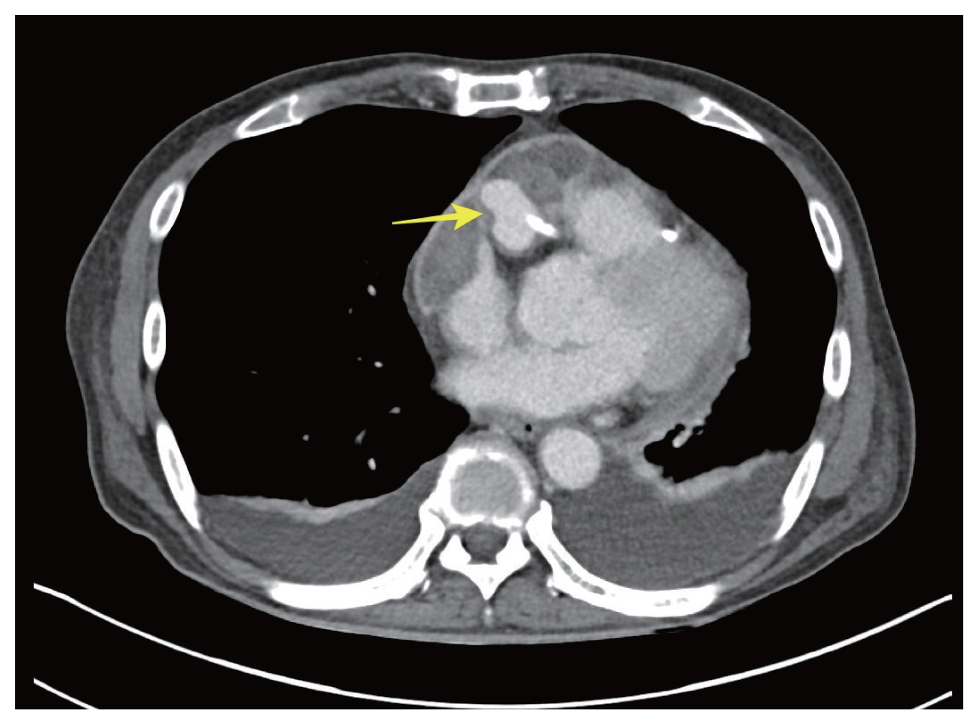
Abdomen computed tomography scan revealed a fresh pericardial effusion with rim enhancement. The yellow arrow indicates a rim enhencement lesion near the right coronary aneurysm.
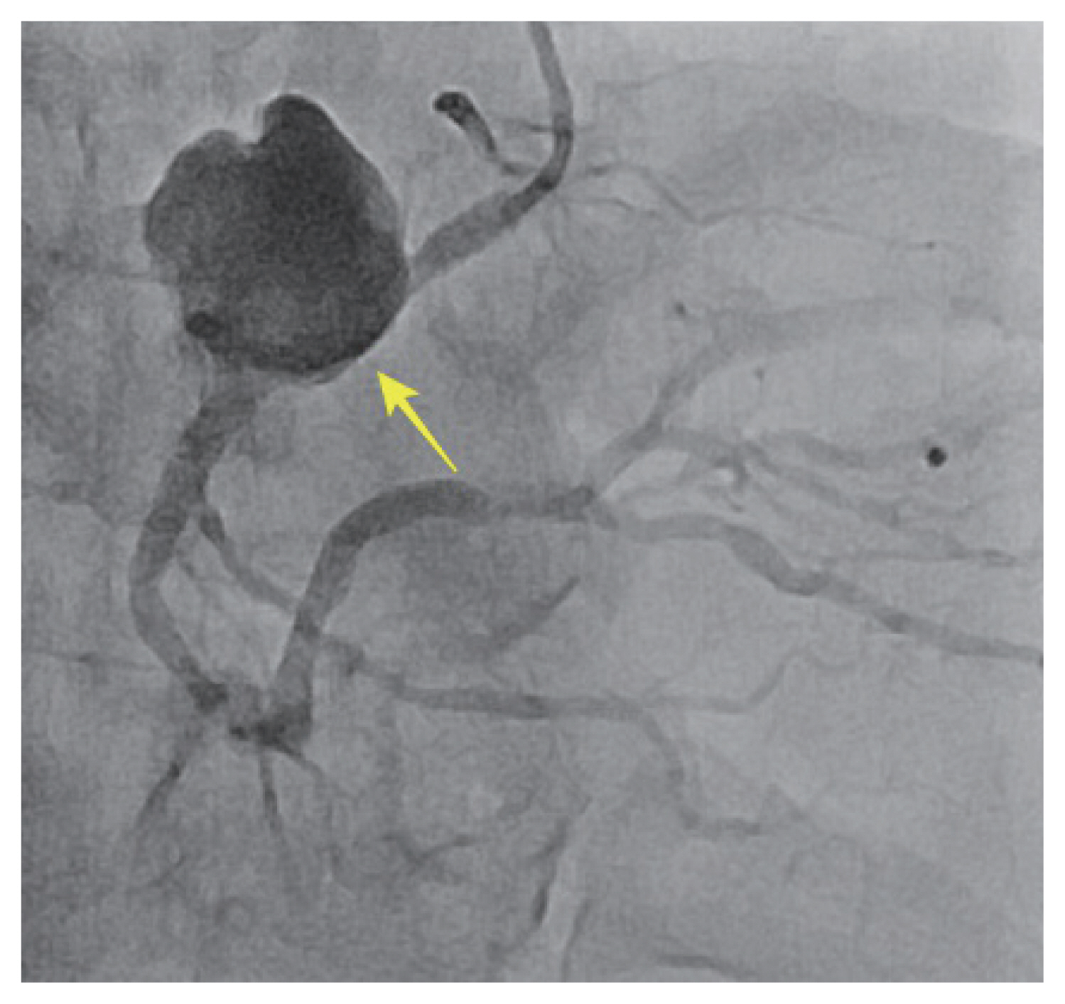
Coronary angiography reveled a huge right coronary aneurysm adjacent to the previously placed stent. The yellow arrow indicates the site of the previous stent.
An infected coronary artery aneurysm is an extremely rare complication of percutaneous coronary intervention (0.3% to 0.6%) [1], and its combination with pericarditis is an even rarer but potentially fatal condition [2]. Interestingly, in this case, the ruptured aneurysm was concealed by massive adhesions, saving the patient from shock or unstable condition. Because lesions > 1 cm in diameter increase risk of aneurysm growth and rupture, coronary artery bypass surgery and aneurysm resection were required to avoid fatal rupture and uncontrolled infection.
Notes
Credit Authorship Contributions
Sook Jung Kim: writing - original draft, writing - review & editing; Yun-Seok Choi: conceptualization, writing - review & editing, supervision; Osung Kwon: conceptualization, writing - original draft, writing - review & editing; Jeong Eun Yi: validation, supervision; Joon-Kyu Kang: supervision
Conflicts of interest
The authors disclose no conflicts.
Funding
None