Leiomyosarcoma at popliteal vein presented as unilateral leg edema in a patient with kidney transplantation
Article information

A 66-year-old female, who underwent kidney transplantation 4 years ago, presented with right lower leg edema. She had been taking prednisolone, tacrolimus, and mycophenolate mofetil as immunosuppressive agents. Her kidney function was stable, with a serum creatinine level of 1.0 mg/dL and an estimated glomerular filtration rate of 59.9 mL/min/1.73 m2. Initially, an ultrasonographic examination detected a mass over 2 cm in size abutting the right popliteal vein, with no evidence of deep vein thrombosis in the lower extremities. Subsequent magnetic resonance imaging revealed a 6 cm long tumor with mild high signal intensity on T1-weighted images, peripheral nodular enhancement, and an internal thrombus. These findings raised suspicions of either leiomyoma or leiomyosarcoma. Mass excision and subsequent tissue next-generation sequencing identified a leiomyosarcoma originating from the popliteal vein wall, with CIC (c.1057C > T) and TP53 (c.785G > T) gene mutations suggesting more aggressive behavior. Moreover, metastatic spread to the liver was confirmed by positron emission tomography scan. For curative purposes, a laparoscopic central bisectionectomy was performed, followed by chemotherapy. Figure 1 is the imaging results described above.
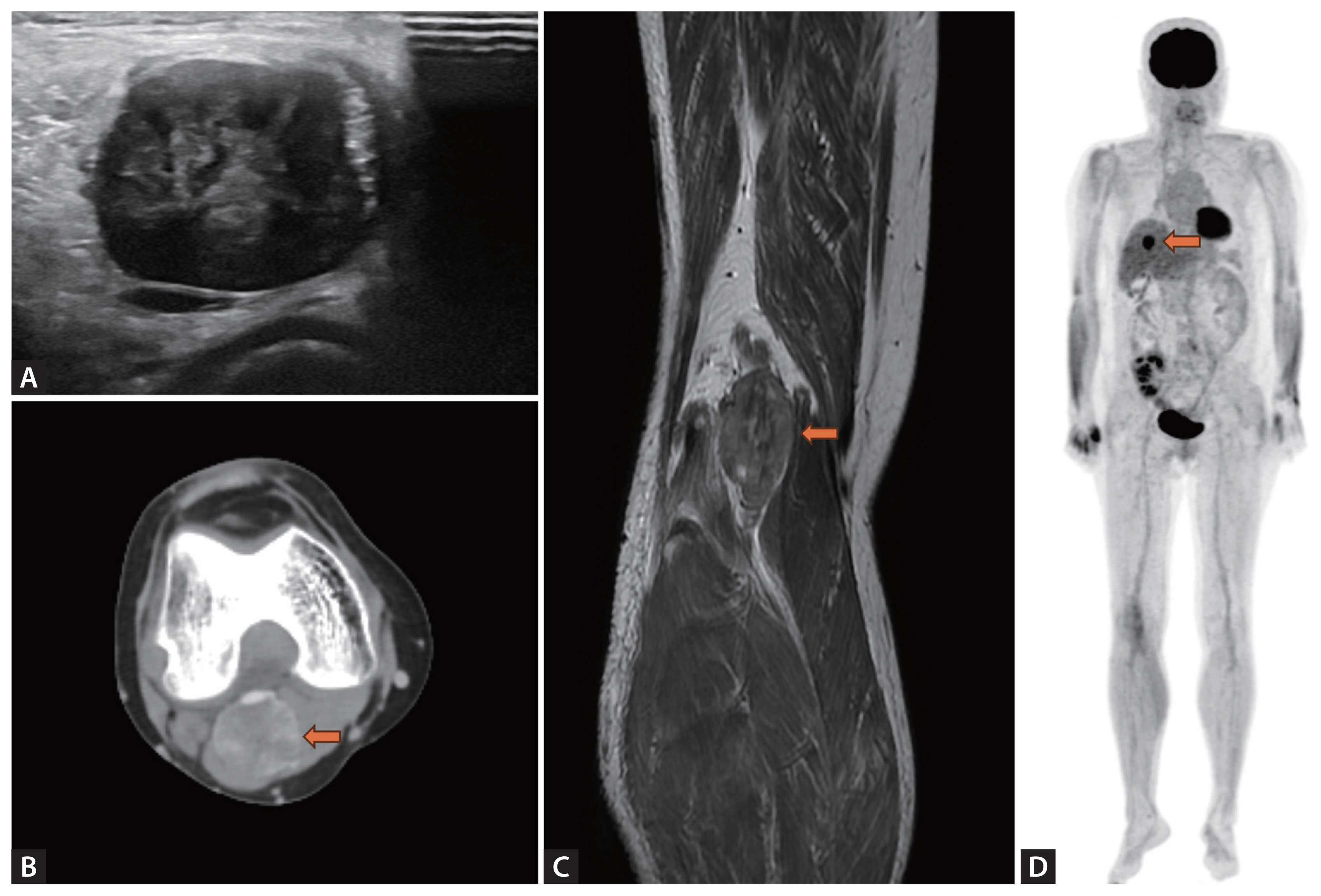
(A) Ultrasonographic finding showing hypoechoic mass at the level of right popliteal vein. (B, C) T1-weighted Magnetic Resonance Imaging showing soft tissue mass with peripheral nodular enhancement, and an internal thrombus at the level of popliteal vein. (D) Positron Emission Tomography Imaging showing metastatic leiomyosarcoma in central portion of liver.
The malignancy significantly impacts survival in kidney transplant patients under long-term immunosuppression. Compared with the general population, patients with transplants are more likely to develop malignant tumors, with sarcomas such as leiomyosarcoma being exceedingly rare yet prone to metastasize to other organs. While leiomyosarcomas may originate from the smooth muscle of vascular walls, this is exceedingly rare, especially in kidney transplant recipients, as far as I am aware. In this case, she remained clinically stable for four years post-transplantation and presented with a swollen right leg at an outpatient clinic. The leiomyosarcoma was detected during the evaluation of her symptoms. Following curative surgery and chemotherapy, we are conducting meticulous follow-up in an outpatient clinic, and currently, there is no further progression of the leiomyosarcoma.
Notes
Conflict of interest
The author discloses no conflicts.
Funding
This work was supported by a clinical research grant from the Pusan National University Hospital in 2023.