Epidemiological and clinical trends of malignant small-bowel tumors in Asia
Article information

Abstract
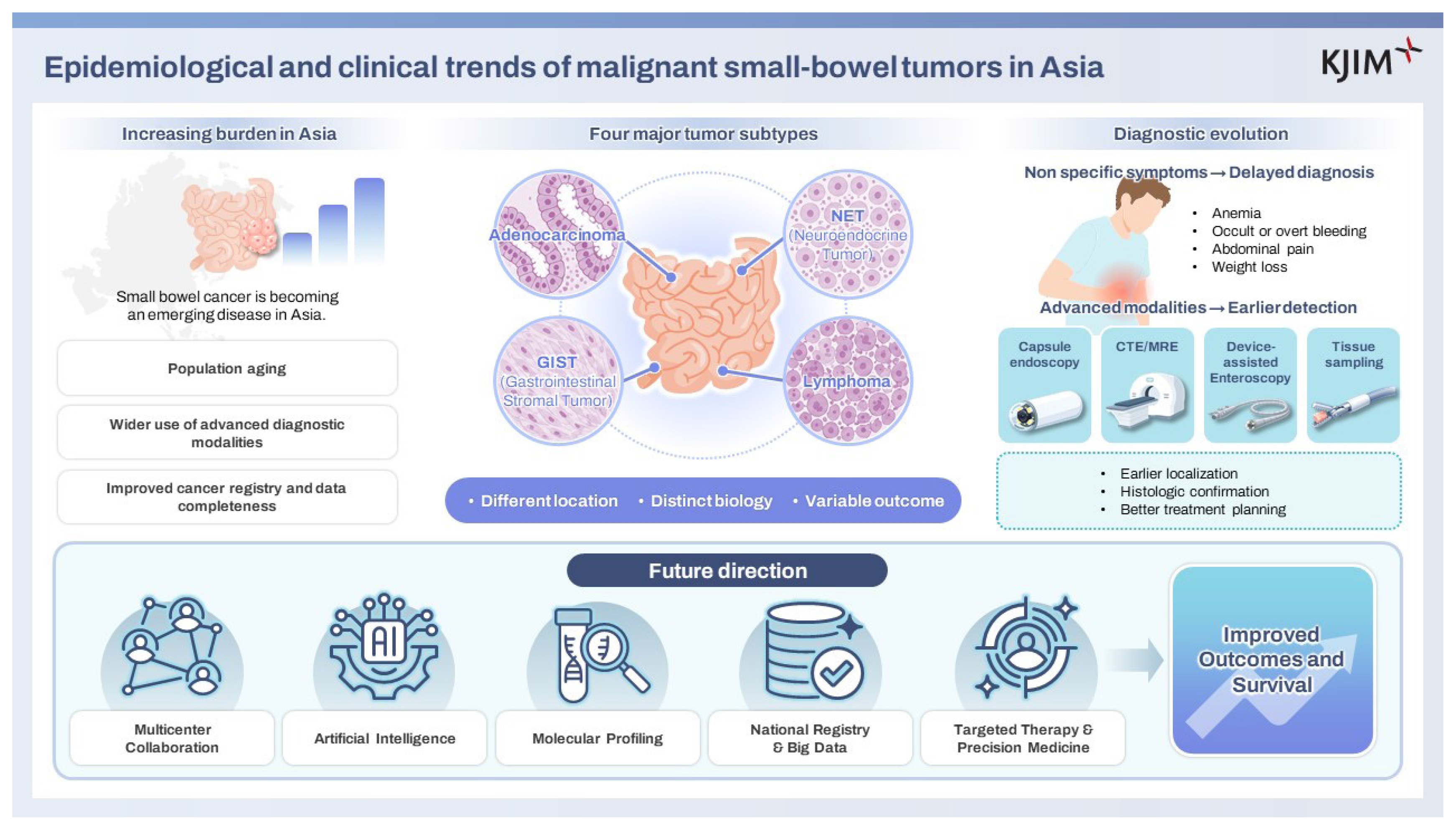
Malignant small-bowel tumors are uncommon, accounting for < 5% of all gastrointestinal cancers, yet their incidence has exhibited a steady rise in both Western and Asian populations. This increase is attributed to expanded use of advanced diagnostic modalities and improved registry completeness. Malignant small-bowel tumors encompass a heterogeneous group—adenocarcinoma, neuroendocrine tumors, gastrointestinal stromal tumors, and lymphoma—each with distinct anatomic predilections, molecular characteristics, and clinical behavior. In Asia, sporadic adenocarcinoma remains the predominant subtype, whereas Crohn’s disease–associated lesions and small-bowel neuroendocrine tumors are relatively less common than in Western cohorts. Early detection continues to be limited by diagnostic delays caused by nonspecific symptoms and the anatomic inaccessibility of the small-bowel. Prognosis varies substantially across subtypes, with adenocarcinoma demonstrating the poorest survival rate despite advances in systemic therapy. Recent nationwide and multicenter studies from Asian countries have highlighted evolving temporal trends, improvements in diagnostic performance, and emerging regional differences in etiology, stage distribution, and outcomes. A clearer understanding of the epidemiologic patterns and clinical characteristics of malignant small-bowel tumors in Asia is essential for developing region-specific diagnostic strategies, optimizing treatment pathways, and prioritizing areas for future collaborative research.
INTRODUCTION
Although the small-bowel accounts for nearly three quarters of the gastrointestinal (GI) tract in length and more than 90% of its mucosal surface area, malignant tumors arising in this segment represent < 5% of all GI cancers [1–4]. This paradox has historically impeded clinical recognition and epidemiologic characterization, as many cases present at advanced stages owing to nonspecific symptoms and the anatomic inaccessibility of the small-bowel [1].
Over the past two decades, however, malignant small-bowel tumors have been increasingly recognized worldwide. Population-based datasets, including SEER and GLOBOCAN, demonstrate a gradual rise in incidence—from approximately 1.0 to > 2.0 per 100,000—primarily driven by adenocarcinoma and neuroendocrine tumors (NETs) [1,5]. Despite substantial improvements in capsule endoscopy (CE), cross-sectional enterography—including computed tomography enterography (CTE) and magnetic resonance enterography (MRE)—and device-assisted enteroscopy (DAE), most patients continue to present with advanced disease, contributing to poorer outcomes compared with other GI malignancies [2,6].
In Asia, the clinical and epidemiologic landscape of small-bowel tumors exhibits distinct characteristics. Although incidence is steadily increasing in parallel with population aging and broader use of advanced diagnostics, survival outcomes remain generally inferior to those reported in Western cohorts [7–9]. Variability in healthcare resources, diagnostic expertise, and registry completeness further complicates direct comparisons across regions.
Recent expansion of national insurance databases and cancer registries in Korea, Japan, and Taiwan has enabled the accumulation of more robust epidemiologic data, providing opportunities for region-specific analyses and comparison with Western populations [7,9,10]. Concurrently, rapid demographic aging and wider adoption of endoscopic and imaging technologies have led to increased detection of small-bowel tumors across Asia. These trends warrant a contemporary synthesis focused on the Asian context.
This review summarizes the epidemiologic and clinicopathological features of malignant small-bowel tumors in Asia, with particular emphasis on subtype-specific patterns, diagnostic challenges, survival outcomes, and priorities for future multicenter collaboration.
EPIDEMIOLOGY AND TEMPORAL TRENDS
The incidence of malignant small-bowel tumors has been steadily increasing worldwide, including in Asia. Global population-based analyses such as SEER and GLOBOCAN indicate that the age-standardized incidence has more than doubled over the past four decades, largely driven by adenocarcinoma and NETs [1,5]. Similar patterns are now evident across Asia, reflecting demographic aging, expanded access to endoscopic evaluation, and improving cancer registry completeness. Nationwide and multicenter studies from Korea, Japan, Taiwan, and Thailand consistently demonstrate a gradual rise in newly diagnosed cases over the past two decades, with longitudinal registry data from Korea and Japan showing steady increases from the mid-2000s to the early 2020s [7–10]. In Korea, analysis of the National Health Insurance Service (NHIS) cohort revealed a steady annual increase from 2005 to 2022, with the most pronounced growth among individuals aged ≥ 65 years [7]. Similarly, Japanese and Taiwanese registry data indicate a predominance among the elderly, while a Thai single-center series reported a mean diagnostic age in the early 60s [8–10]. Collectively, these findings suggest that the increasing burden of malignant small-bowel tumors in Asia parallels the region’s rapid demographic shift toward an aging population.
Although advances in CE, CTE/MRE, and DAE have improved case detection, population aging remains the principal driver of incidence growth. A comparative overview of incidence, age distribution, male predominance, and subtype patterns across Asian and Western populations is summarized in Table 1. Although crude rates and age-standardized incidence rates are not directly comparable, the inclusion of both metrics reflects differences in data availability and reporting practices across regions. Accordingly, Table 1 is intended to provide a descriptive overview of regional disease burden rather than a strict quantitative comparison. Compared with Western populations, Asian countries have slightly lower absolute incidence but broadly similar demographic characteristics, with a predominance in the sixth to seventh decades of life. The absolute incidence in Asia remains modest—approximately 1.0–1.8 versus 2.0–2.4 per 100,000—but the relative rate of increase is comparable to that in Western populations, or even steeper in some regions [5,7,10]. Distinct etiologic patterns are also evident. Unlike Western countries, where Crohn’s disease– associated small-bowel adenocarcinoma (SBA) accounts for 15–30% of cases, this subtype remains uncommon (< 5%) in Asia, mirroring the lower regional prevalence of inflammatory bowel disease (IBD) [6,7,10,11]. Sporadic, inflammation-unrelated adenocarcinoma therefore constitutes the majority of cases in Asian registries. In certain subregions such as China and Southeast Asia, a slightly younger age at onset has been reported, possibly due to demographic differences and uneven healthcare access [8,9].
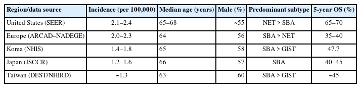
Comparative epidemiology of malignant small-bowel tumors: Asian vs. Western populations
Overall, the rising incidence of malignant small-bowel tumors in Asia reflects a convergence of demographic aging and improved diagnostic capability, accompanied by distinct regional patterns in histology, sex distribution, and etiologic background. Further increases in disease burden are anticipated as Asian societies continue to age, underscoring the need for tailored surveillance strategies targeting older, atrisk populations.
HISTOLOGIC SUBTYPES OF MALIGNANT SMALL-BOWEL TUMORS
Malignant small-bowel tumors comprise a heterogeneous group, with adenocarcinoma, NETs, gastrointestinal stromal tumors (GISTs), and lymphoma accounting for more than 95% of cases. These subtypes differ in anatomic predilection, molecular behavior, and clinical trajectory, resulting in distinct patterns of presentation and prognosis. The major clinicopathologic characteristics, typical anatomic distribution, and prognostic features of these subtypes are summarized in Table 2.

Summary of major histologic subtypes of malignant small-bowel tumors
Adenocarcinoma
SBA is the most common histologic subtype in Asia, accounting for 40–60% of malignant cases [7–10]. Histologically, SBA resembles colorectal adenocarcinoma but commonly arises at mucosal transition zones, most frequently in the duodenum and proximal jejunum. Large registry and multicenter datasets report involvement of the duodenum in 50–65% of cases, followed by the jejunum (20–30%) and ileum (10–20%) [12]. In contrast to Western cohorts— where Crohn’s disease–associated SBA represents up to 30% of cases—Asian studies consistently report that < 5% of SBA cases are associated with Crohn’s disease, reflecting the lower Asian IBD prevalence [13,14]. Most Asian SBAs are sporadic and are diagnosed at advanced stages because of nonspecific symptoms and delayed investigation. Molecularly, KRAS and TP53 mutations predominate, while microsatellite instability–high tumors constitute < 10% of cases [12]. Prognosis remains poor, with reported 5-year overall survival (OS) ranging from 20% to 40%, largely dependent on stage and resectability [7,10].
NETs
NETs are relatively common in Western populations, comprising 30–40% of malignant small-bowel tumors according to SEER analyses and large institutional series [3,15,16]. In contrast, NETs account for only 10–20% of malignant small-bowel tumors in most Asian cohorts [8–10]. Small-bowel NETs typically arise in the ileum and are frequently identified incidentally or during evaluation for anemia or small-bowel bleeding. Most are well-differentiated (Grade 1–2) tumors expressing chromogranin A and synaptophysin. Localized NETs have an excellent prognosis, with 5-year OS exceeding 80%, although outcomes worsen in the presence of liver metastasis or high Ki-67 index [6]. Recent data from Japan and Korea suggest a gradual increase in the detection of small-bowel NETs, potentially attributable to wider clinical use of CE and DAE [17–19].
GISTs
GISTs account for 10–20% of malignant small-bowel tumors and most commonly arise in the jejunum, followed by the ileum. Small-bowel GISTs tend to exhibit more aggressive biological behavior than their gastric counterparts, with higher mitotic activity and a greater likelihood of recurrence [20,21]. These tumors originate from the interstitial cells of Cajal and typically express KIT (CD117) and DOG1, driven by activating mutations in KIT or PDGFRA. The introduction of tyrosine kinase inhibitor treatment—particularly imatinib—has markedly improved outcomes and remains the therapeutic backbone [20,21]. In the Korean NHIS cohort, the 5-year OS for small-bowel GIST exceeded 70%, substantially higher than that observed for SBA [7]. Reports from Southeast Asia describe a relatively low frequency of diagnosed GISTs, although this likely reflects differences in diagnostic access and histopathologic capability rather than true epidemiologic variation [8].
Lymphoma
Primary small-bowel lymphoma accounts for approximately 5–10% of malignant small-bowel tumors in Asia and demonstrates notable geographic variation [4,22]. The ileum is the most frequently involved site, reflecting its high concentration of gut-associated lymphoid tissue. Diffuse large B-cell lymphoma (DLBCL) and mucosa-associated lymphoid tissue (MALT) lymphoma are the predominant subtypes, whereas enteropathy-associated T-cell lymphoma (EATL) is rare in Asian populations because of the low prevalence of celiac disease [4,22–24]. Other entities such as follicular lymphoma, mantle cell lymphoma, and NK/T-cell lymphoma are also encountered.
Clinical presentation is often nonspecific—abdominal pain, anemia, obstruction—mimicking IBD or intestinal tuberculosis, leading to diagnostic delay. Advances in immunohistochemistry, endoscopic biopsy, and molecular pathology have refined diagnostic accuracy and improved subtype classification and risk stratification [25,26].
TRENDS IN DIAGNOSTIC TOOL UTILIZATION & CHALLENGES
The diagnosis of malignant small-bowel tumors remains challenging because clinical manifestations are often nonspecific— including bleeding, anemia, or intermittent abdominal pain—leading to delayed diagnosis and advanced-stage presentation in many cases [4,27]. Cross-sectional imaging, particularly CTE and MRE, has become the cornerstone of initial evaluation, providing detailed assessment of mural and extramural involvement [28]. CE has revolutionized the non-invasive visualization of the entire small-bowel, markedly improving the detection of mucosal and early-stage lesions [29]. DAE further enhances diagnostic accuracy by permitting direct visualization, targeted biopsy, and therapeutic intervention, thereby complementing the diagnostic pathway [19,30,31]. Reported diagnostic performance varies across modalities, with CTE demonstrating approximately 80% sensitivity for mass-forming lesions [32], CE yielding 50–70% detection rates for small-bowel tumors [29], and DAE providing diagnostic yields of 60–80%, particularly when guided by prior imaging [19,30]. The comparative performance of these modalities is summarized in Table 3, which outlines their strengths, limitations, and representative diagnostic yields. CTE provides excellent sensitivity for mass-forming lesions, CE is superior for mucosal evaluation, and DAE remains essential for histologic diagnosis and therapeutic intervention (Table 3). Understanding the complementary roles of these modalities is particularly important in Asian clinical settings, where availability of advanced imaging and enteroscopy expertise may differ substantially among institutions. Beyond differences in diagnostic tools and availability, the clinical scenarios that trigger evaluation for malignant small-bowel tumors may also differ between Asia and Western populations, largely reflecting differences in subtype distribution and tumor location. In many Asian cohorts, where SBA constitutes a larger proportion of malignant small-bowel tumors and duodenal or proximal jejunal involvement is common, patients frequently enter the diagnostic pathway through investigation of iron-deficiency anemia, occult GI bleeding, or nonspecific upper abdominal symptoms, often prompting upper endoscopy followed by cross-sectional enterography and/or CE [7–10]. By contrast, in Western cohorts with a higher relative burden of ileal NETs, evaluation is more often initiated by intermittent bleeding, abdominal pain, obstructive symptoms, or incidental findings on imaging, with subsequent CE or DAE used for localization and confirmation [3,15,16,19]. In addition to biological differences, region-specific practice factors— such as FIT-based screening uptake, referral thresholds for iron-deficiency anemia, and institutional availability of advanced enteroscopic techniques—may further shape real-world diagnostic pathways across Asian countries.

Comparison of diagnostic modalities for malignant small-bowel tumors: utility, limitations, and diagnostic yield
Beyond improving detection, the expanding use of CE, high-quality enterography, and DAE has also reshaped treatment pathways in routine practice [29]. Earlier localization and histologic confirmation facilitate timely referral to surgery and enable more accurate operative planning, particularly for SBA and localized lymphoma [19]. Cross-sectional enterography increasingly supports risk-adapted decisions regarding resectability and extent of lymph-node dissection, while DAE provides tissue diagnosis and, in selected cases, endoscopic hemostasis or temporizing interventions in bleeding tumors [19,28]. Importantly, improved subtype recognition through immunohistochemistry and molecular testing has strengthened the role of multidisciplinary management, allowing earlier initiation of subtype-specific systemic therapy—such as tyrosine kinase inhibitors for GIST and somatostatin receptor-based strategies for NETs— thereby contributing to gradual improvements in outcomes reported in recent Asian series [6,33].
Despite these technological advances, significant disparities in diagnostic access, procedural expertise, and interpretation persist across Asian regions. Overlapping features with Crohn’s disease or intestinal tuberculosis may further delay definitive histologic confirmation. A recent Korean nationwide study demonstrated that increased utilization of CE and DAE was associated with earlier-stage diagnosis and gradual improvement in survival over the past decade [7]. Beyond conventional imaging and endoscopic techniques, artificial intelligence (AI)–based image analysis is increasingly being explored to enhance the detection of subtle mucosal lesions in CE and cross-sectional imaging. Deep-learning algorithms have shown promising sensitivity in retrospective validation studies, although their clinical applicability remains to be established [34].
PROGNOSIS AND SURVIVAL OUTCOMES
Prognosis varies markedly with malignant small-bowel tumors, depending on histologic subtype and stage at diagnosis, which are the two most powerful determinants of survival.
Among malignant small-bowel tumors, NETs tend to have the most favorable outcomes, with Western population-based studies reporting 5-year OS rates of 70–90% [35,36]. A landmark analysis of 3,911 small-bowel NETs showed that, despite rising incidence, diagnostic delay and limited stage migration persisted over three decades [3]. More recent Nordic data suggest improving outcomes, with 5-year survival increasing from 63.5% to 83.5% across diagnostic periods [37]. Expert reviews likewise emphasize metastatic burden, mesenteric fibrosis, and delayed diagnosis as major prognostic determinants [6]. In Asia, however, NETs account for only a small proportion of small-bowel malignancies, limiting the availability of robust, subtype-specific survival data.
In contrast, SBA carries the poorest prognosis among the major histologic subtypes of malignant small-bowel tumors, with Western studies reporting 5-year OS of 20–35% [27,38]. Most patients present with advanced-stage disease, and even after curative resection, locoregional recurrence remains common, reflecting the aggressive tumor biology and the limited benefit of current systemic therapies. Established prognostic factors—including tumor stage, lymphnode involvement, margin status, lymphovascular invasion, and histologic grade—have been consistently identified across large retrospective and population-based studies [39–41]. Asian institutional cohorts similarly report predominantly late-stage presentation and poor long-term outcomes, underscoring the need for earlier detection pathways and region-specific therapeutic strategies [2,10,42].
GISTs represent the second most common malignant small-bowel tumor subtype in many Asian cohorts and typically demonstrate more favorable outcomes compared with SBA. Reported 5-year OS commonly exceeds 70%, largely attributable to the availability of effective molecular-targeted therapy with tyrosine kinase inhibitors such as imatinib [20,33]. Prognosis is strongly influenced by tumor size, mitotic index, and primary site, with jejunum and ileum locations associated with a higher recurrence risk than gastric GISTs [21]. Despite therapeutic advances, resistance to imatinib and progression to metastatic disease remain major clinical challenges, highlighting the need for improved genomic stratification and second-line targeted treatment options.
Primary small-bowel lymphoma shows heterogeneous survival outcomes largely determined by histologic subtype, stage, and response to chemo-immunotherapy. DLBCL generally demonstrates poorer survival than MALT lymphoma, while EATL carries the worst prognosis [4,43]. In a recent Chinese cohort of 94 patients, the 5-year OS was approximately 60%, with subtype and endoscopic morphology serving as key prognostic indicators [44]. A Taiwanese series similarly reported 5-year overall and disease-free survival of 59% and 54%, respectively, in non-IPSID small-intestinal lymphoma treated with combination chemotherapy [45]. Larger Asian GI lymphoma datasets further show inferior outcomes with advanced stage and T-cell lineage, although small-intestinal cases are often analyzed within broader GI lymphoma cohorts [43,46,47]. Overall, survival in Asian populations remains limited by late presentation and scarce subtype-specific evidence.
UNMET NEEDS AND FUTURE DIRECTIONS
Despite growing awareness of malignant small-bowel tumors in Asia, substantial gaps remain in epidemiologic characterization, early detection, and therapeutic optimization. Under-recognition and under-reporting continue to limit accurate burden estimation, particularly in regions with fragmented referral pathways or restricted access to advanced endoscopic and radiologic modalities. With most available data derived from small institutional series or retrospective studies, the scarcity of prospective cohorts and population-based registries has hindered robust subtype-specific survival analyses.
To address these limitations, multicenter collaboration and dedicated rare-tumor registries are essential to support adequately powered studies, harmonized data collection, and longitudinal outcome tracking. In parallel, big-data platforms such as nationwide insurance databases (e.g., NHIS Korea) offer an effective means to overcome rarity by providing population-level insights into incidence, diagnostic patterns, treatment utilization, and real-world outcomes [7]. Looking forward, several developments have the potential to reshape the clinical trajectory of malignant small-bowel tumors in Asia. Advances in molecular profiling and genomic stratification may refine therapeutic selection and identify candidates for targeted or immunologic therapies. Improvements in minimally invasive diagnostic technologies—including enhanced enteroscopy, high-resolution cross-sectional imaging, and AI-assisted lesion detection—could facilitate earlier diagnosis and more accurate risk assessment. Collectively, these efforts represent crucial opportunities to strengthen early detection strategies, reduce regional disparities in care, and support the development of evidence-based, regionally appropriate management pathways for malignant small-bowel tumors.
CONCLUSION
Malignant small-bowel tumors represent an increasingly important clinical and epidemiologic challenge in Asia, with their incidence rising in parallel with population aging and broader deployment of advanced diagnostic modalities. Subtype-specific differences in epidemiology, anatomic distribution, and prognosis highlight the need for tailored diagnostic pathways and individualized treatment approaches rather than a uniform strategy.
Persistent gaps—including delayed diagnosis, under-recognition in routine practice, and the limited availability of prospective or registry-based data—underscore the importance of coordinated multicenter research initiatives and the establishment of robust rare-tumor registries. Future efforts that integrate big-data analytics, risk-stratified early detection strategies, molecular characterization, and equitable access to advanced diagnostic technologies will be essential to improve outcomes and support the development of regionally optimized care models for Asian patients with malignant small-bowel tumors.
Notes
CRedit authorship contributions
Jung Rock Moon: conceptualization, writing - original draft, writing - review & editing; Seong Ran Jeon: conceptualization, writing - review & editing, supervision, funding acquisition
Conflicts of interest
The authors disclose no conflicts.
Funding
This work was supported by the National Research Foundation (NRF) of Korea grant funded by the Korea government (MSIT) (RS-2021-NR066198) and the Soonchunhyang University Research Fund.