Castleman disease-like mesenteric lymphadenopathy in a Korean adult ultimately diagnosed with familial Mediterranean fever
Article information

A 47-year-old Korean man was referred for evaluation of recurrent febrile episodes accompanied by abdominal pain. Since his early 30s, he had experienced attacks every two to three months, with fevers up to 38.4°C lasting three days and periumbilical abdominal pain persisting about a week. Each episode was associated with elevated inflammatory markers, including C-reactive protein up to 20.03 mg/dL and erythrocyte sedimentation rate up to 46 mm/h, while symptom-free intervals showed no abnormalities. He reported no arthralgia, rash, or serositis.
Repeated abdominal computed tomography (CT) scans revealed multiple enlarged mesenteric lymph nodes with homogeneous enhancement, engorged mesenteric vessels, and thickened mesenteric folds (Fig. 1A, B), raising suspicion for Castleman disease. An excisional biopsy at an outside hospital was initially interpreted as “consistent with Castleman disease,” leading to a provisional diagnosis of idiopathic multicentric Castleman disease and a recommendation for siltuximab therapy. However, the patient’s periodic febrile attacks were atypical for Castleman disease. In addition, fluorodeoxyglucose positron emission tomography–CT obtained during a symptom-free interval demonstrated only minimal metabolic activity in the mesenteric lymph nodes (Fig. 1C), prompting reconsideration of the diagnosis.
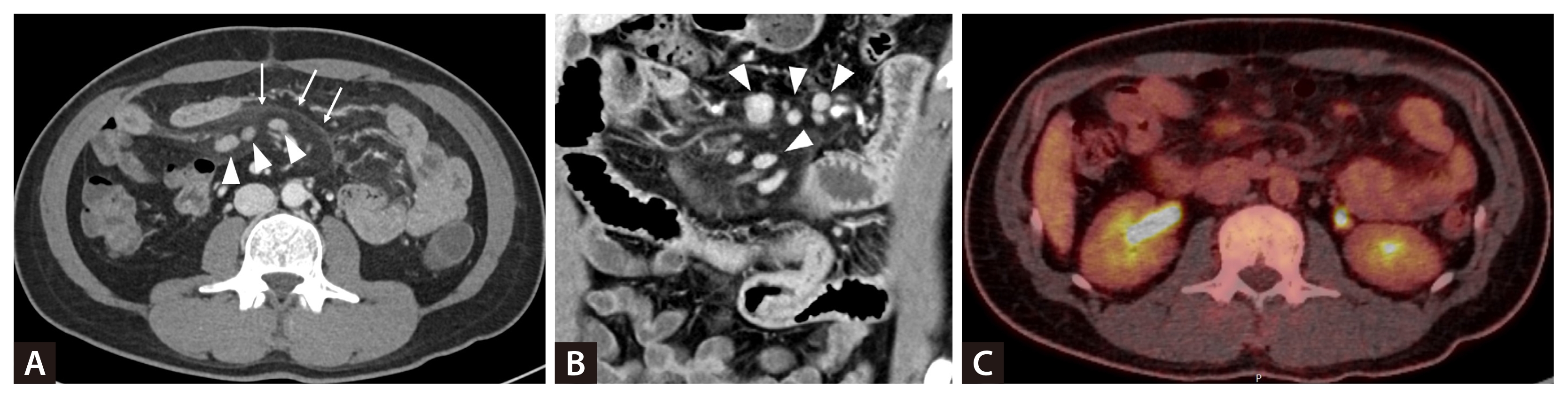
Radiologic findings of mesenteric lymphadenopathy. Axial (A) and coronal (B) images from contrast-enhanced abdominal computed tomography (CT) reveal multiple homogeneously enhancing enlarged mesenteric lymph nodes (arrowheads) measuring up to 1.2 cm in short-axis diameter, accompanied by engorged mesenteric vessels and thickened mesenteric folds (white arrows). (C) Fluorodeoxy-glucose (FDG) positron emission tomography–CT obtained during a symptom-free interval shows only minimal FDG uptake in the mesenteric lymph nodes (SUVmax 3.1–3.4), indicating low metabolic activity outside of acute inflammatory attacks.
Pathologic re-evaluation indicated reactive lymphoid hyperplasia with focal Castleman-like features, including atretic germinal centers with penetrating hyalinized vessels and thickened mantle zones exhibiting an onion-skin appearance (Fig. 2). Given the long-standing relapsing inflammatory episodes, periodic fever syndromes were considered [1]. Next-generation sequencing identified heterozygous P369S and R408Q variants in the MEFV gene, both classified as variants of uncertain significance [2]. A diagnosis of familial Mediterranean fever (FMF) was established by integrating the pattern of recurrent fever, abdominal pain, reactive mesenteric lymphadenopathy, and MEFV variants [3]. Colchicine therapy led to complete remission of attacks and normalization of inflammatory markers within six months. This case illustrates that FMF, although rare in Korea, can radiologically and pathologically resemble Castleman disease.
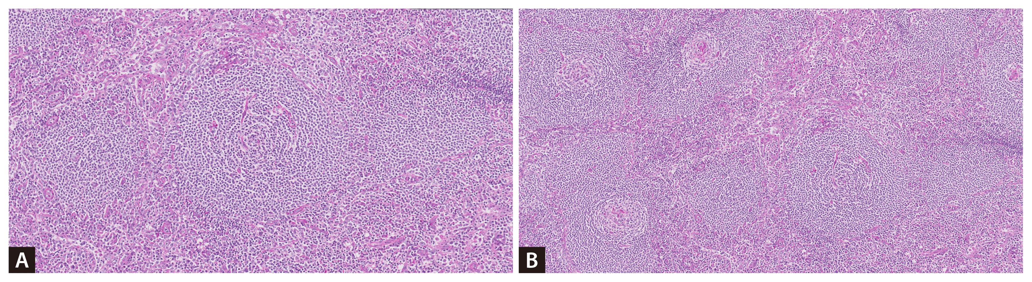
Histological findings of the mesenteric lymph node. The lymph node biopsy reveals atretic germinal centers infiltrated by hyalinized vessels, resulting in the formation of “lollipop follicles” (A) (H&E, ×100). Additionally, the mantle zones are thickened, exhibiting an onion-skin appearance (B) (H&E, ×40).
Notes
CRedit authorship contributions
Hee Won Park: resources, investigation, writing - original draft, writing - review & editing, visualization; Seo Yeon Youn: resources, investigation, writing - original draft, writing - review & editing, visualization; Gyeongsin Park: resources, investigation, validation; Youngjae Park: conceptualization, resources, investigation, writing - original draft, writing - review & editing, visualization, supervision, project administration
Conflicts of interest
The authors disclose no conflicts.
Funding
None
Ethical statement
This case was approved by the Institutional Review Board of Seoul St. Mary’s Hospital (IRB No. KC25ZISI0903). Written informed consent was waived.