Optimizing multiple diagnostic modalities for small bowel evaluation: a strategic approach
Article information

Abstract
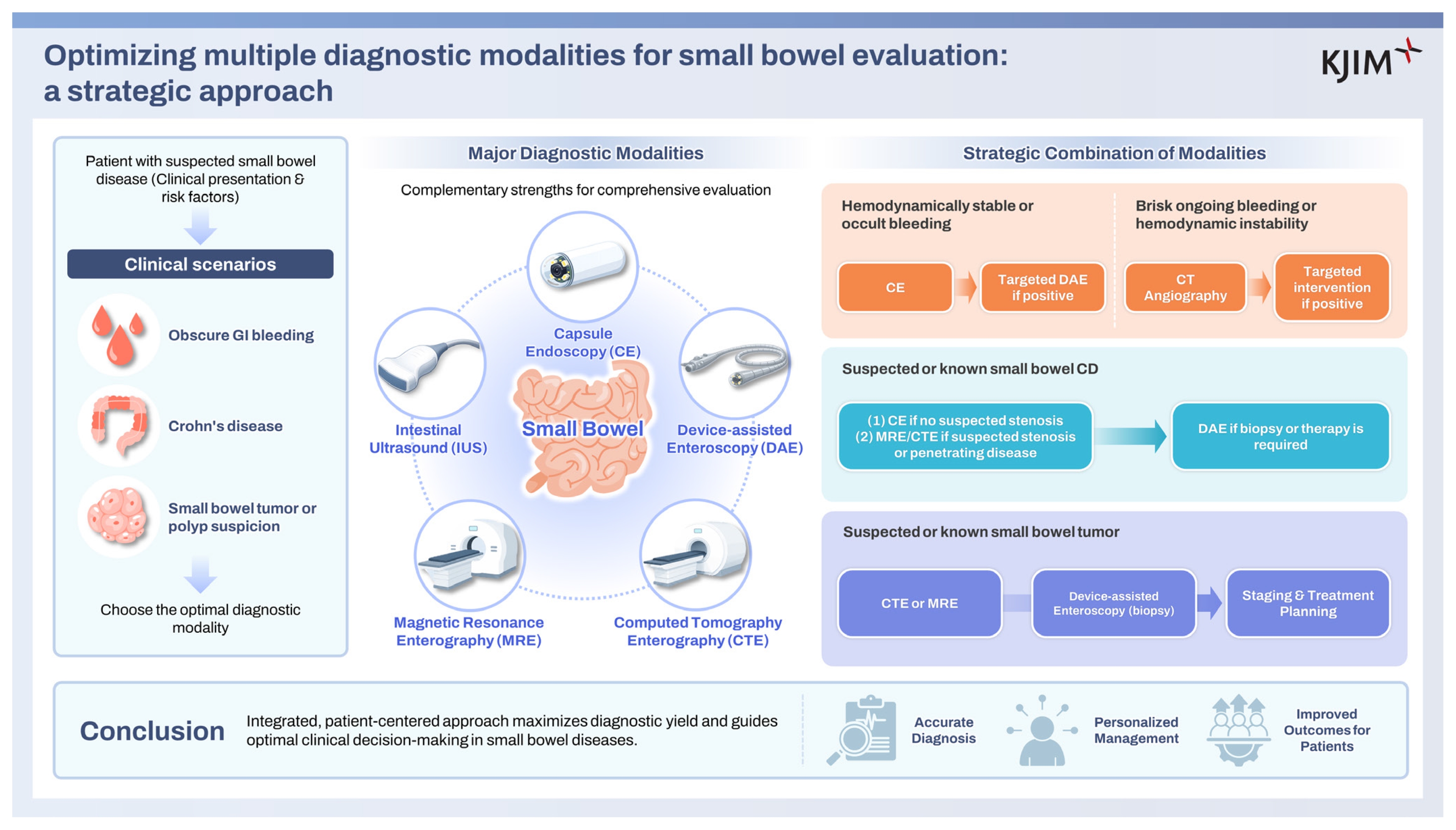
Over the past two decades, the small bowel, once considered a diagnostic “blind spot,” has become fully and comprehensively accessible. This review summarizes recent advances in the main diagnostic modalities: capsule endoscopy (CE), device-assisted enteroscopy (DAE), computed tomography enterography (CTE), magnetic resonance enterography (MRE), and intestinal ultrasound (IUS). CE provides a noninvasive assessment of mucosal abnormalities, whereas DAE offers the added benefits of tissue sampling and therapeutic intervention. CTE and MRE provide complementary cross-sectional information on mural inflammation, strictures, and extraintestinal involvement. IUS has emerged as a valuable, radiation-free technique for repeated bedside monitoring. We review the diagnostic performance, strengths, and limitations of each modality in key clinical scenarios, including suspected small bowel bleeding, Crohn’s disease, and tumors. Finally, we highlight the importance of an integrated, patient-centered approach to optimize the evaluation of small bowel disease and to guide clinical decision-making.
INTRODUCTION
The small bowel, which constitutes approximately 75% of the gastrointestinal (GI) tract, has historically been regarded as a diagnostic “blind area” because of its length and anatomical complexity [1]. However, the past two decades have seen remarkable advances in both endoscopic and imaging technologies. The introduction of capsule endoscopy (CE) and device-assisted enteroscopy (DAE) allowed for direct and comprehensive mucosal visualization. Simultaneously, cross-sectional imaging techniques have also greatly improved. These imaging modalities include computed tomography enterography (CTE), magnetic resonance enterography (MRE), and the rapidly emerging intestinal ultrasound (IUS). These tools provide critical data about the bowel wall and extraintestinal involvement [2,3].
These diagnostic modalities represent a major advance, but the abundance of available options also makes it more challenging for clinicians to select the optimal diagnostic strategy. Clinical decisions require careful consideration, balancing diagnostic accuracy with patient safety, cost-effectiveness, and availability. Given that other articles in this special issue address specific small bowel pathologies, this review is structured to present a modality-based overview of recent diagnostic advances and their practical implications for clinical decision-making. We primarily emphasize the diagnostic performance, strengths, and limitations of the main modalities (CE, DAE, CTE, MRE, and IUS). Our primary aim is to present an integrated framework for clinical decision-making and to provide algorithm-based suggestions for strategically selecting endoscopic and imaging modalities to maximize the diagnostic yield in small bowel disease.
SMALL BOWEL DIAGNOSTIC MODALITIES
To implement the strategic approach, optimal diagnostic tool selection is vital, requiring a thorough understanding of the specific roles, strengths, and limitations of each diagnostic modality based on the suspected pathology and required findings (Table 1).
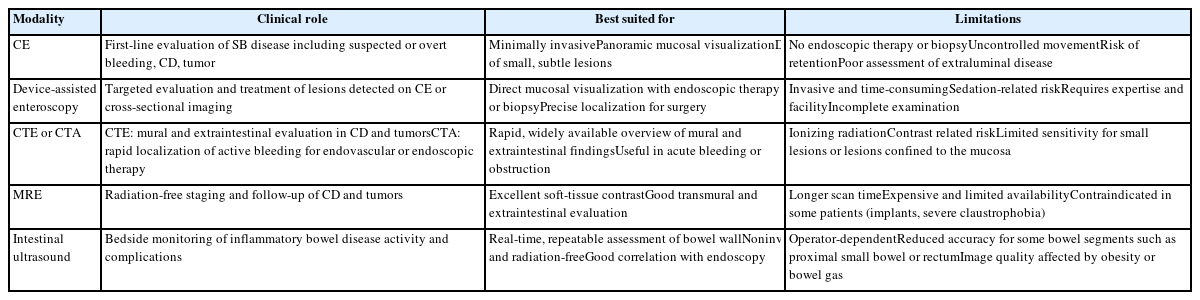
Roles of major diagnostic modalities in small bowel diseases
CE
CE, introduced in 2000, is a noninvasive visualization method for diagnosing small bowel disease. The capsule is typically about 30 mm × 10 mm and contains a miniature camera, LED light source, battery supply, and wireless transmitter. Following swallowing, the capsule travels passively through the GI tract, propelled by peristalsis and gravity. It captures images at a rate of 1–3 frames per second and transmits them wirelessly to an external receiver [4]. Recent technological advancements include higher image resolution and wider-angle lenses. In addition, adaptive framerate technology adjusts the image capture speed according to capsule transit [5,6].
Clinical utility
CE plays a crucial role in the diagnostic evaluation of various small bowel diseases, and its strategic use is supported by several key advantages and clinical roles [7–9].
For obscure GI bleeding (OGIB) or small bowel bleeding, CE is a valuable first-line tool for identifying the source of small bowel bleeding. Because the small bowel is the most common source of OGIB, CE plays a pivotal role after negative findings on upper endoscopy and colonoscopy (conventional endoscopy). Several studies show that CE identifies the bleeding source in about 60–70% of OGIB patients [10,11].
For Crohn’s disease, CE provides panoramic mucosal visualization. In suspected small bowel Crohn’s disease, it is the preferred modality for detecting subtle lesions and diffuse mucosal disease, such as aphthous or deep ulcers, with a reported diagnostic yield of about 50–70% [12]. Network meta-analysis demonstrated that CE has superior diagnostic performance for small bowel Crohn’s disease compared with other imaging modalities, with a sensitivity of 90%, specificity of 86%, and overall diagnostic accuracy of 72% [13]. CE is also useful for assessing drug-induced small bowel injuries such as nonsteroidal anti-inflammatory drug enteropathy [14].
For small bowel tumors, CE contributes to the diagnosis of lesions, which are detected in about 8%–10% of patients. The reported specificity and positive predictive value for tumor detection have been notably as high as 100% [15,16]. Beyond these major indications, CE can also be used for small bowel polyp surveillance in patients with Peutz-Jeghers syndrome [17].
Future directions
Despite its numerous benefits, clinicians must recognize the limitations of CE, which often guide the subsequent choice of modality. These limitations include the lack of therapeutic capabilities or the ability to perform a tissue biopsy, time-consuming image interpretation, uncontrolled capsule movement, the significant risk of capsule retention in patients with known or suspected strictures, and the need for thorough bowel preparation for optimal image quality [18,19]. Capsule retention is defined as the capsule remaining within the GI tract for at least 2 weeks or requiring directed intervention for removal. According to meta-analyses, the overall retention rate is approximately 2%, but can reach 8.2% in patients with established inflammatory bowel disease (IBD). Small bowel patency assessment using a patency capsule or CTE/MRE can reduce this risk [20]. In most cases, asymptomatic retention can be managed conservatively. However, symptomatic or prolonged cases may require endoscopic or surgical retrieval [21].
Current research efforts aim to modify these drawbacks, primarily focusing on two areas. The first aims for enhanced maneuverability through the development of magnetically assisted CE systems that can be externally controlled. The second seeks optimized reading efficiency via artificial intelligence (AI)-assisted CE reading systems designed to improve diagnostic accuracy and reduce interpretation time [22–24]. While several prototype systems and commercial products have demonstrated promising performance, further validation studies in real-world clinical settings remain essential before widespread adoption [6,25].
Enteroscopy
The evolution of enteroscopy techniques has focused on increasing the depth of insertion into the deep small bowel. Enteroscopy systems generally consist of a long, flexible endoscope specifically designed to access the deep small bowel, and an overtube that facilitates endoscope insertion and stabilization. These devices are essential for overcoming the diagnostic and therapeutic limitations of CE, such as the inability to perform a tissue biopsy or therapeutic interventions. Enteroscopy systems are primarily classified into push enteroscopy and DAE (Fig. 1). DAE techniques include double-balloon enteroscopy (DBE), single-balloon enteroscopy (SBE), and spiral enteroscopy (SE) [26].
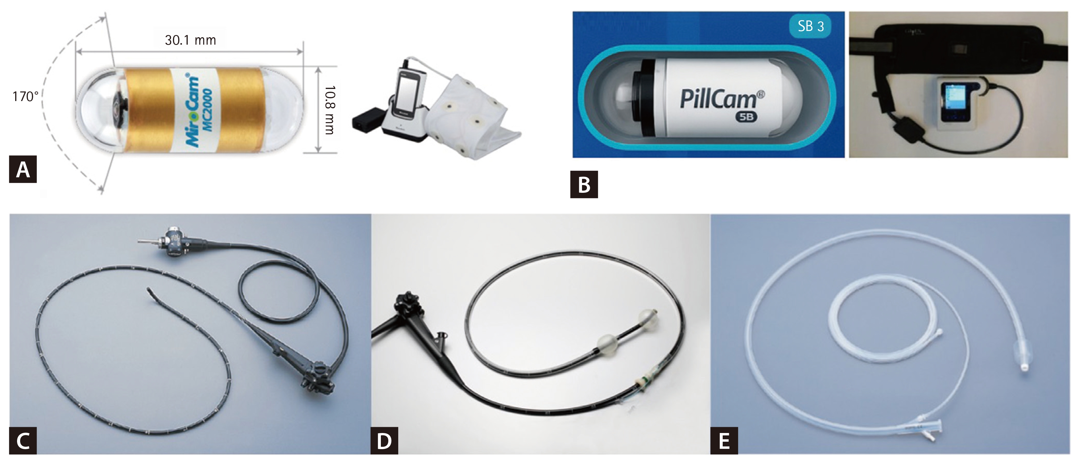
Representative capsule endoscopy and device-assisted enteroscopy systems currently available in Korea. (A) MiroCam system (Intromedic). (B) PillCam system (Medtronic). (C) Small intestinal videoscope (Olympus). (D) Assembled double-balloon enteroscopy system (Fujifilm). (E) Single-use single-balloon splinting tube (Olympus). Images are provided and used with the permission of each manufacturer.
Push enteroscopy, first introduced in the mid-1990s, typically allows access only to the proximal jejunum. These limitations have contributed to its decline in use and its replacement by newer modalities, such as DAE and CE, for comprehensive small bowel evaluation [11,26]. DBE was introduced in 2001 to overcome the limitations of CE and push enteroscopy. DBE remains the leading technique for deep enteroscopy [11,27–29]. SBE was introduced in 2007 to simplify the structurally complex DBE system, making the procedure easier to set up and manipulate [26,30,31]. SE was introduced around 2009, and motorized SE (MSE) was introduced in 2016 to reduce operator burden. SE/MSE achieves similar or deeper insertion depths in a shorter duration of about 40–50 minutes compared to balloon-assisted methods (60–100 minutes for DBE and 40–70 minutes for SBE) [31–35].
Clinical utility
A major advantage of enteroscopy is its capability to perform both diagnostic and therapeutic procedures, including tissue biopsy and endoscopic therapy, in a single session [11]. The diagnostic yield of enteroscopy techniques varies markedly based on the technique and the underlying small bowel pathology. Therefore, the strategic selection among enteroscopy systems is primarily guided by the desired depth of reach and expected diagnostic or therapeutic performance, though the final choice often depends on institutional equipment availability and the endoscopist’s expertise [36–38].
For OGIB or small bowel bleeding, push enteroscopy records a diagnostic yield ranging from about 20% to 60% due to its inherent limitation, which restricts access primarily to the proximal jejunum [39]. DBE demonstrates a high diagnostic yield in OGIB, generally reported around 70% [28,40,41]. SBE reports an OGIB diagnostic yield in the range of approximately 40–70% [42,43]. SE/MSE shows comparable performance, recording about 65–80% [44–46].
For Crohn’s disease, DAE is indicated when tissue sampling, evaluation of strictures, or therapeutic intervention is needed. Push enteroscopy has limited utility for comprehensive evaluation of Crohn’s disease. DBE is useful for Crohn’s disease, reporting a diagnostic yield of about 65– 85% [28,40,41]. SBE demonstrates a comparably high diagnostic yield for Crohn’s disease, reported at around 80% [42,43,47]. Notably, SE/MSE records the highest reported yield for suspected Crohn’s disease, ranging from 75–95%, positioning it as a powerful strategic option [38,44–47]. However, since July 2023, MSE is not currently available as a routine clinical option for safety concerns.
For small bowel tumor, the reported diagnostic yield of DAE is about 7–15%. While these diagnostic yields appear relatively low compared with small bowel bleeding or Crohn’s disease, the primary role of DAE is therapeutic and confirmatory rather than initial screening [28,40–43,45].
Regarding the complete small bowel examination, the reported rate of complete enteroscopy using DBE is about 66% [28,48]. However, SBE generally achieves lower completion rates, typically ranging from 20% to 30% [48]. The diagnostic success rate of MSE was found to be 78%, and therapeutic interventions were successful in 98% of cases [44]. Moreover, MSE tended to achieve higher technical success in the deeper segments than SBE [38].
Future directions
Despite its high diagnostic yield, DAE remains an invasive and relatively time-consuming procedure, typically requiring 1–2 hours and specialized training. Major complications, including perforation, bleeding, acute pancreatitis, and aspiration pneumonia, occur in about 1% of cases. Nevertheless, DAE remains indispensable as it provides the critical benefits of tissue sampling and endoscopic therapy [40,41,44,49,50]. The future directions of enteroscopy focus on enhancing depth of insertion, improving maneuverability, integrating real-time diagnostic technologies, and combining these advances with minimally invasive therapeutic capabilities.
CTE
CTE is a widely used cross-sectional imaging technique for evaluating small bowel disease. CTE examination requires the patient to ingest 1–1.5 L of oral contrast followed by intravenous (IV) contrast administration to acquire multiplanar images [51]. CTE is generally preferred over standard CT of the abdomen and pelvis with IV contrast because optimized bowel distention improves diagnostic performance and allows for detection of more subtle inflammatory findings. However, in urgent settings, standard CT or CT angiography (CTA) remains a suitable option, particularly for conditions such as obstruction or massive bleeding. Clinicians should appropriately utilize standard CT, CTE, or CTA depending on the clinical indication and patient condition [52,53].
Clinical utility
CTE is indicated for assessing various small bowel diseases and for further evaluation of abnormal findings from other imaging studies. CTE has demonstrated reliable diagnostic performance for small bowel diseases, with a reported sensitivity of 76%, specificity of 95%, and accuracy of 88% [11,54]. CTE may also be the preferred choice for patients in whom CE is contraindicated, such as those with suspected strictures, a history of abdominal surgery, or dysphagia [3,51].
For patients with OGIB or small bowel bleeding, CTE is the most rapid tool available for assessing the entire small bowel. CTE is also valuable for identifying the underlying cause of bleeding such as vascular lesion and tumors [55]. CTA can promptly and accurately localize the site of active bleeding by demonstrating contrast extravasation, while also providing crucial information for subsequent management [56]. A meta-analysis reported that CTE has a sensitivity of 47% and specificity of 94% for detecting suspected small bowel bleeding. In comparison, other meta-analyses have shown that CTA is more sensitive (85–90%), specific (92%), and accurate (94–95%) for detecting and localizing overt GI bleeding [3,57].
For Crohn’s disease, CTE is essential for assessing transmural inflammation and extraintestinal complications such as bowel wall thickening, mural enhancement, fistulas, abscesses, and extraintestinal lymphadenopathy. CTE often reveals mesenteric hypervascularity, known as the “comb sign,” along with inflammatory changes in the surrounding mesenteric fat. The diagnostic yield of CTE is reported to be about 70–90% [11,12,54,58]. Because of its shorter acquisition time compared to MRE, it may be preferred when strictures or acute abdominal complications are suspected. Moreover, quantitative assessment of CTE findings has shown a significant correlation with the degree of histopathologic inflammation in Crohn’s disease [12,59].
For small bowel tumors, CTE is indispensable for the staging of tumors, providing critical information on the tumor’s size, location, extramural invasion, and lymph node metastasis (Fig. 2) [60]. A recent meta-analysis reported that CTE has a sensitivity of 93% and specificity of 83% for tumor diagnosis, which is comparable to the performance of MRE [61]. Unlike CE or DAE, CTE is critical for characterizing the full mural component of a tumor and its relationship with surrounding structures. CTE is significantly more sensitive than CE (93% vs. 30%) in detecting mural-based masses, subepithelial tumors, and lesions with mesenteric extension, which are findings that are often challenging to identify endoscopically. CTE is widely recommended as a firstline imaging modality for the diagnosis and localization of small bowel tumors and for guiding endoscopic or surgical management [62]. However, the diagnostic yield decreases substantially for very small lesions (< 1 cm), with reported detection rates of 0% for epithelial tumors and 24% for subepithelial tumors [63].
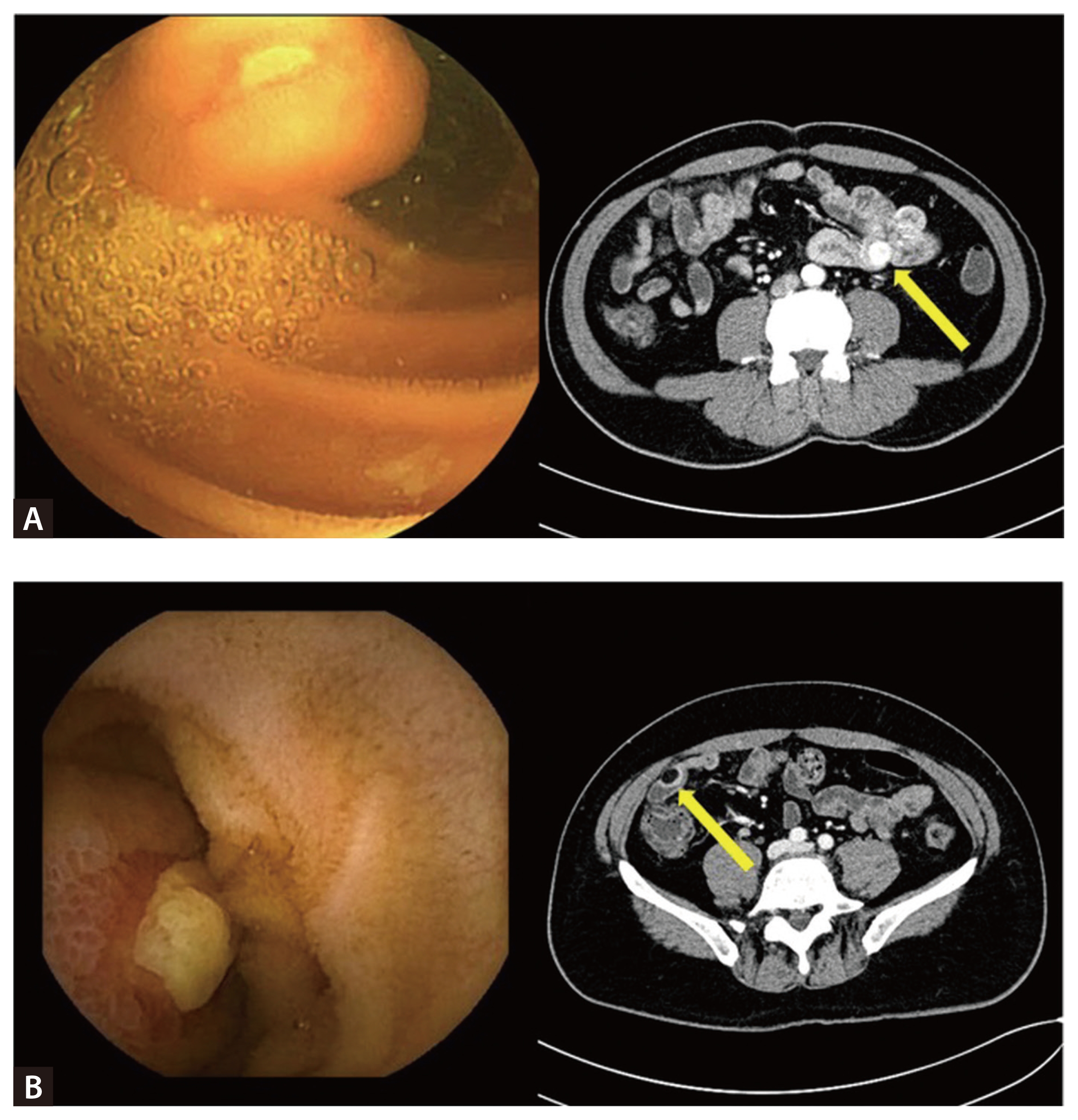
Case examples of small bowel tumors presenting with obscure gastrointestinal bleeding. (A) A 62-year-old man with anemia and melena. CE shows a 2-cm subepithelial mass with overlying ulceration, and CTE demonstrates a well-defined, strongly enhancing submucosal lesion in the jejunoileal junction (arrow). Segmental resection confirmed a very low-risk gastrointestinal stromal tumor. (B) A 42-year-old man with anemia and melena. CE reveals a 2-cm subepithelial mass with ulceration, and CTE shows a well-defined hypodense submucosal lesion with peripheral enhancement (arrow). Segmental resection confirmed a complicated submucosal lipoma. CE, capsule endoscopy; CTE, CT enterography.
Future directions
The main drawbacks of CTE include exposure to ionizing radiation and limited sensitivity for small, subtle mucosal lesions [54,63]. Future directions focus on addressing current limitations through technological advances, including low-dose imaging protocols to reduce cumulative radiation [12]. In addition, newer techniques such as dual-energy CT and photon-counting CT can enhance lesion detection capability. Furthermore, ongoing research aims to integrate AI-assisted image interpretation and develop functional imaging methods to expand clinical indications [64].
MRE
MRE is a cross-sectional imaging technique that provides detailed anatomical and functional information about the small bowel without using ionizing radiation. It uses T1-weighted, T2-weighted, and Diffusion-Weighted Imaging sequences to assess bowel wall inflammation, edema, fibrosis, vascular changes, and bowel motility [65]. Similar to CTE, MRE requires patients to ingest a large volume of oral contrast. Antispasmodic agents may be administered to reduce bowel motility and optimize image quality. MRE is primarily used for diagnosing and monitoring small bowel Crohn’s disease and other forms of IBD, as well as for evaluating small bowel tumors, strictures, fistulas, and abscesses. Its major advantage is the absence of ionizing radiation, making it especially suitable for children and patients who require repeated imaging studies. However, MRE is limited by its longer scan time, higher cost, and more limited availability compared to CTE, and is contraindicated in patients with certain metallic implants or severe claustrophobia [66,67].
Clinical utility
MRE is often selected because it provides high-quality soft-tissue contrast and can be safely repeated, making it particularly suitable for long-term monitoring of Crohn’s disease or for diagnosing small bowel tumors.
For OGIB or small bowel bleeding, MRE is primarily used to assess underlying structural pathologies rather than to directly localize an active bleeding source. Its longer scan time and lower diagnostic performance limit its ability to rapidly localize active bleeding compared to CTE/CTA. Current guidelines generally recommend CT-based imaging as the first-line modality [3,11,54,68].
For Crohn’s disease, MRE is considered the cornerstone for noninvasive, radiation-free staging and follow-up of the disease. It is highly accurate in assessing both mural activity (e.g., wall thickening, edema, and deep ulcerations) and penetrating complications (e.g., perianal disease, fistulas, and abscesses). MRE is strategically the preferred choice for monitoring disease activity and therapeutic response over time in young patients due to its radiation-free nature. MRE demonstrates high diagnostic performance in both pediatric and adult small bowel Crohn’s disease, with reported accuracy of about 93%, sensitivity of 83–92%, and specificity of 81–95% [12,58,69,70].
MRE is valuable for the radiation-free staging of small bowel tumors, especially when assessing the depth of mural invasion and the involvement of adjacent structures. While CTE offers faster acquisition, MRE’s superior soft-tissue resolution can be strategically utilized for detailed characterization of mass lesions, exhibiting high diagnostic performance with a reported sensitivity of 93–96%, specificity of 96–99%, and an overall accuracy of about 96%. Comparative studies suggest that MRE performs similarly to, or may even outperform, CTE in diagnostic accuracy [54,61,71,72].
Future directions
Future research in MRE is increasingly focused on three key areas: quantitative imaging, simplified activity indices, and AI-assisted analysis. Quantitative imaging techniques are being developed to better differentiate inflammation from fibrosis. These multiparametric approaches aim to provide objective, biologically relevant biomarkers. There is also ongoing research into simplifying MRE protocols and reducing scan times. Recent studies suggest that essential MR sequences alone may be sufficient in selected clinical settings. Furthermore, AI and radiomics are expected to transform MRE clinical workflows. These tools enable automated bowel segmentation, objective scoring, and prognostic modeling. Such advances may improve the prediction of treatment response and long-term outcomes in small bowel diseases. Ultimately, by reducing observer dependency, AI-assisted analysis will improve the accuracy and efficiency of MRE in guiding patient management [73–75].
IUS
IUS uses high-resolution ultrasound equipment with both low-frequency (convex) and high-frequency (linear) probes to perform transabdominal scanning. This technique enables real-time visualization of the bowel wall, blood flow, and surrounding structures. Characteristic IUS findings include increased bowel wall thickness and Doppler hyperemia (increased blood flow), which serve as key markers of active inflammation. IUS can also identify structural and penetrating complications such as strictures, fistulas, and abscesses [76,77].
Clinical utility
The strategic strength of IUS lies in its safety profile and utility for monitoring IBD. Because IUS is noninvasive and does not involve ionizing radiation, it is especially valuable for children, pregnant women, and patients who require repeated assessment. Recent meta-analyses have confirmed its diagnostic efficacy in IBD, reporting a sensitivity of 68–100%, specificity of 57–100%, and overall accuracy of 72–91%. Current guidelines recommend IUS as an objective tool to support disease assessment and treatment decisions in patients with IBD [76,78].
In Crohn’s disease, IUS demonstrates high diagnostic performance, with a reported sensitivity of 80–95% and specificity of 81–98%. For terminal ileal and colonic involvement, its accuracy is often comparable to that of MRE and CTE. For detecting complications such as strictures, fistulas, and abscesses, sensitivity of 81–94% and specificity of 90–97% have been reported. However, its accuracy may be reduced for lesions located in the mid-to-proximal small bowel, for very long disease segments (> 10 cm), and for rectal involvement [76,77,79]. In ulcerative colitis, IUS also shows high diagnostic accuracy for assessing disease activity and predicting remission. Recent studies indicate that combining IUS with fecal calprotectin further improves diagnostic performance, achieving a sensitivity of 88% and specificity of 80% for detecting active disease [80–82]. Overall, IUS serves as a highly practical, real-time imaging tool that effectively guides clinical decision-making, particularly in the outpatient setting.
Future directions
Future perspectives in IUS include the integration of advanced modalities, including contrast-enhanced ultrasound, elastography, and AI-based analysis. These technologies are expected to enable more accurate differentiation between active inflammation and chronic fibrosis. When combined with predictive modeling for treatment response, these advances will play a key role in clinical decision-making. This is particularly relevant as novel therapeutic strategies, such as anti-fibrotic agents, continue to emerge. However, several challenges must be addressed to realize this potential. These include the development of validated disease activity scores, expansion of large-scale multicenter trials, and establishment of standardized global educational frameworks. Ultimately, international standardization will allow IUS to evolve from a supportive tool into a cornerstone of objective, quantitative disease assessment in IBD management [73,83–85].
STRUCTURED APPROACH TO DIAGNOSTIC MODALITIES FOR SMALL BOWEL DISEASES
Patients who present with nonspecific symptoms (OGIB, iron-deficiency anemia, abdominal pain, or unexplained weight loss) and have negative findings on conventional endoscopy typically require further evaluation for small bowel disease. Therefore, clinicians should adopt an individualized diagnostic strategy and may combine CE, DAE, CTE, MRE, and IUS depending on the suspected pathology (Table 2).
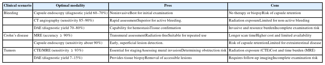
Optimal diagnostic modalities for small bowel diseases
Suspected or overt small bowel bleeding
The initial approach depends on the patient’s hemodynamic status. In hemodynamically stable patients with negative findings on conventional endoscopy, CE is generally preferred as the first-line test because of its high diagnostic yield. However, in patients with known or suspected strictures or a high risk of capsule retention, CTE or MRE should be performed before CE. Conversely, in patients with brisk or ongoing bleeding, CTE or CTA is prioritized to localize the active bleeding source rapidly and to guide subsequent angiographic embolization or DAE. DAE is then used for targeted diagnosis and endoscopic hemostasis [3,11,56].
Suspected small bowel Crohn’s disease
For suspected cases of small bowel Crohn’s disease, ileocolonoscopy with biopsies remains the diagnostic cornerstone, and clinical assessment of inflammatory markers and fecal calprotectin is also necessary (Fig. 3). CE offers excellent sensitivity for detecting early, superficial inflammatory lesions when strictures are unlikely. Conversely, CTE and MRE are preferred in patients with suspected strictures or penetrating disease because they provide detailed information on mural inflammation, stenosis, fistulas, and abscesses. Additionally, as emphasized in the strategy, IUS can be incorporated as a radiation-free, bedside tool for monitoring disease activity [12,86].
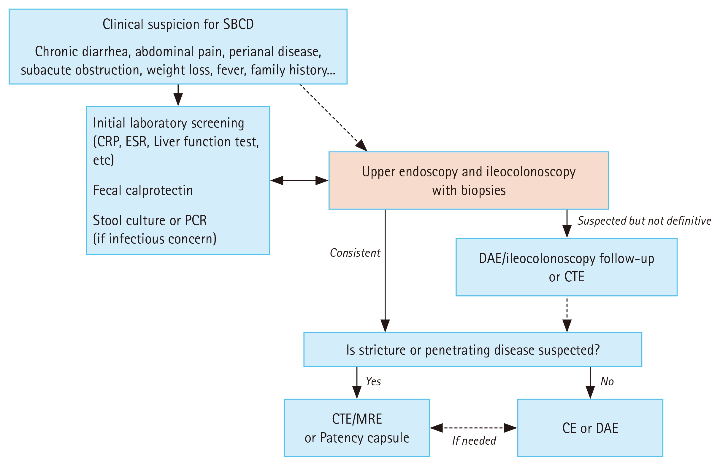
Suggested diagnostic algorithm for suspected small bowel Crohn’s disease. SBCD, small bowel Crohn’s disease; CRP, C-reactive protein; ESR, erythrocyte sedimentation rate; PCR, polymerase chain reaction; DAE, device-assisted enteroscopy; CTE, computed tomography enterography; MRE, magnetic resonance enterography; CE, capsule endoscopy.
Suspected small bowel tumors
The initial step in evaluating suspected small bowel tumors is to assess the likelihood of obstruction or mural/extraluminal disease. As outlined in the diagnostic algorithm (Fig. 4), when obstruction or a mural mass is suspected, CTE or MRE should be performed first to obtain a comprehensive overview of the entire abdomen. If CTE or MRE identifies a tumor, DAE is then used for histologic confirmation, tattooing, and endoscopic resection of accessible lesions. If CTE or MRE reveals lesions that are clearly malignant, obstructing, or inaccessible, surgical planning with additional staging is required. If initial imaging results are negative but clinical suspicion remains high, CE can be performed to detect polypoid lesions, followed by DAE for intervention as needed. For the specific evaluation of neuroendocrine tumors, combined CTE/MRE and positron emission tomography are recommended [62,87,88].
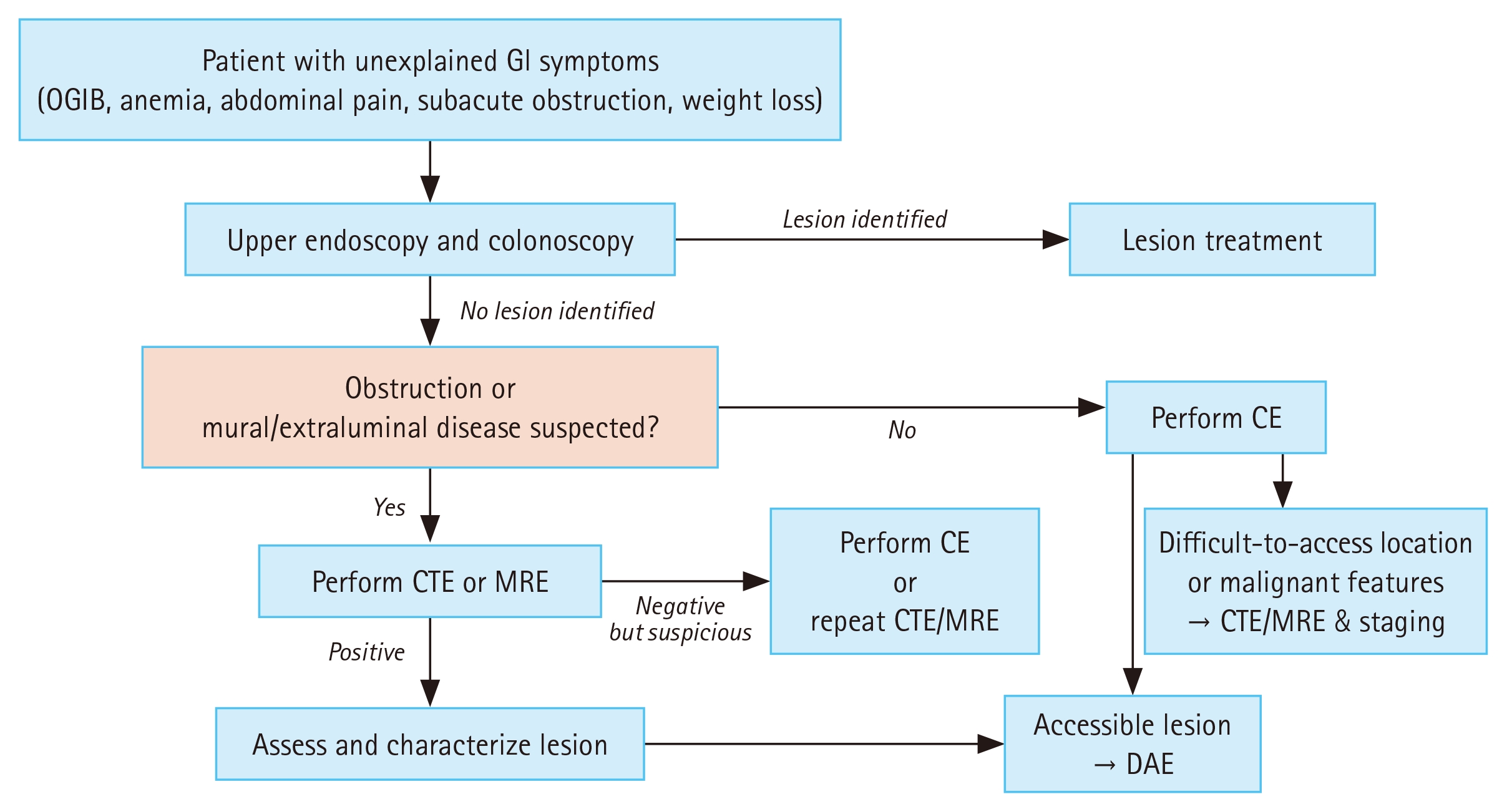
Suggested diagnostic algorithm for suspected small bowel tumors. GI, gastrointestinal; OGIB, obscure gastrointestinal bleeding; CE, capsule endoscopy; CTE, computed tomography enterography; MRE, magnetic resonance enterography; DAE, device-assisted enteroscopy.
CONCLUSION
The diagnostic landscape for small bowel diseases has been profoundly transformed over the past two decades by major advancements in endoscopic and imaging technologies. CE and DAE enable direct mucosal visualization, tissue confirmation, and therapeutic intervention. CTE and MRE provide essential cross-sectional information on structural changes and inflammation extent. IUS further complements these modalities with its noninvasive, radiation-free capability for real-time bedside assessment and monitoring. In the future, the integration of AI-assisted image analysis and functional imaging is expected to further optimize diagnostic pathways. Therefore, achieving an accurate and efficient diagnosis requires the strategic integration of these tools. Clinicians must customize the diagnostic approach by aligning the selection and combination of modalities with the patient’s clinical presentation, suspected pathology, and available resources.
Notes
CRedit authorship contributions
Dong Jun Oh: conceptualization, visualization, writing - original draft; Yun Jeong Lim: conceptualization, validation, supervision, writing - review & editing
Conflicts of interest
The authors disclose no conflicts.
Funding
None