Prognostic implications of functional significance, high-risk plaque and vessel characteristics in patients with diabetes mellitus
Article information

Abstract
Background/Aims
We investigated the prognostic value of high-risk plaque (HRP) and high-risk vessel (HRV) characteristics on coronary computed tomography angiography (CCTA) alongside fractional flow reserve (FFR) in patients with diabetes mellitus (DM).
Methods
We analyzed 307 vessels from 184 diabetic patients who underwent both CCTA and invasive FFR. HRP was defined as the presence of ≥ 3 high-risk features (minimal lumen area < 4 mm2, plaque burden ≥ 70%, low-attenuation plaque, positive remodeling, spotty calcification, or napkin-ring sign). HRV was defined as the presence of ≥ 1 quantitative volumetric parameter above the optimal cutoff. The primary endpoint was target vessel failure (TVF). HRs were adjusted for FFR, HRP, HRV, and the number of clinical risk factors.
Results
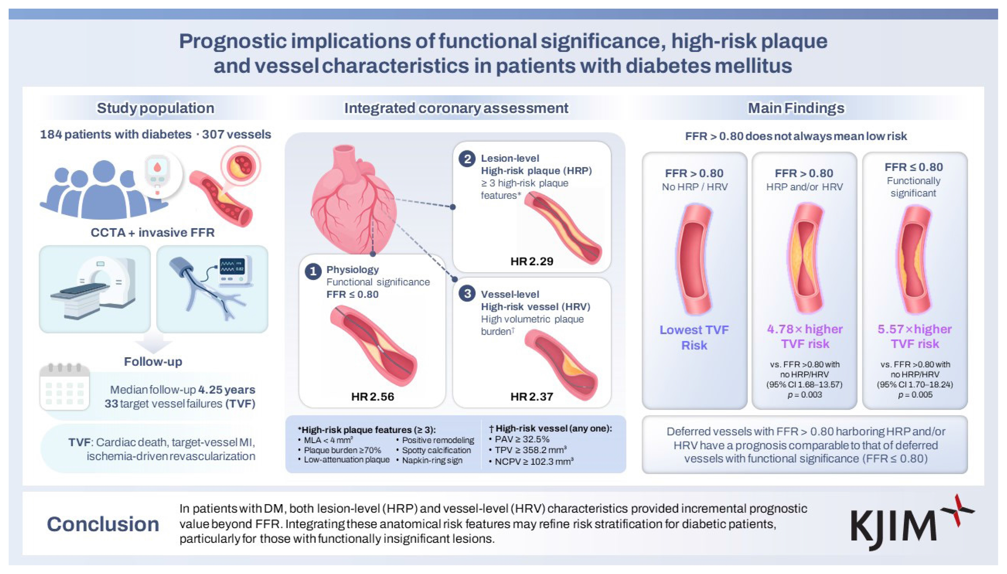
Functional significance (FFR ≤ 0.80; adjusted HR 2.56; 95% CI 1.07–6.12; p = 0.034), HRP (adjusted HR 2.29; 95% CI 1.10–4.77; p = 0.028), and HRV (adjusted HR 2.37; 95% CI 1.12–5.01; p = 0.025) were independent predictors of TVF. Notably, deferred vessels with FFR > 0.80 harboring HRP and/or HRV had a significantly higher risk of TVF compared to the deferred low-risk group (HR 4.78; 95% CI 1.68–13.57; p = 0.003), and their prognosis was comparable to that of deferred vessels with functional significance (FFR ≤ 0.80; HR 5.57; 95% CI 1.70–18.24; p = 0.005).
Conclusions
In patients with DM, both lesion-level (HRP) and vessel-level (HRV) characteristics provided incremental prognostic value beyond FFR. Integrating these anatomical risk features may refine risk stratification for diabetic patients, particularly for those with functionally insignificant lesions.
INTRODUCTION
Diabetes mellitus (DM) is one of the major prognostic factors in patients with coronary artery disease (CAD) conferring a two- to fourfold higher risk of adverse cardiovascular events compared with individuals without diabetes [1–3]. Diabetic patients exhibit a more extensive burden of coronary atherosclerosis and a rapid plaque progression, as well as a greater prevalence of high-risk plaque (HRP) features, relative to non-diabetic patients [4–7]. Notably, endothelial dysfunction, microvascular impairment, and reduced coronary flow reserve have been demonstrated to occur in diabetic patients even before the development of significant epicardial coronary stenosis [8–10]. Despite advances in contemporary percutaneous coronary intervention (PCI) strategies, including intravascular imaging or coronary physiological assessment, diabetic patients remain at higher residual risk of adverse cardiac events [11,12], which underscores the need for refined risk stratification in diabetic patients with CAD. We therefore hypothesized that an integrated approach combining lesion- and vessel-level plaque characteristics with functional significance could provide incremental prognostication, which has not yet been fully elucidated. Accordingly, the present study aimed to evaluate the comprehensive prognostic implications of coronary computed tomography angiography (CCTA)-based assessment, incorporating lesion-level plaque characteristics and overall vessel-level disease burden, and functional significance determined by fractional flow reserve (FFR) in patients with DM.
METHODS
Study population and design
The study population was derived from the CCTA-FFR registry (NCT04037163), which comprises a total of 872 vessels in 538 patients and has been described elsewhere [13]. In brief, the registry enrolled patients with suspected CAD who underwent CCTA followed by invasive FFR measurement within 90 days. In the current study, only vessels in which invasive FFR was measured and for which CCTA analysis by the core laboratory (Severance Cardiovascular Hospital, Seoul, Korea) was feasible were included. Patients were excluded if they had a reduced ejection fraction (< 35%), presented with an acute ST-elevation myocardial infarction (MI) within 72 hours, had a prior history of coronary artery bypass graft surgery, had chronic kidney disease, showed thrombolysis in MI flow < 3, or were scheduled to undergo coronary artery bypass graft surgery after angiography. For the current analysis, a total of 307 vessels from 184 patients with DM were included. Decisions regarding PCI were made at the discretion of the treating physician. The study was conducted in accordance with the principles of the Declaration of Helsinki, and the study protocol was approved by the institutional review board of each participating center, including Seoul National University Hospital (IRB No. H-1907-094-1048).
CCTA analysis and plaque characterization
Qualitative and quantitative plaque assessment was performed on a per-lesion and per-vessel basis using CCTA data analyzed at an independent core laboratory by an independent investigator blinded to all clinical characteristics and invasive angiographic findings, and FFR data. All CCTA acquisitions followed the Society of Cardiovascular Computed Tomography guidelines. Quantitative plaque analysis was conducted using semi-automated software (QAngioCT Research Edition version 2.1.9.1; Medis Medical Imaging Systems, Leiden, the Netherlands), with manual adjustments applied as needed. When multiple lesions were present in a vessel, the most stenotic lesion was designated as the index lesion for plaque characterization. Plaque burden was defined as the ratio of plaque area to vessel area at the segment with the minimum lumen area (MLA). For vessel-level analysis, volumetric quantitative parameters were measured across the entire vessel, encompassing all atherosclerotic lesions within the vessel. Per-vessel volumetric plaque analysis included measurement of total plaque volume (TPV), percent atheroma volume (PAV), calculated as plaque volume normalized to vessel volume, and non-calcified plaque volume (NCPV; ≤ 130 Hounsfield units [HU]).
Definitions of HRP characteristics (HRPC) and high-risk vessel characteristics (HRVC)
For lesion-level assessment, HRPC were defined as the presence of any of the following features: (1) MLA < 4 mm2, (2) plaque burden ≥ 70%, (3) low attenuation plaque (mean density ≤ 30 HU), (4) positive remodeling (remodeling index ≥ 1.1), (5) spotty calcification (average density > 130 HU with diameter < 3 mm in any direction, calcium length < 1.5 times the vessel diameter, and width < two-thirds of the vessel diameter), or (6) napkin-ring sign (ring-like attenuation with peripheral high-density tissue encircling a central low-density core). A lesion was classified as HRP if ≥ 3 HRPC were present, in line with prior literature [14]. For vessel-level assessment, HRVC was defined as (1) PAV ≥ 32.5%, (2) TPV ≥ 358.2 mm3, and (3) NCPV ≥ 102.3 mm3, based on binary classification using optimal thresholds for predicting the primary endpoint. A vessel was defined as a high-risk vessel (HRV) if any one of the HRVC were present [4].
Invasive coronary angiography and FFR measurement
Invasive coronary angiography and FFR measurements were conducted using standard protocols. Following engagement of a guide catheter, a pressure-monitoring guidewire was advanced distal to the target lesion. Maximal hyperemia was induced by continuous intravenous infusion of adenosine (140 μg/kg/min) or adenosine triphosphate (160 μg/kg/min). FFR was defined as the ratio of mean distal coronary pressure to mean aortic pressure during hyperemia. All FFR tracings were independently validated by a core laboratory (Seoul National University Hospital, Seoul, Korea). For patients undergoing PCI, post-PCI FFR was obtained and considered the final FFR value for the corresponding vessel.
Clinical outcomes and event adjudication
Clinical follow-up data were collected through scheduled outpatient visits or telephone interviews. All clinical events were adjudicated by an independent committee blinded to clinical, angiographic, and physiologic information. The primary endpoint was target vessel failure (TVF), a vessel-oriented composite endpoint consisting of cardiac death, target-vessel MI, or ischemia-driven target vessel revascularization. Event definitions followed the criteria of the Academic Research Consortium. All deaths were considered cardiac unless a clearly documented non-cardiac cause was established. MI was defined according to the Third Universal Definition of MI [15], with periprocedural MI excluded from outcome assessment. Ischemia-driven revascularization was defined as any repeat revascularization prompted by at least one of the following: (1) recurrent angina symptoms, (2) a positive non-invasive test, or (3) a positive invasive physiologic test [15].
Statistical analysis
Categorical variables were expressed as counts and percentages, while continuous variables were reported as mean ± standard deviation or median with interquartile range (Q1–Q3), depending on their distribution assessed by the Kolmogorov–Smirnov test. All analyses were conducted on a per-vessel basis. For between-group comparisons of clinical outcomes, event rates were estimated using the Kaplan–Meier method and presented as cumulative incidence. In assessment of the significance of trends, the chi-square test for trend in proportions was conducted. Hazard ratios (HRs) with 95% confidence intervals (CIs) were calculated using marginal Cox proportional hazards regression by accounting for per-vessel correlation within the patients. To prevent model overfitting given the limited number of events, multivariable models were adjusted using the number of clinical risk factors calculated as the sum of age ≥ 65 years, DM, hypertension, dyslipidemia, and smoking. Optimal cut-off values of quantitative plaque parameters for predicting TVF were determined using maximally selected log-rank statistics within the DM population. The plots for determining these cut-off values are presented in Supplementary Figure 1. In all outcome analyses, the final post-PCI FFR value was applied. All statistical tests were two-sided, with p < 0.05 considered statistically significant. Analyses were performed using R software, version 4.3.3 (R Foundation for Statistical Computing, Vienna, Austria).
RESULTS
Baseline characteristics
Among a total of 872 vessels in 538 patients, 307 vessels from 184 patients with DM were included, and these showed a higher risk of TVF compared with the non-DM group (15.9% vs. 5.2%) as shown in Supplementary Figure 2. Baseline patient and lesion characteristics are detailed in Table 1. The mean age was 66.2 ± 9.5 years, and 74.5% of patients were male. The majority presented with chronic coronary syndrome (59.8%), with a mean FFR of 0.83 ± 0.11. In plaque analysis, the mean plaque burden was 63.6 ± 18.3%, MLA was 3.3 ± 2.2 mm2, and HRP was present in 32.2% of lesions. On a per-vessel basis, the mean PAV was 23.4 ± 14.4%, TPV was 177.5 ± 158.2 mm3, and NCPV was 37.1 ± 59.3 mm3, with HRV observed in 30.3% of vessels. The prevalence of HRP and HRV increased progressively with decreasing FFR values: 10.3%, 30.7%, 50.7%, and 54.3% for HRP and 20.7%, 28.1%, 39.4%, and 42.9% for HRV in FFR categories of > 0.90, 0.81–0.90, 0.71–0.80, and ≤ 0.70, respectively (p for trend < 0.001 for HRP and p for trend = 0.002 for HRV) (Fig. 1). There was a positive correlation between the number of HRPC and PAV (r = 0.378; p < 0.001), TPV (r = 0.261; p < 0.001), and NCPV (r = 0.286; p < 0.001) (Supplementary Fig. 3).
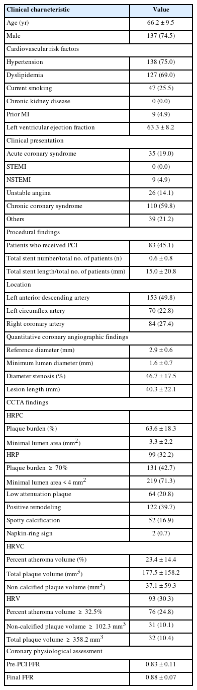
Baseline patient and lesion characteristics
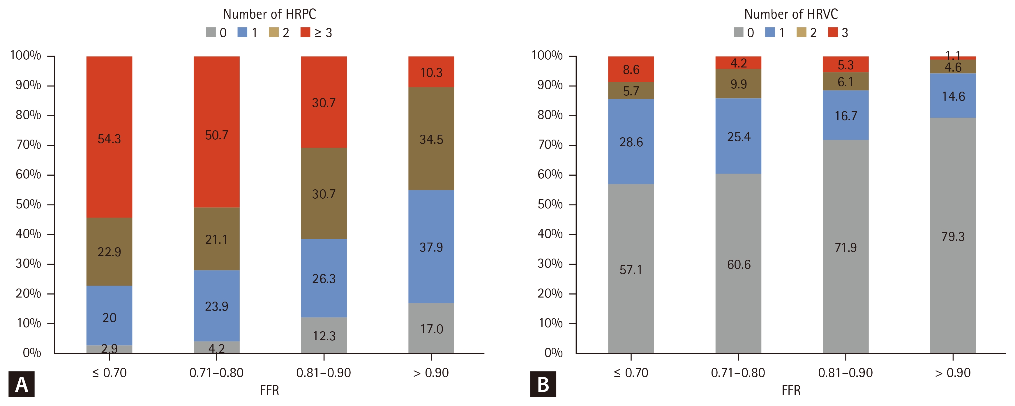
Association of FFR with number of HRPC and HRVC. Bar graphs showing the distribution of the number of (A) HRPC and (B) HRVC stratified by FFR categories. FFR, fractional flow reserve; HRPC, high-risk plaque characteristics; HRVC, high-risk vessel characteristics.
Prognostic implications of functional significance, HRP and HRV
During a median follow-up of 4.25 years (IQR, 2.38–4.82), 33 TVF events were observed (Kaplan–Meier cumulative incidence, 15.9%) (Supplementary Table 1). The cumulative incidence of TVF according to physiological and plaque characteristics is shown in Figure 2. The presence of FFR ≤ 0.80, HRP or HRV was each associated with a higher risk of TVF (13.3% vs. 35.8% for FFR > 0.80 vs. FFR ≤ 0.80; p = 0.008, 25.3% vs. 12.0% for HRP vs. non-HRP; p = 0.014, 29.3% vs 10.7% for HRV vs. non-HRV; p = 0.005, respectively) (Fig. 2). In multivariable analysis, FFR ≤ 0.80 (adjusted HR 2.56, 95% CI 1.07–6.12; p = 0.034), HRP (adjusted HR 2.29, 95% CI 1.10–4.77; p = 0.028), and HRV (adjusted HR 2.37, 95% CI 1.12–5.01; p = 0.025) were each identified as independent predictors of TVF (Table 2).
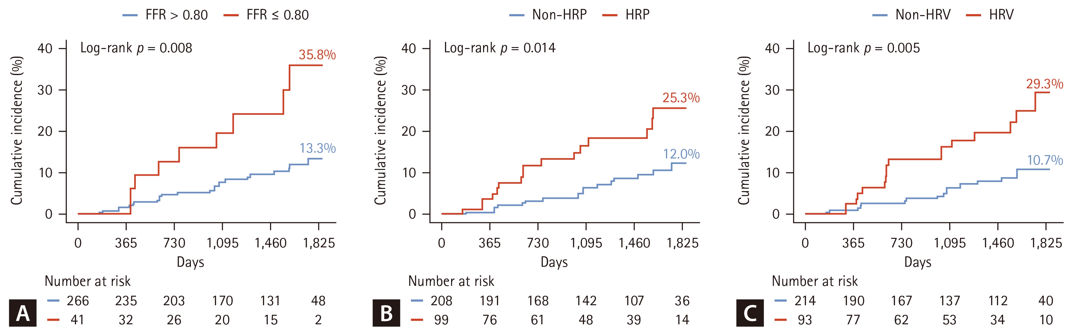
Cumulative TVF rate according to functional significance, HRP, and HRV. Kaplan–Meier curves for TVF. The curves are stratified by (A) functional significance (FFR ≤ 0.80 vs. FFR > 0.80), (B) presence of HRP, and (C) presence of HRV. HRP was defined as the presence of ≥ 3 HRPC, and HRV was defined as the presence of ≥ 1 HRVC. TVF, target vessel failure; FFR, fractional flow reserve; HRP, high-risk plaque; HRV, high-risk vessel.

Independent relationship of functional significance, HRP, and HRV with TVF
Association of HRP and HRV with TVF in vessels with high FFR
When the prognostic impact of HRP and HRV was examined in 266 vessels with FFR > 0.80, the presence of HRP and HRV remained associated with a higher risk of TVF (22.8% vs. 9.2% for HRP vs. non-HRP; p = 0.009, 28.3% vs. 8.0% for HRV vs. non-HRV; p = 0.007). In multivariable analysis, HRP (adjusted HR 2.80, 95% CI 1.15–6.79; p = 0.023) and HRV (adjusted HR 2.95, 95% CI 1.21–7.20; p = 0.017) were independent predictors of TVF (Table 3). A stepwise increase in the cumulative rate of TVF was observed across groups with neither, either, and both HRP and HRV (p for trend < 0.001) (Fig. 3). In a sensitivity analysis restricted to deferred vessels to minimize the confounding effect of revascularization, deferred vessels with FFR > 0.80 accompanied by HRP and/or HRV (HR 4.78, 95% CI 1.68–13.57; p = 0.003) and deferred vessels with FFR ≤ 0.80 (HR 5.57, 95% CI 1.70–18.24; p = 0.005) were associated with a higher risk of TVF as compared to deferred vessels with FFR > 0.80 without HRP or HRV (Supplementary Fig. 4).

Prognostic significance of HRP and HRV in vessels with FFR > 0.80
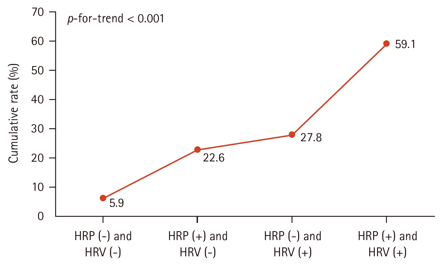
TVF trends according to the presence of HRP and HRV in vessels with FFR > 0.80. Kaplan–Meier curves for TVF in the subgroup of vessels with FFR > 0.80. Patients were classified based on the combined presence of HRP and HRV (neither, either, or both). TVF, target vessel failure; FFR, fractional flow reserve; HRP, high-risk plaque; HRV, high-risk vessel.
Clinical outcomes according to functional significance, HRP, HRV, and treatment types
Vessels stratified according to the presence of HRP, HRV, FFR ≤ 0.80 and treatment types (deferred or revascularized) showed the cumulative event TVF rate of 10.6%, 5.9%, 32.6%, and 31.9% in revascularized vessels achieving post-PCI FFR > 0.80, deferred vessels with FFR > 0.80 and without HRP or HRV, deferred vessels with FFR > 0.80 and with HRP and/or HRV, and deferred vessels with FFR ≤ 0.80, respectively. Using revascularized vessels with post-PCI FFR > 0.80 as the reference, deferred vessels with FFR > 0.80 that harbored HRP and/or HRV had a significantly higher risk of TVF (HR 2.55, 95% CI 1.00–6.49; p = 0.049), comparable to that of deferred vessels with FFR ≤ 0.80 (HR 3.00, 95% CI 0.94–9.51; p = 0.062). In contrast, deferred vessels with FFR > 0.80 and without HRP or HRV showed the lowest risk of TVF (Fig. 4).
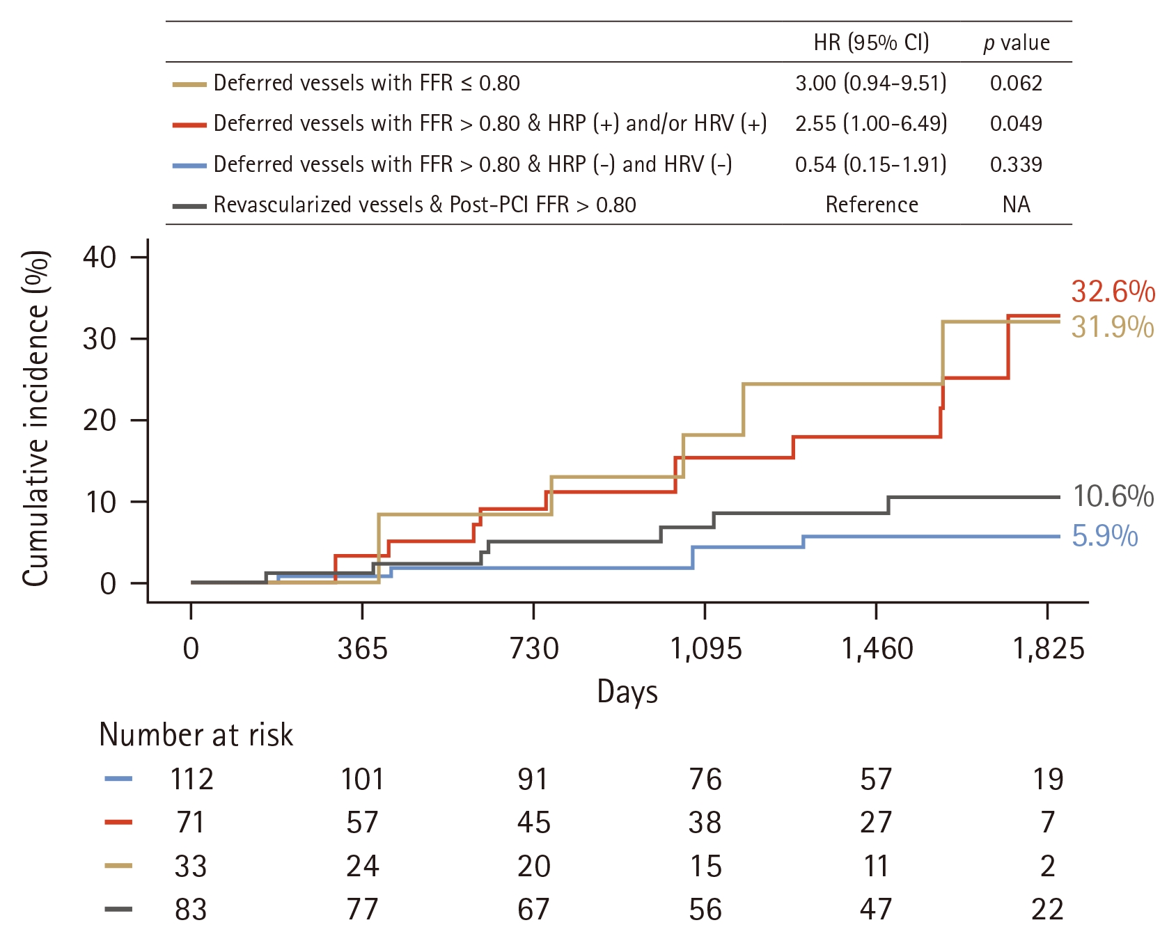
Cumulative TVF rate according to functional significance, HRP, HRV, and treatment types. Kaplan–Meier curves for TVF stratified into four groups according to treatment strategy and plaque characteristics: (1) Revascularized vessels (post-PCI FFR > 0.80; reference group); (2) Deferred vessels with FFR > 0.80 without HRP or HRV; (3) Deferred vessels with FFR > 0.80 with HRP and/or HRV; and (4) Deferred vessels with FFR ≤ 0.80. TVF, target vessel failure; FFR, fractional flow reserve; PCI, percutaneous coronary intervention; HRP, high-risk plaque; HRV, high-risk vessel; HR, hazard ratio; CI, confidence interval.
DISCUSSION
The present study evaluated the prognostic significance of coronary lesion-and vessel-level plaque in conjunction with functional significance among patients with DM and CAD. The main findings can be summarized as follows. First, although both HRPC and HRVC were correlated with FFR, each demonstrated an independent association with an increased risk of TVF. Second, even among vessels with FFR > 0.80, the presence of HRP and HRV remained a significant and independent predictor of TVF. Third, medically treated non-ischemic vessels with HRP and/or HRV exhibited a higher risk of TVF compared with revascularized vessels achieving post-PCI FFR > 0.80.
Independent prognostic contributions of physiological and plaque characteristics in diabetes
DM is a major risk factor for CAD and cardiovascular events [16,17]. Coronary arteries in patients with DM are typically characterized by diffuse narrowing and multivessel disease [18,19]. Moreover, patients with DM exhibit extensive coronary artery calcium [20] along with higher non-calcified, low attenuation and total plaque burdens compared with non-diabetic patients [5,7]. CCTA studies investigating the natural history of diabetic coronary atherosclerosis have also shown that DM is associated with accelerated plaque progression, particularly involving the accumulation of adverse plaque features [4]. Simultaneously, non-invasive physiological studies have consistently demonstrated impaired coronary flow reserve and coronary microvascular dysfunction in this population [8,9,21]. Furthermore, a comparison with the non-diabetic cohort (Supplementary Fig. 2) demonstrated that the cumulative incidence of TVF was significantly higher in the DM group compared to the non-DM group. Thus, multiple distinct aspects of CAD may be attributed to adverse cardiac events in patients with DM [6]. Given that substantial residual risk persists despite optimal medical therapy and contemporary PCI strategies [10–12], whether an integrated assessment combining physiological severity, lesion-specific plaque characteristics, and vessel-level plaque burden can provide superior risk stratification remains to be further elucidated. Building upon the CCTA-FFR registry, which showed the prognostic implications of comprehensive CCTA-based plaque characteristics and FFR on future coronary events [13], we sought to evaluate the independent and incremental prognostic value of HRPC, HRVC, and FFR specifically within a diabetic cohort. In this analysis, FFR values tended to decrease as the number of HRPC and HRVC increased, consistent with previous studies [14,22–24]. Despite this correlation, functional significance, HRP, and HRV were each independently associated with a higher risk of TVF in diabetic patients. These findings are supported by the complex interaction among plaque quantity, plaque quality, and hemodynamics to adverse cardiac events in patients with CAD [13,25], and our results further confirm that this distinct prognostic contribution of each domain remains valid specifically within the high-risk diabetic population. Thus, integrating functional significance with lesion- and vessel-level plaque characteristics may allow for refined risk stratification, capturing high-risk phenotypes that remain undetected when relying on a single domain alone.
Prognostic value of plaque characteristics in functionally insignificant lesions in diabetes
Diabetic patients in whom revascularization were deferred based on high FFR have shown a higher incidence of adverse cardiac events compared with non-diabetic patients [26–29]. In particular, the presence of adverse plaque characteristics, including positive remodeling and plaque burden ≥ 70% on intravascular ultrasound (IVUS), was associated with a higher risk of adverse cardiac events in patients with high FFR, beyond clinical risk factors like DM [30]. Given that diabetic coronary arteries are characterized by diffuse atherosclerosis, greater lipid burden, and a higher prevalence of thin-cap fibroatheromas (TFCA) across the entire vessel tree independent of focal stenosis severity [31,32], we explored the prognostic implications of HRP and HRV in functionally insignificant vessels in diabetic patients. In the current study, the presence of HRP was independently associated with an approximately 2.8-fold higher risk of TVF in vessels with FFR > 0.80, paralleling the results of the COMBINE OCT-FFR trial, which showed that optical coherence tomography (OCT)-detected TFCA was related to an increased risk of adverse events in diabetic patients with FFR-negative lesions [6]. Moreover, in our study, HRV also emerged as an independent predictor, and a synergistic prognostic impact between HRP and HRV was evident: the TVF risk was lowest in vessels with neither characteristic, intermediate in those with either, and highest when both HRP and HRV were present. Expanding the prognostic relevance of adverse plaque characteristics in non-flow limiting lesions, our findings illustrate the additive prognostic value of focal HRPC as well as whole atherosclerotic burden across the vessel in diabetic patients with functionally insignificant lesions. Therefore, in patients with DM, comprehensive plaque assessment at both the lesion and vessel level may provide better risk stratification, even in vessels without functional significance.
Clinical implications of an integrated physiological and plaque assessment along with treatment types
Although FFR-guided deferral of revascularization is generally associated with favorable outcomes [33], plaque characterization-based treatment decision-making also holds clinical relevance, as shown in the PREVENT trial, which showed that PCI for non-flow limiting vulnerable plaques may reduce future cardiac events compared with medical treatment alone [34]. In this context, we stratified vessels according to functional significance, HRP, and HRV in conjunction with treatment types, and we observed that deferred vessels with HRP and/or HRV had more than a 2.5-fold higher risk of TVF compared with revascularized vessels achieving post-PCI FFR > 0.80 and a TVF risk comparable to that of deferred vessels with FFR ≤ 0.80. These findings align with prior observations that deferred vessels with FFR > 0.80 with HRPC portended a numerically higher risk than revascularized vessels with FFR ≤ 0.80 [14]. While meticulous guideline-directed medical therapy remains the cornerstone of management for non-flow limiting lesions, our results suggest that substantial residual risk persists despite standard medical therapy in morphologically HRP and HRV profiles, in some cases exceeding the risk observed in revascularized vessels with high post-PCI FFR. This result is consistent with a subgroup analysis of the PREVENT trial that confirmed that PCI for vulnerable plaques with high FFR was associated with a lower cardiac event in patients with DM [35]. To expand upon these findings, future large-scale studies are needed to determine how the presence of HRP and HRV influences the optimal treatment strategy between PCI and medical therapy, as HRP may favor plaque sealing with PCI whereas HRV may attenuate the clinical benefit of focal revascularization. Collectively, these findings imply that in carefully selected patients in whom an optimal PCI result and high post-PCI FFR can reasonably be anticipated, a hybrid strategy combining intensive medical therapy with lesion-specific revascularization of morphologically high-risk but physiologically insignificant lesions might be beneficial to improve clinical outcomes in diabetic patients with CAD.
Limitations
Several limitations of the current study should be noted. First, this is a retrospective analysis focusing exclusively on a specific subgroup of patients with DM. While this selection allows for a focused analysis of a high-risk phenotype, it may limit generalizability of our result to the broader population, and the findings should be considered hypothesis-generating. Second, the study population included both revascularized and deferred vessels based on real-world clinical practice. Although we adjusted for treatment strategies and performed sensitivity analyses, the observational nature of the registry implies that treatment decisions were influenced by both physiological severity and plaque morphology. Consequently, the observed outcomes may reflect the combined effect of plaque biology and therapeutic interventions rather than the true natural history of coronary atherosclerosis. Third, detailed diabetes-specific characteristics, including hemoglobin A1c levels, duration of DM, and insulin usage, were not available in the registry. Fourth, the cut-off values for quantitative plaque parameters (HRV) were derived from our specific study cohort using maximally selected log-rank statistics. This data-driven approach carries an inherent risk of circular reasoning and overfitting. Furthermore, because the definition of HRVs relies on the observed outcomes, a potential for reverse causation exists, and a definitive causal relationship cannot be fully established. Therefore, these thresholds should be considered exploratory, and external validation in an independent cohort is mandatory to confirm the biological relevance of HRV. Fifth, the number of clinical events is relatively small, which may limit statistical power for some comparisons. Sixth, FFR values were not blinded to physicians during follow-up, and might have caused a potential bias for revascularization events. However, all events were adjudicated by a blinded, independent committee. Finally, invasive imaging modalities like IVUS or OCT were not included, and external validation incorporating intravascular imaging is warranted.
Conclusion
HRPC and HRVC provided independent prognostic value beyond invasive FFR in patients with DM and CAD. In functionally non-ischemic vessels, the presence of HRP and/or HRV was associated with a higher TVF risk, exceeding that of revascularized vessels. These findings support an integrated approach that combines plaque characteristics and physiological assessment to improve risk stratification and optimize treatment selection in patients with DM.
KEY MESSAGE
1. CCTA-derived HRP and HRV characteristics provided independent prognostic value for TVF beyond invasive FFR in patients with DM.
2. Even in diabetic patients with deferred vessels with FFR > 0.80, the presence of HRP and HRV remained independent predictors of TVF.
3. Deferred vessels with FFR > 0.80 harboring HRP and/or HRV had a significantly worse prognosis than revascularized vessels achieving optimal post-PCI FFR (> 0.80), suggesting that a comprehensive plaque assessment is crucial for risk stratification in this high-risk population.
Notes
CRedit authorship contributions
Jin-Eun Song: conceptualization, methodology, investigation, data curation, formal analysis, writing - original draft, visualization; Seokhun Yang: conceptualization, methodology, investigation, data curation, formal analysis, writing - original draft, writing - review & editing, visualization; Masahiro Hoshino: resources, investigation, validation, writing - review & editing; Taishi Yonetsu: resources, investigation, validation, writing - review & editing; Jinlong Zhang: resources, investigation, validation, writing - review & editing; Eun-Seok Shin: resources, investigation, validation, writing - review & editing; Joon-Hyung Doh: resources, investigation, validation, writing - review & editing; Chang- Wook Nam: resources, investigation, validation, writing - review & editing; Jianan Wang: resources, investigation, validation, writing - review & editing; Shaoliang Chen: resources, investigation, validation, writing - review & editing; Nobuhiro Tanaka: resources, investigation, validation, writing - review & editing; Hitoshi Matsuo: resources, investigation, validation, writing - review & editing; Takashi Kubo: resources, investigation, validation, writing - review & editing; Hyuk-Jae Chang: resources, investigation, validation, writing - review & editing; Tsunekazu Kakuta: resources, investigation, validation, writing - review & editing; Bon-Kwon Koo: conceptualization, methodology, resources, formal analysis, writing - review & editing, visualization, supervision
Conflicts of interest
Dr. Koo received an Institutional Research Grant from Abbott, Philips, and HeartFlow Inc. Dr. Nam received Institutional research grants from Abbott and Genoss. All other authors declare that there is no conflict of interest relevant to the submitted work.
Funding
This study was supported by grants from the Patient-Centered Clinical Research Coordinating Center (HI19C0481 and HC19C0305) funded by the Ministry of Health and Welfare and from the Ministry of Food and Drug Safety (RS-2023-00215667), Republic of Korea.