Aspergillus positivity in lower respiratory specimens as a predictor of poor prognosis in invasive pulmonary aspergillosis
Article information

Abstract
Background/Aims
In patients with invasive pulmonary aspergillosis (IPA), the clinical significance of culture positivity is unclear. Here we evaluated whether Aspergillus isolation from lower respiratory tract (LRT) specimens was associated with greater disease severity and higher mortality among patients with IPA.
Methods
This retrospective cohort study included patients with proven or probable IPA cases. Patients were divided into a culture-positive group with Aspergillus species detected in LRT samples, and a control group with fungus-negative bronchoalveolar lavage (BAL) samples. Clinical characteristics and outcomes were compared between these groups, with propensity score weighting to adjust for baseline differences. The primary end-point was 6-week mortality. Secondary outcomes included galactomannan optical density index (ODI) values, radiologic severity, and 12-week mortality.
Results
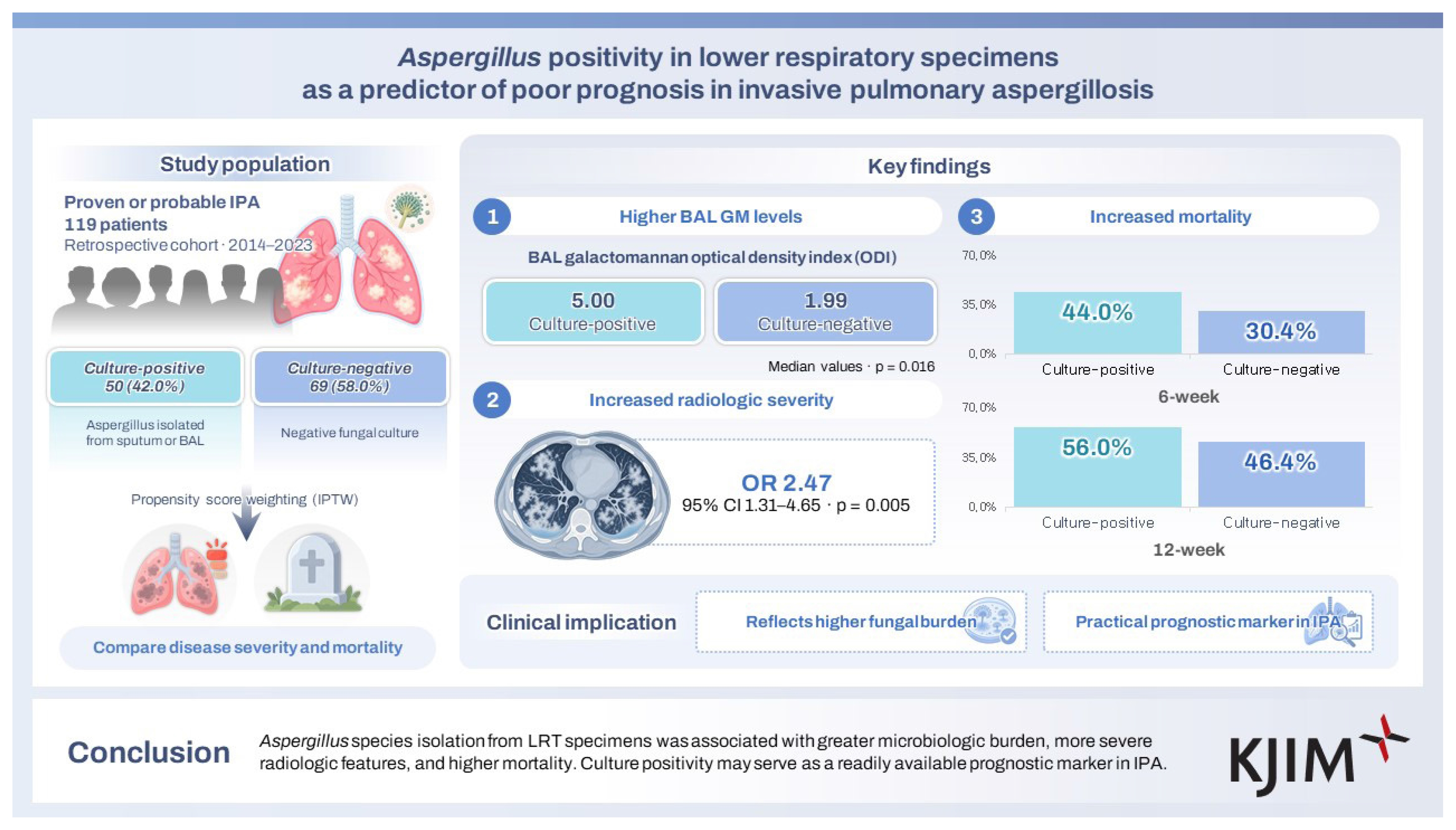
A total of 119 patients were included—50 (42.0%) with Aspergillus species isolated, and 69 (58.0%) with negative cultures. Compared to the control group, patients in the culture-positive group exhibited significantly higher BAL galactomannan ODI values (5.00 vs. 1.99; p = 0.016), more frequent high-grade radiologic findings (odds ratio [OR], 2.47; 95% confidence interval [CI], 1.31–4.65; p = 0.005), higher 6-week mortality (OR, 2.20; 95% CI, 1.24–3.92; p = 0.007), and higher 12-week mortality (OR, 1.83; 95% CI, 1.03–3.27; p = 0.041). Multivariable analysis confirmed that culture positivity was independently associated with worse radiologic severity and higher 6-week mortality.
Conclusions
Aspergillus species isolation from LRT specimens was associated with greater microbiologic burden, more severe radiologic features, and higher mortality. Culture positivity may serve as a readily available prognostic marker in IPA.
INTRODUCTION
Invasive pulmonary aspergillosis (IPA) is a serious lung disease caused by Aspergillus spp. inhalation, which primarily affects immunocompromised individuals [1,2]. IPA has attracted increasing attention due to recent advances in medical care—including immunosuppressive drugs, hematopoietic stem cell transplantation, and solid-organ transplantation [3]. Moreover, viral epidemics, such as COVID-19 and influenza, have highlighted the burden of IPA among patients lacking typical risk factors [4].
Isolation of Aspergillus spp. from the lower respiratory tract (LRT) may indicate a higher fungal burden. Previous studies among patients with IPA suggest that positive fungal cultures are associated with lower treatment success [5] and increased mortality, particularly when combined with bronchoalveolar lavage (BAL) galactomannan (GM) positivity [6]. However, another study shows that Aspergillus culture results are not associated with mortality, highlighting the ongoing controversy regarding the prognostic value of LRT cultures [7]. Notably, these studies have categorized patients mainly based on survival status, and none has directly compared culture-positive patients with a rigorously defined culture-negative control group.
In the present study, we aimed to investigate whether Aspergillus culture positivity in LRT specimens was associated with disease severity and mortality in patients with IPA. The included patients were categorized into a culture-positive group and a culture-negative control group, and propensity score (PS) weighting was applied to adjust for potential confounding factors.
METHODS
Study design
This retrospective cohort study included patients diagnosed with, and treated for, IPA at Seoul National University Hospital between January 1, 2014, and December 31, 2023. Eligible cases were those in which patients had probable or proven IPA, as well as available sputum or BAL culture results, and chest computed tomography (CT) performed at the time of diagnosis. Cases of disseminated aspergillosis and aspergillus tracheobronchitis were included if pulmonary infiltrations were present. Cases of possible IPA, chronic pulmonary aspergillosis, aspergilloma, or isolated tracheobronchitis were excluded.
Patients were assigned to the culture-positive group if Aspergillus spp. was isolated from sputum or BAL specimens. The control group comprised patients with probable or proven IPA, who had negative fungal cultures from both sputum and BAL specimens, or who had negative BAL fungal cultures without available sputum specimens.
This study was approved by the Seoul National University Hospital Institutional Review Board (IRB No. H-2408-104-1562). The requirement for informed consent was waived due to the retrospective study design. All methods were conducted in accordance with the approved guidelines.
Laboratory assays for IPA
Sputum and BAL specimens were collected as part of routine clinical care, at the discretion of the treating physicians, and based on clinical indications. BAL specimens were obtained by bronchoscopy, which was performed by experienced pulmonologists using a flexible video bronchoscope, with the patient under propofol or midazolam sedation. During BAL, normal saline was instilled in aliquots, and then aspirated. The sampling site was determined based on the radiologic location of the lesion. BAL fluid was immediately sent to the laboratory for microbiological testing and GM assays.
Serum and BAL GM were measured using an enzyme-linked immunosorbent assay (Platelia Aspergillus kit; Bio-Rad Laboratories) in accordance with the manufacturer’s instructions. The optical density index (ODI) cut-off for Aspergillus GM positivity was 0.5.
Definitions of potential confounders and outcomes
Proven and probable IPA were defined based on the criteria of the 2020 European Organisation for Research and Treatment of Cancer/Mycoses Study Group Education and Research Consortium (EORTC/MSGERC) [8]. Corticosteroid use was defined as a prednisone dose of ≥ 0.3 mg/kg/day, for ≥ 3 weeks, within 2 months prior to IPA diagnosis [8]. Neutropenia was defined as an absolute neutrophil count of < 500 cells/μL, lasting for at least 10 days, within 1 month prior to diagnosis [8]. An immunosuppressed state was defined by the receipt of chemotherapy within the past month, or ongoing use of immunosuppressive agents.
The primary outcome was all-cause mortality at 6 weeks after starting antifungal therapy, which was selected because mortality at later time-points may be influenced more by underlying disease than by IPA itself. Secondary outcomes included GM positivity, absolute GM ODI values, radiologic severity of IPA, and all-cause mortality at 12 weeks. As there is currently no consensus-based criteria for rating the radiologic severity of IPA, an in-house classification scheme was developed based on previous literature. Several studies demonstrate that more severe disease is associated with multiple consolidations, bilateral pulmonary involvement, and pleural effusion [9–14]. Accordingly, patients were categorized into two groups: low severity (presence of none or one of the three severe radiologic features), or high severity (presence of two or three features). Radiologic findings were reviewed by a single infectious disease specialist who was not blinded to clinical information.
Clinical data collection
Clinical data regarding demographics, comorbidities, immunosuppressive treatments, radiologic findings, laboratory findings, antifungal agents, surgical biopsy results, and mortality were collected from the patients’ electronic medical records. The date of IPA diagnosis was defined as the date on which the patient first met the criteria for proven or probable IPA. Baseline variables collected at diagnosis included age, comorbidities, immune status, isolated Aspergillus species, radiologic features, and serum or BAL GM values. Comorbidity burden was assessed using the Charlson comorbidity index. The antifungal agents administered after diagnosis were identified. Patients were followed-up for 12 weeks after initiation of IPA treatment. Analyses included the chest CT findings, GM values, and mortality recorded at 6 and 12 weeks after treatment initiation. To assess the changes in radiologic disease burden on chest CT, the findings at diagnosis were compared with those at weeks 6 and 12.
Statistical analysis
The culture-positive and control groups were compared after adjustment for confounding factors using inverse probability of treatment weighting (IPTW) based on PS. Covariates included age at diagnosis, hematologic malignancy, hematopoietic stem cell transplantation, solid organ transplantation, lung cancer, chronic pulmonary disease, corticosteroid use, immunosuppressed status, and neutropenia. PS were estimated using gradient boosting machine, and IPTW weights were winsorized at the 1st and 99th percentile. The covariate balance was assessed using standardized mean differences, with a value of < 20% considered acceptable [15,16].
Analyses of serum or BAL GM values at diagnosis and survival status were performed only in patients with available data for each variable. Radiologic severity, GM positivity, and mortality were analyzed using univariable logistic regression. GM ODIs were compared using the Mann–Whitney U-test. IPTW-weighted multivariable logistic regression analyses were also implemented to evaluate whether LRT culture positivity was independently associated with high-grade radiologic severity and 6-week mortality. The model included culture positivity, and the same confounding variables used in the PS weighting analysis.
Survival was assessed using Kaplan–Meier curves, and hazard ratios (HRs) with 95% confidence intervals (CIs) were obtained from an IPTW-adjusted Cox proportional hazards model. Follow-up duration was defined as the interval from the date of antifungal treatment initiation to the date of death, 12 weeks after treatment initiation, or the last documented date on which the patient was confirmed to be alive, whichever came first. Odds ratios (ORs) and 95% CIs were reported.
Prespecified subgroup analyses evaluated whether the prognostic impact of LRT culture positivity varied according to host immune and pulmonary status. Patients were stratified into two subgroups based on the presence or absence of (1) lung cancer and/or chronic pulmonary disease (lung cancer and/or chronic pulmonary disease subgroup) and (2) hematologic diseases and/or neutropenia (hematologic disease and/or neutropenia subgroup). Within each subgroup, the association between culture positivity and disease severity or clinical outcomes was assessed using IPTW-weighted logistic regression. IPTW weights were derived from the overall cohort and applied consistently across subgroup analyses. Kaplan–Meier survival curves and IPTW-adjusted Cox proportional hazards models were also performed for each hematologic/neutropenia subgroup. Interaction tests were conducted by including a multiplicative interaction term in the logistic regression model.
An exploratory analysis was conducted to compare outcomes among three culture-source groups—culture-negative controls, sputum-positive, and BAL-positive (including cases of concurrent positivity)—using IPTW-weighted logistic regression with the control group as the baseline. Kaplan–Meier survival curves were also created for the three groups based on the culture source.
Additionally, a sensitivity analysis was performed to evaluate the association between culture positivity, disease severity, and clinical outcomes in patients with respiratory specimens obtained before antifungal initiation and without prior prophylactic antifungal exposure. After excluding patients with prior prophylactic antifungal exposure, those whose specimens (either sputum or BAL) were obtained before the initiation of IPA treatment were included in the analysis. IP-TW-adjusted logistic regression analysis was performed, and the ORs, 95% CIs, and p values were reported. All statistical analyses were performed using R software, version 4.5.1 (R Foundation for Statistical Computing).
RESULTS
Baseline characteristics of the study population
A total of 516 patients with suspected IPA had available LRT culture results. Among them, 119 patients with proven or probable IPA had available LRT culture results—revealing Aspergillus spp. isolation from 50 patients (42.0%), and negative results from 69 patients (58.0%) (Fig. 1). Among the culture-positive patients, 26 had culture-positive sputum, 22 had culture-positive BAL, and 2 had positive results from both tests; the timing of specimen collection relative to antifungal treatment initiation is detailed in Supplementary Table 1.
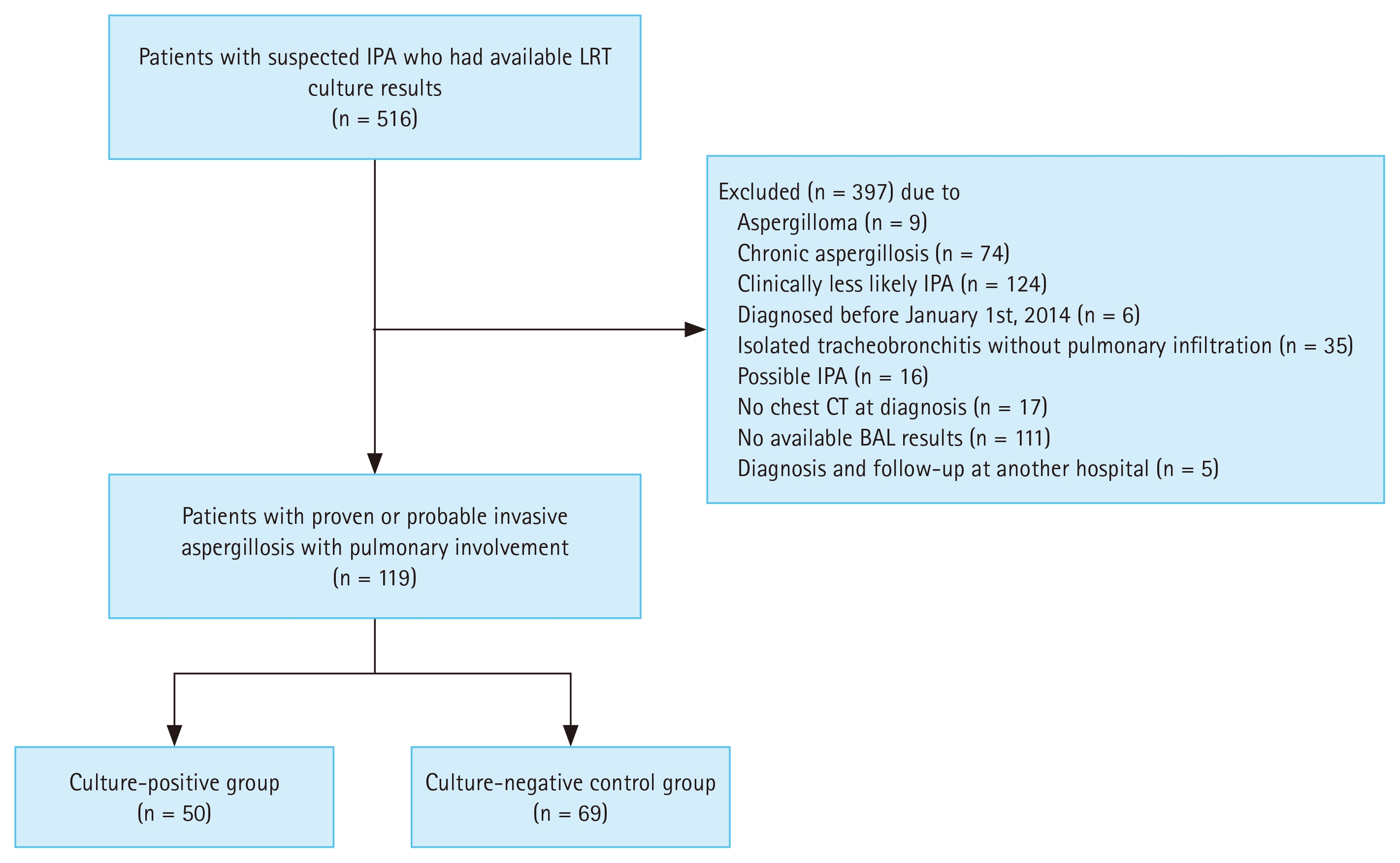
A flow diagram of patient selection. IPA, invasive pulmonary aspergillosis; LRT, lower respiratory tract; CT, computed tomography; BAL, bronchoalveolar lavage.
Table 1 summarizes the baseline characteristics before and after PS weighting. After PS weighting, the overall covariate balance substantially improved, although small residual imbalances remained for a few variables—including hematologic disease, lung cancer, solid organ transplantation, and steroid use. Among culture-positive patients, A. fumigatus was the organism most frequently isolated from both sputum and BAL cultures (29.14% and 21.50%, respectively), followed by non-speciated Aspergillus (16.25% and 18.04%, respectively).

Baseline characteristics before and after propensity score weighting
Disease severity of culture-positive IPA
BAL GM-positivity was found in 26 patients (52.0%) of the culture-positive group vs. 32 patients (46.4%) of the control group, yielding an OR of 2.86 (95% CI, 1.31–6.23; p = 0.008) (Table 2). Serum GM positivity was similar between the groups (72.0% vs. 66.7%; OR, 1.11; 95% CI, 0.58–2.13; p = 0.753). The median BAL GM ODI was also significantly higher in the culture-positive group (5.00 vs. 1.99; p = 0.016). On the other hand, the two groups did not significantly differ in serum GM ODI (1.46 vs. 1.14; p = 0.144).

IPTW-adjusted analysis of clinical features based on fungal culture status
With regards to radiologic findings, the groups did not significantly differ in the halo sign (OR, 0.87; 95% CI, 0.49–1.57; p = 0.655), but severe radiologic patterns were significantly more common among culture-positive patients (OR, 2.47; 95% CI, 1.31–4.65; p = 0.005) (Table 2). Multivariable logistic regression analysis revealed that severe radiologic patterns were independently predicted by culture positivity (OR, 2.79; 95% CI, 1.45–5.58; p = 0.003), as well as by hematologic malignancy (OR, 4.58; 95% CI, 1.96–11.40; p = 0.001) and steroid use (OR, 2.18; 95% CI, 1.04–4.67; p = 0.041) (Table 3).

IPTW-adjusted multivariable logistic regression analysis of factors associated with radiologic severity
Exploratory subgroup analyses were performed to examine the association between culture positivity and radiologic severity according to host immune and pulmonary status (Supplementary Table 2). In the lung cancer and/or chronic pulmonary disease subgroup, culture positivity was statistically significant for radiologic severity (OR, 3.86; 95% CI, 1.63–9.10; p = 0.002) in patients without lung disease, with no significance in those with lung disease. Nevertheless, no significant interaction was observed between lung disease status and culture positivity for radiological severity (p for interaction > 0.05). In the hematologic disease and/or neutropenia subgroup, culture positivity was significantly associated with greater radiologic severity in patients with hematologic disease and/or neutropenia than in those without (OR, 5.62 vs. 1.88; 95% CI, 1.48–21.32 vs. 0.86–4.12; p = 0.011 vs. 0.115); however, no significant interaction was observed (p for interaction > 0.05). An analysis was also conducted to explore whether radiologic severity varied according to culture source. The distribution of specimens across the three groups is shown in Supplementary Table 3. Both the sputum-positive and BAL-positive groups had higher rates of high-grade radiologic severity than the control group (OR, 1.68 vs. 4.08; 95% CI, 0.79–3.54 vs. 1.62–10.28; p = 0.177 vs. 0.003) (Supplementary Table 4).
Clinical outcomes
After PS weighting, 6-week mortality was significantly higher in the culture-positive group than in the control group (44.0% vs. 30.4%; OR, 2.20; 95% CI, 1.24–3.92; p = 0.007) (Table 2). Multivariable logistic regression analysis revealed that culture positivity was independently associated with 6-week mortality (Table 4). Consistently, Kaplan–Meier analysis demonstrated poorer survival in the culture-positive group, compared to the control group (Fig. 2). In an IPTW-adjusted Cox regression model, compared to the control group, the culture-positive group had a significantly higher hazard of death (HR, 2.041; 95% CI, 1.037–4.017; p = 0.039).

IPTW-adjusted multivariable logistic regression analysis of factors associated with mortality at 6 weeks
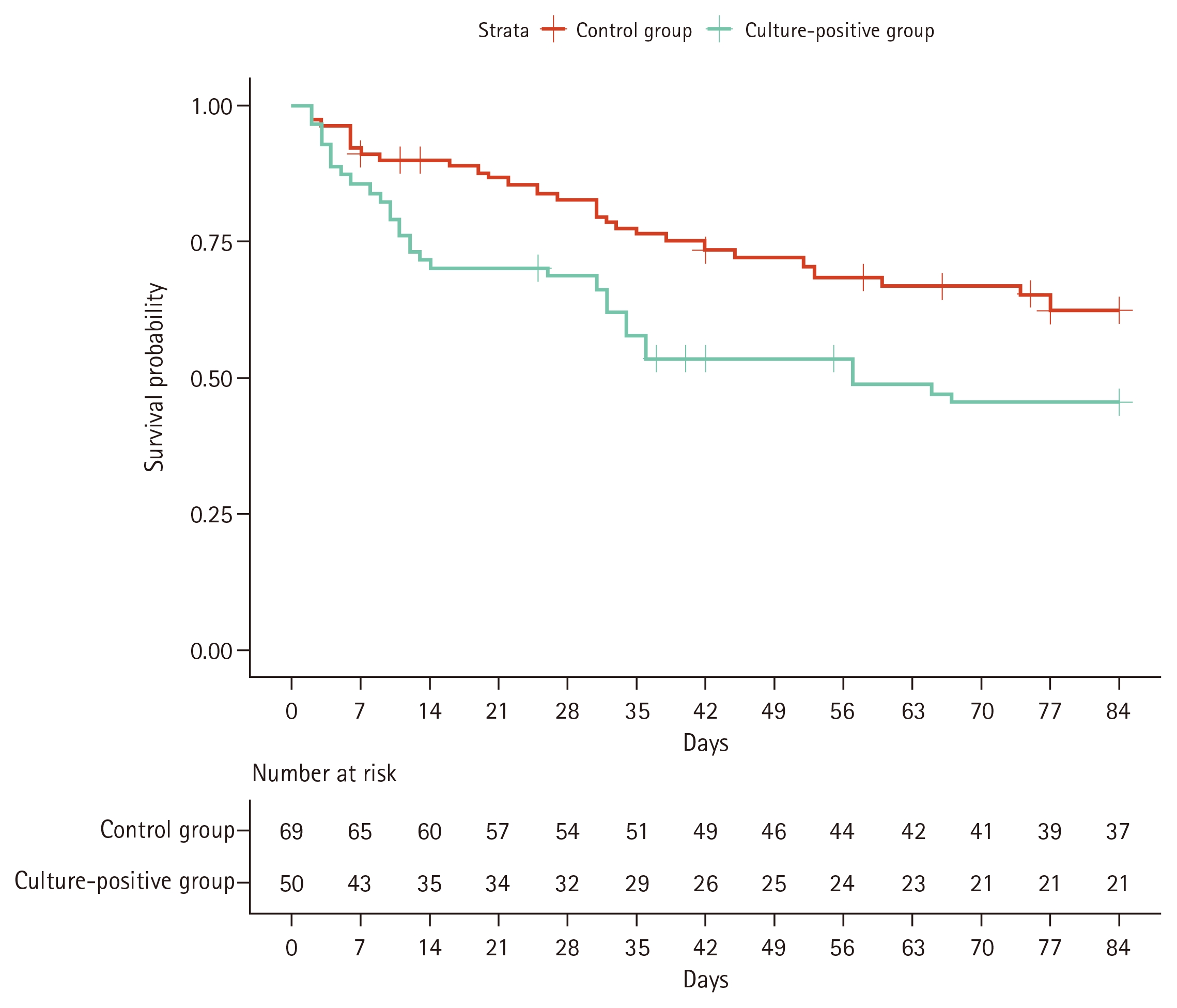
Kaplan–Meier survival analysis of the culture-positive and control groups, assessed over 12 weeks. The number of “at risk” patients in each group is presented below the time axis.
At week 12, mortality remained higher in the culture-positive group, compared to the control group (56.0% vs. 46.4%; OR, 1.83; 95% CI, 1.03–3.27; p = 0.041). The culture-positivity group continued to show poorer survival on Kaplan–Meier analysis through week 12, and the IPTW-adjusted Cox regression model showed a similar tendency (HR, 1.783; 95% CI, 0.985–3.229; p = 0.056).
In the exploratory subgroup analysis by lung status (Supplementary Table 5), culture positivity was statistically significant for 6-week mortality (OR, 2.06; 95% CI, 1.08–3.90; p = 0.027) in patients without lung disease, with no significance in those with lung disease, and no significant interaction was observed (p for interaction > 0.05). For the hematologic disease and/or neutropenia subgroup analysis (Supplementary Tables 5, 6), culture positivity was significantly associated with higher 6-week mortality (OR, 3.23 vs. 1.57; 95% CI, 1.39–7.53 vs. 0.70–3.49; p = 0.007 vs. 0.274) and 12-week mortality (OR, 2.49 vs. 1.19; 95% CI, 1.05–5.92 vs. 0.56–2.54; p = 0.039 vs. 0.656) in patients with hematologic disease and/or neutropenia, while no significant associations were observed in those without. Interaction testing revealed no statistically significant effect modification by hematologic disease and/or neutropenia status for 6- and 12-week mortality (both p for interaction > 0.05). The Kaplan–Meier survival curves in both subgroups showed a trend toward a lower survival probability in the culture-positive group compared to controls (Supplementary Fig. 1). IPTW-adjusted Cox regression analysis demonstrated higher HRs for 6-week mortality in patients with hematologic disease and/or neutropenia than in those without (6-week HR, 2.19 vs. 1.57; 95% CI, 0.97–4.98 vs. 0.62–3.93; p = 0.060 vs. 0.340). The analysis according to culture source revealed higher 6- and 12-week mortality than the control group for both the sputum-positive and BAL-positive groups (6-week OR, 2.39 vs. 2.03; 95% CI, 1.19–4.83 vs. 1.00–4.09; p = 0.015 vs. 0.049; 12-week OR, 1.96 vs. 1.36; 95% CI, 0.97–3.95 vs. 0.69–2.70; p = 0.062 vs. 0.375) (Supplementary Table 4). Kaplan–Meier survival curves also demonstrated a trend toward a lower survival probability in both the sputum-positive and BAL-positive groups compared with controls (Supplementary Fig. 2).
Sensitivity analysis
Sensitivity analysis was conducted to examine the associations between culture positivity, disease severity and clinical outcomes, restricted to patients whose respiratory specimens were obtained prior to IPA treatment initiation and who had no prior prophylactic antifungal exposure (Supplementary Table 7). Culture positivity demonstrated higher odds of high-grade radiologic severity (OR, 2.17; 95% CI, 0.79–5.95; p = 0.130) and 6-week (OR, 2.22; 95% CI, 0.83–5.97; p = 0.112) and 12-week mortality (OR, 1.49; 95% CI, 0.59–3.78; p = 0.398).
DISCUSSION
In this retrospective cohort study, we compared culture-positive IPA patients with strictly defined culture-negative IPA patients, after adjustment for confounding factors using PS weighting. The culture-positive group exhibited significantly more severe disease and higher mortality, suggesting that culture positivity may serve as a prognostic marker in IPA.
The present study demonstrated that culture positivity was independently associated with 6-week mortality. Importantly, our study design allowed us to evaluate the prognostic impact of culture positivity against a rigorously defined BAL-negative control group, rather than simply comparing survivors versus non-survivors. In this context, our findings are broadly consistent with previous reports from other high-risk populations [6,17,18]. Notably, in a study of critically ill patients with COVID-19, 90-day mortality was higher among patients with both positive BAL GM and positive Aspergillus cultures [6]. Likewise, among lung transplant recipients, mortality was highest in those with positive results for both BAL GM and Aspergillus cultures [18]. In a study of patients requiring ventilator care in intensive care units, BAL GM > 1.0 or Aspergillus culture positivity was associated with increased in-hospital mortality [17]. Overall, these findings suggest that fungal culture positivity may reflect a poorer prognosis. Although all-cause mortality does not necessarily indicate IPA-related death, the significant difference observed after PS weighting indicates that fungal culture positivity may reflect greater disease burden and a poorer prognosis.
Our results showed that culture positivity was associated not only with higher mortality but also with markers of greater disease burden or severity—including higher BAL GM positivity, higher BAL GM ODI values, and more severe radiologic findings. Higher BAL GM ODI values reportedly may reflect a greater fungal burden in the LRT, and previous studies have linked higher BAL GM indices to increased mortality among kidney transplant recipients and non-neutropenic patients [11,19]. BAL GM has limitations with regards to standardization; however, it may serve as a complementary indicator of disease severity in selected settings.
We also found that culture positivity was independently associated with radiologic severity in IPA. Prior studies have identified radiologic risk factors for mortality, including multiple consolidations, pleural effusion, and bilateral lung involvement [9–14]. In this context, our present findings suggest that the higher mortality in the culture-positive group may be partly explained by more severe radiologic features. Although the halo sign has been linked to better outcomes in immunocompromised hosts [20], we found that halo sign prevalence did not differ between groups in our study, possibly because our cohort contained relatively few immunocompromised patients.
We also found that serum GM positivity did not differ between the patient groups, although the median index was higher in the culture-positive group. Serum GM is thought to reflect angioinvasion, and has been previously reported to be associated with increased mortality; however, its performance is affected by host immune status, particularly neutrophil function [21]. It is possible that the lack of statistical significance in our study is due to our inclusion of both neutropenic and non-neutropenic patients.
Subgroup analyses were conducted to further examine the prognostic relevance of culture positivity based on the patient’s underlying pulmonary and immune status. In the culture source subgroup analysis, both sputum- and BAL-positive patients showed higher odds of radiological severity and mortality, with notably comparable 6-week mortality risks between the two groups. Although concurrent BAL positivity could not be confirmed in sputum-positive patients, the comparable ORsand survival curve trends between the sputum- and BAL-positive groups suggest that colonization in sputum culture was less likely to be included in the study. However, in the lung cancer and/or chronic pulmonary disease subgroup analysis, culture positivity was significantly associated with increased disease severity and 6-week mortality in patients without underlying lung cancer and/or chronic lung disease. Conversely, no significant association was observed in patients with lung disease. The subgroup analyses were exploratory and the limited sample size precluded statistical correction for multiple comparisons. Therefore, the prognostic impact of culture positivity according to underlying lung disease status needs to be confirmed in future studies.
In the subgroup with hematologic disease and/or neutropenia, culture positivity was associated with increased disease severity and higher mortality than that in patients without these conditions. This may reflect the fact that in immunocompromised hosts with impaired defense mechanisms, a positive culture more likely represents a progressive, uncontrolled infection with a higher fungal burden. These findings suggest that respiratory culture positivity may have prognostic relevance particularly in patients with hematologic disease and/or neutropenia, although further studies are needed to confirm this observation.
To address potential misclassification bias due to prior antifungal exposure, a sensitivity analysis was conducted among patients whose respiratory specimens were obtained prior to IPA treatment initiation and who had no prior prophylactic antifungal exposure. The analysis showed a tendency similar to that of the main analysis, with higher odds of disease severity and mortality. These results suggest that potential misclassification due to prior antifungal exposure may not substantially affect the primary conclusions of this study.
The present study had several limitations. One key limitation when analyzing respiratory specimens is the difficulty in distinguishing true infection from colonization, particularly in patients with chronic lung disease [22]. However, the risk of misclassifying colonized Aspergillus as a true infection was likely minimized in our study, as we included only patients with proven or probable IPA., Subgroup analysis revealed that sputum-positive patients—who are inherently at a higher risk of colonization—showed results consistent with the main analyses. Another limitation is that the use of prophylactic or empirical antifungal agents before IPA diagnosis may have affected fungal growth in LRT specimens. Although our sensitivity analysis demonstrated a similar tendency similar to that of the main analysis, the restricted sample size made it difficult to determine statistical significance, suggesting that the potential influence of prior antifungal exposure on culture results cannot be entirely excluded. Nevertheless, the collection of respiratory specimens after antifungal initiation reflects real-world clinical practice, in which obtaining cultures prior to antifungal initiation is not always feasible, particularly for BAL. In this context, our findings may offer practical value by revealing the clinical significance of a positive culture in real-world clinical settings. Additionally, our study did not account for several acute illness-related factors that may contribute to increased mortality in patients with IPA such as ICU admission; mechanical ventilation; renal replacement therapy; organ dysfunction; treatment delay; and concomitant bacterial, viral, or opportunistic pulmonary infections [23]. Therefore, the possibility of residual confounding from these unmeasured acute severity factors cannot be excluded. Finally, the relatively small sample size and heterogeneous cohort in our study limit the generalizability of the results. The manifestations of IPA vary depending on the underlying disease; therefore, future studies should include stratified analyses to better account for heterogeneity.
In conclusion, the present study showed that among patients with IPA, Aspergillus culture positivity was associated with higher BAL GM values, more severe radiologic findings, and higher mortality. As respiratory cultures are ubiquitously used in clinical settings, culture positivity offers a practical and highly feasible prognostic marker for IPA in actual clinical practice.
KEY MESSAGE
1. Aspergillus culture positivity from LRT specimens was associated with higher BAL GM levels, more severe radiologic findings, and increased 6-week mortality.
2. Culture positivity may serve as a practical and readily available prognostic marker in patients with IPA.
Notes
Acknowledgments
The authors used ChatGPT (OpenAI, version 5.4) solely for English language editing and improvement of readability. The authors reviewed and approved the final manuscript and take full responsibility for its scientific content, interpretations, and conclusions.
CRedit authorship contributions
Ka Eun Kim: conceptualization, data curation, formal analysis, investigation, methodology, writing - original draft, writing - review & editing, visualization; Jung Soo Son: formal analysis, writing - review & editing; Hyeon Jae Jo: conceptualization, methodology, writing - review & editing; Chan Mi Lee: conceptualization, methodology, writing - review & editing; Jae Hyeon Park: data curation; Chang Kyung Kang: conceptualization, methodology, writing - review & editing; Taek Soo Kim: data curation; Pyoeng Gyun Choe: conceptualization, methodology, writing - review & editing; Wan Beom Park: conceptualization, formal analysis, methodology, writing - review & editing, supervision, project administration; Nam Joong Kim: conceptualization, methodology, writing - review & editing
Conflicts of interest
The authors disclose no conflicts.
Funding
This work was supported by the Creative-Pioneering Researchers Program through Seoul National University (to W. B. Park), and by the Bio & Medical Technology Development Program of the National Research Foundation (NRF), funded by the Korean government (MSIT) (grant number: 2021M3A9I2080498 to W. B. Park).