Masked Hydronephrosis
Article information
A 77-year old woman visited the emergency room (ER) because of abdominal pain and severe diarrhea. She had undergone radiation therapy for cervical cancer 30 years earlier. Pertinent initial laboratory data included a serum urea nitrogen level of 90.9 mg/dL and a creatinine level of 4.1 mg/dL. Sigmoidoscopy showed circular ulceration with whitish exudates consistent with ischemic colitis in the mid rectum. When she came to the ER, she showed oliguria with no evidence of hydronephrosis or indication of urinary tract obstruction on unenhanced computed tomography (CT) (Fig. 1). She was treated aggressively with fluid and antibiotics for dehydration and ischemic colitis. Her condition seemed to be improving, with diminished diarrhea and increased urine output. Serum creatinine level dropped to 0.6 mg/dL. However, on the 5th day in the hospital, she suddenly developed a spiking fever and flank pain, and there was bilateral costovertebral angle tenderness. Follow-up CT scan demonstrated newly emerged bilateral hydronephrosis and diffuse thickening of the bladder wall without stone, mass, or stricture (Fig. 2). Cystoscopic biopsy showed many chronic inflammatory cells, especially plasma cells and some acute inflammatory cells, throughout the bladder wall.
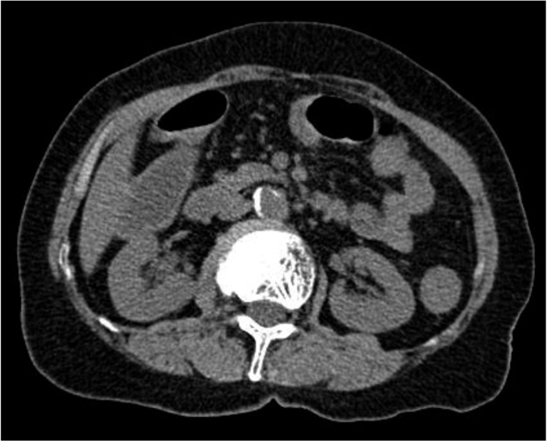
Initial CT scan. There was no evidence for hydronephrosis or urinary obstruction on admission.

Follow-up CT scan. Note marked bilateral hydronephrosis, which was not detected on early CT.
The urinary bladder is irradiated incidentally in patients with cancer involving other pelvic structures. Late-phase symptoms may occur years after the initial radiation therapy. Ischemia and fibrosis may result from radiation, leading to contracted bladders, ulcer formation, fistulas, and dysfunction of the bladder. Decreased bladder capacity followed by chronic inflammation is one of the causes of urinary tract obstruction. Urinary tract obstruction can be diagnosed by the presence of hydronephrosis on ultrasound or CT scan. However, in the following situations hydronephrosis may not be apparent: 1) Within the first three days after the obstruction event when the collecting system is relatively incompliant and less likely to dilate; 2) When the collecting systems are encased by retroperitoneal tumor or fibrosis. In this setting, hydronephrosis may be present in the absence of ureteral dilatation; 3) With mild obstruction, there is usually no impairment in renal function; 4) With volume depletion, particularly in the elderly, partial obstruction may be obscured.
In this case, urinary flow was partially reduced without the need for compensation because of the radiation-induced cystitis. The concomitant colitis caused severe diarrhea and volume depletion, decreased the renal function, and masked the presence of hydronephrosis at first. When the patient recovered from decreased renal function, with hydration by forced diuresis, the hydronephrosis manifested fully.
Notes
No potential conflict of interest relevant to this article was reported.