A Large, Free-Floating Right Atrial Thrombus Evoking Periodic Dizziness
Article information
A 62-year-old man suffered from exertional dyspnea and repetitive momentary dizziness that began occurring just after forceful chest beating to end an episode of acute shortness of breath 3 days prior. His blood pressure was 102/66 mmHg. His D-dimer level (435 µg/L) and N-terminal pro-brain natriuretic peptide level (2,863 pg/mL) were elevated. Electrocardiography exhibited atrial fibrillation with a regular junctional rhythm and a rate of 56 bpm. An intracardiac mass and acute thromboembolism in both pulmonary arteries (APTE) were diagnosed by computed tomography (Fig. 1A and 1B). Echocardiography revealed a very large 60 × 50 mm deformable mass f loating freely in the markedly enlarged right atrium (RA), plugging the right ventricle through the tricuspid valve and periodically occupying the entire cavity (Fig. 1C). Emergent surgery was performed to remove the mass and the APTE. The RA mass was a Chinese moon cake-like thrombus (65 × 55 × 20 mm) (Fig. 2). Two remnant stalks were found on the RA wall. Pathology confirmed that the mass was an organized thrombus and that the stalks were degenerative muscular tissue.
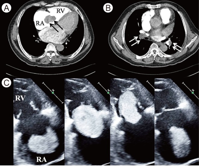
(A, B) Computerized tomographic images showing a mass floating in the right heart (A, black arrows) and embolism in both pulmonary arteries (B, white arrows). (C) A series of echocardiographic images of the right heart. A huge deformable echogenic mass in the enlarged right atrium (RA) is floating and plugging the right ventricle (RV) during diastole (the first three images) and ejected out during systole (the last).
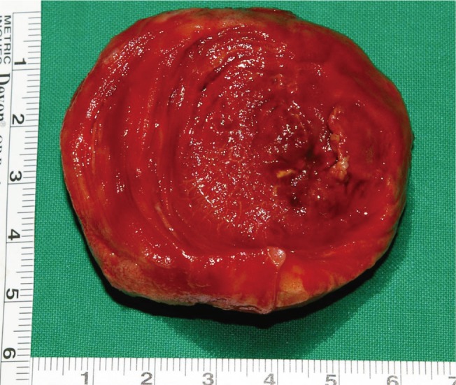
An easily deformable huge thrombus (65 × 55 × 20 mm) removed from the right atrium.
Notes
No potential conflict of interest relevant to this article is reported.