Massive small bowel bleeding in an immunocompromised patient who underwent steroid treatment
Article information
A 79-year-old man was referred to our hospital with hematochezia and anemia (Hb level: 8.9 g/dL). He had several comorbidities including diabetes, hypertension, dyslipidemia, and Parkinson’s disease. In addition, he had developed COVID-19 infection one month before his referral to our hospital. At the time of his admission to our hospital, generalized edema and skin petechiae were prominent, and hematochezia, diarrhea, and azotemia were gradually worsened. Abdominopelvic computed tomography showed massive ascites and diffuse bowel edema; however, a definite gastrointestinal bleeding lesion was not detected. Because of a high suspicion of COVID-19-associated systemic vasculitis, empirical intravenous methylprednisolone was administered and hemodialysis was initiated; however, massive hematochezia and anemia persisted (Hb level: 5.7 g/dL).
Emergent endoscopy revealed fresh blood from the terminal ileum in the lower endoscopy, whereas there was no sign of recent bleeding in the upper endoscopy. Considering a high possibility of overt small bowel bleeding and a high risk of aspiration during bowel preparation, peroral single-balloon enteroscopy was performed. It revealed multiple ulcers, mucosal friability, and hyperemia in the proximal and middle jejunum, suggesting cytomegalovirus (CMV) enteritis (Fig. 1). Thus, intravenous ganciclovir therapy was started, and a diagnosis of CMV enteritis was finally made based on histopathologic findings (Fig. 2). However, recurrent bleeding with hypotension persisted despite additional angiographic embolization for the proximal jejunum. Thus, surgical resection and anastomosis of the proximal jejunum were performed. The surgical specimen showed findings corresponding to CMV enteritis. However, pancytopenia and azotemia gradually aggravated, and the patient finally died one month after the surgery.
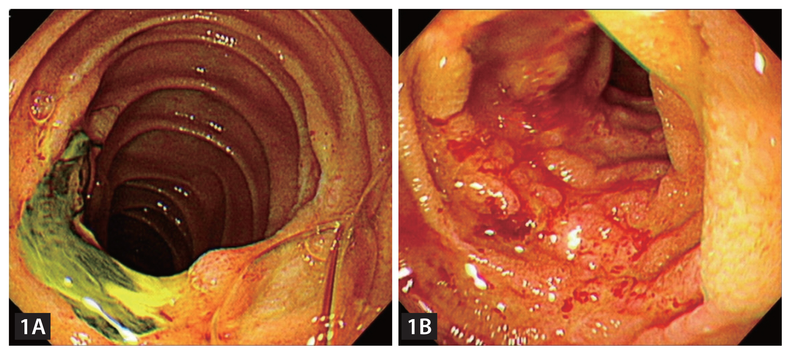
Enteroscopic findings. (A, B) Multiple erosions and ulcers with hyperemic friable mucosa was noted between proximal and middle jejunum. Other segments showed normal-looking mucosa.
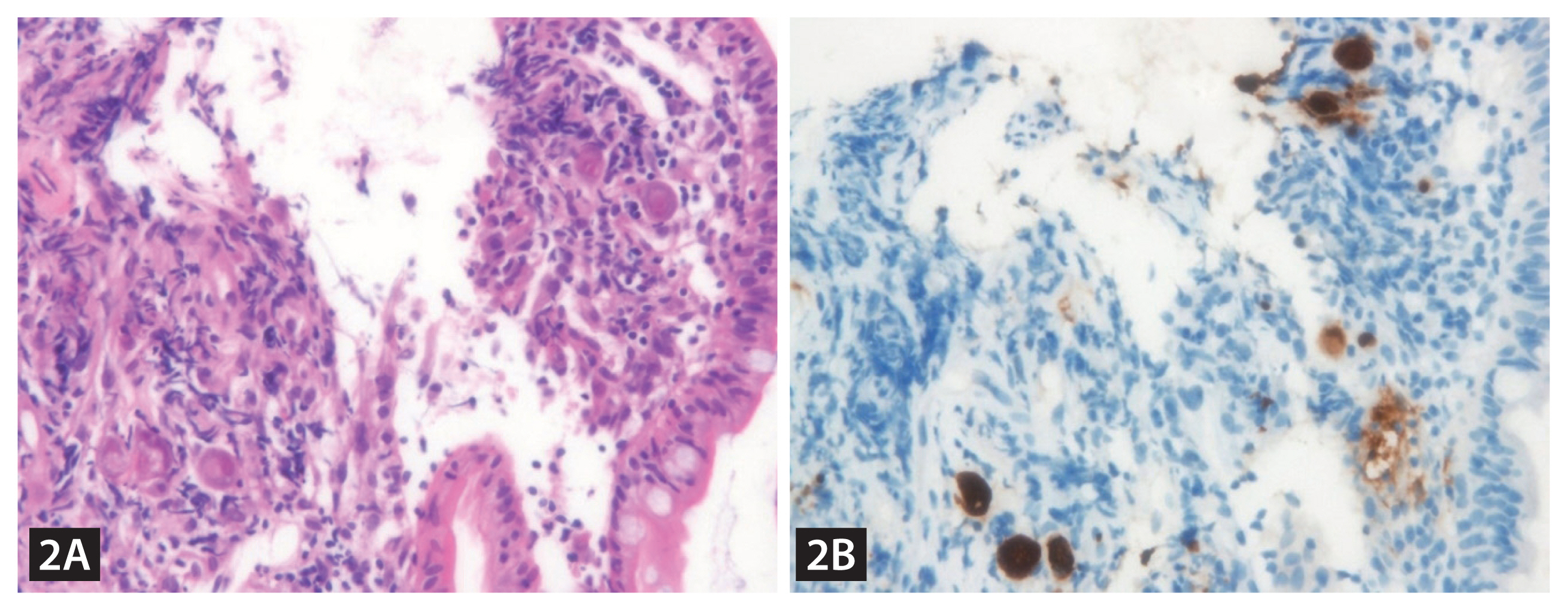
Histopathologic findings of enteroscopic biopsies, (A) H&E ×100, (B) immunohistochemistry ×100. Characteristic owl eye inclusion body and corresponding immunohistochemistry findings of cytomegalovirus enteritis were noted.
CMV enteritis could manifest as gastrointestinal bleeding in patients with marked ulceration. However, massive bleeding as in this case is relatively rare. The diagnosis of CMV enteritis is challenging owing to the difficulty in performing a biopsy. However, enteroscopy can facilitate early diagnosis and proper treatment.
Acknowledgments
The patient’s legal guardian provided informed consent and agreed to publication of the images.
Notes
Conflicts of interest
The authors disclose no conflicts.
CRedit authorship contributions
Jinmo Kim: conceptualization, methodology, investigation, writing - original draft; Kwangwoo Nam: conceptualization, investigation, data curation, writing - original draft, supervision; Eun Kyoung Lee: investigation, data curation, writing - review & editing, visualization; Dong-Wook Kim: investigation, data curation, writing - review & editing, visualization; Won-Ae Lee: investigation, data curation, writing - review & editing, visualization
Funding
None