Pectus excavatum: a rare cause of gastric subepithelial lesion
- Sung Yong Han, Gwang Ha Kim
- Received October 30, 2016; Revised November 19, 2016; Accepted November 25, 2016;
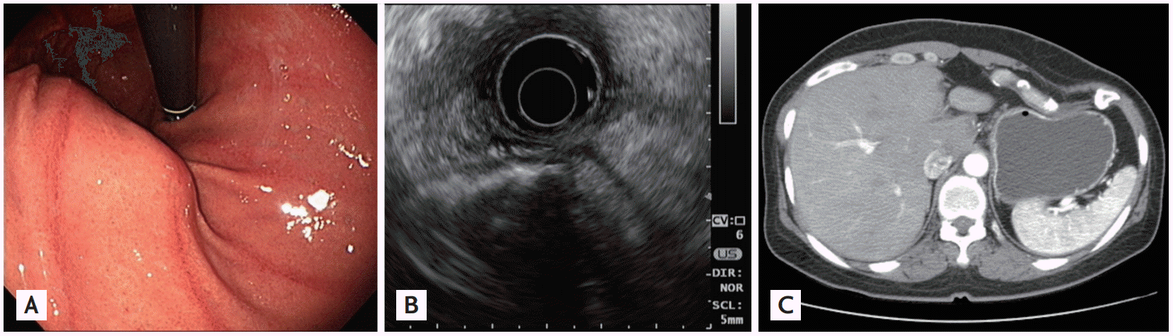
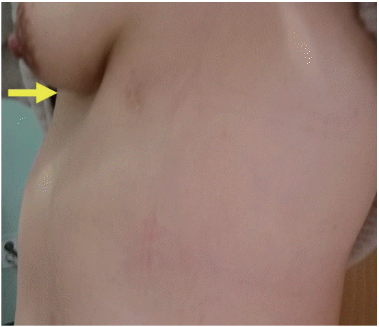
A 52-year-old woman presented with a gastric subepithelial lesion (SEL) on routine endoscopy. Upper endoscopy revealed a 5-cm SEL at the anterior wall of the upper body of the stomach (Fig. 1A). Neither a cushion sign nor a rolling sign appeared. Endoscopic ultrasonography revealed an extragastric lesion with sharp hyperechoic margins with posterior acoustic shadowing, which interrupted the detailed examination to observe inner structures (Fig. 1B). Abdominal computed tomography revealed a deformity in the left chest wall, with resultant compression of the stomach (Fig. 1C). Further inspection showed a sunken deformity in the left chest wall (Fig. 2). Final diagnosis was pectus excavatum, a congenital deformity of the anterior thoracic wall in which the sternum and rib cage grow internally.
- Conflicts of Interest
- Conflicts of Interest
No potential conflict of interest relevant to this article was reported.
Figure 1.
Image modality findings. (A) Upper endoscopy showing a 5-cm subepithelial lesion at the anterior wall of the upper body of the stomach. (B) Endoscopic ultrasonography showing an extragastric lesion with sharp hyperechoic margins with posterior acoustic shadowing. (C) Cross sectional scan of abdomen computed tomography showing a deformity in the left chest wall, with resultant compression of the stomach.