Current insights into the diagnosis and management of small bowel bleeding
-
Sang Hyun Kim1,†, Ja Seol Koo2

- Received January 7, 2026; Revised February 17, 2026; Accepted March 23, 2026;
- Abstract
-
Small bowel bleeding accounts for 5–10% of gastrointestinal (GI) hemorrhage and remains a diagnostic challenge because of its diverse etiologies and subtle clinical presentation. Advances in small bowel imaging have refined the diagnostic approach and have led to the replacement of the term obscure GI bleeding with suspected small bowel bleeding (SSBB) when findings on upper and lower endoscopy are unrevealing. The initial evaluation requires careful assessment of clinical history, comorbidities, medication use, and physical findings, which may help direct suspicion toward specific etiologies such as inflammatory bowel disease, Meckel’s diverticulum, angioectasia, or small bowel neoplasms. Small bowel capsule endoscopy (SBCE) is currently recommended as the first-line investigation in stable patients because it offers high diagnostic accuracy, facilitates subsequent deep enteroscopy, and significantly influences clinical management. Device-assisted enteroscopy, including double-balloon, single-balloon, and spiral enteroscopy, provides both diagnostic confirmation and therapeutic intervention, and its diagnostic yield is optimized when performed within 48–72 hours of bleeding. Cross-sectional imaging modalities, such as computed tomography (CT) and CT angiography, serve complementary roles, particularly in hemodynamically unstable patients or when SBCE is negative or contraindicated. Endoscopic therapy, most commonly argon plasma coagulation, remains the primary treatment for small bowel angioectasia; however, rebleeding is common, and long-term outcomes remain suboptimal. Supportive measures, including iron supplementation and blood transfusion, are essential components of care. Pharmacologic therapies, such as thalidomide or octreotide, show potential benefit; however, they are not routinely recommended. A structured, multimodal diagnostic and therapeutic strategy is therefore essential for the optimal management of SSBB.
- Graphical abstract
- Graphical abstract
- BACKGROUND AND DEFINITIONS
- BACKGROUND AND DEFINITIONS
Small bowel bleeding refers to gastrointestinal (GI) hemorrhage originating from the segment between the ampulla of Vater and the ileocecal valve [1]. Clinically, it is classified as either overt or occult [2]. Overt bleeding presents as hematochezia or melena, whereas occult bleeding is characterized by persistent iron-deficiency anemia, regardless of fecal occult blood test results [3]. Historically, this condition was termed obscure GI bleeding (OGIB), a designation used for bleeding of undetermined origin that persisted or recurred despite negative findings on initial upper endoscopy and colonoscopy [4]. Recent advances in small bowel evaluation— including small bowel capsule endoscopy (SBCE), device-assisted enteroscopy (DAE), and improved radiologic imaging—have enabled identification of the bleeding source in 40–75% of patients previously labeled as having OGIB [5–7]. In accordance with these developments, the American College of Gastroenterology (ACG) and the American Society for Gastrointestinal Endoscopy (ASGE) recommend the term ‘suspected small bowel bleeding (SSBB)’ when both upper and lower endoscopic evaluations are negative [4,7]. The term OGIB is now reserved for patients in whom no bleeding source is identified anywhere in the GI tract despite comprehensive small bowel evaluation using contemporary imaging techniques. This review therefore focuses on currently available diagnostic modalities and outlines a practical approach to the evaluation of patients with SSBB.
- DIAGNOSIS
- DIAGNOSIS
- Causes of small bowel bleeding
- Causes of small bowel bleeding
Small bowel bleeding represents an uncommon source of GI hemorrhage, accounting for 5–10% of all GI bleeding events [8,9]. The diagnostic evaluation should begin with a detailed medical history and a thorough assessment of the patient’s presenting symptoms. Because small bowel bleeding may arise from a wide spectrum of lesions, the underlying etiology often varies according to patient age, comorbidities, and medication exposure [10–12]. In patients younger than 40 years, common causes include inflammatory bowel disease and Meckel’s diverticulum. In older adults, angioectasias and non-steroidal anti-inflammatory drug (NSAID)-associated ulcers are more frequently encountered [4]. Dieulafoy lesions and small bowel neoplasms may occur across all age groups. During the evaluation of patients with SSBB, several clinical predictors may help identify the underlying cause. Patients with chronic kidney disease have an increased risk of bleeding from small bowel angioectasias, whereas ulcerative lesions are more commonly observed in individuals receiving proton pump inhibitors or low-dose aspirin [3,4]. Overt bleeding occurs more frequently in patients undergoing hemodialysis and in those with liver cirrhosis [13]. In addition, acquired von Willebrand disease is often present in patients with cardiovascular disorders, such as aortic stenosis or in individuals managed with left ventricular assist devices, thereby predisposing them to angioectasia formation and recurrent bleeding [2]. Common causes of small bowel bleeding are listed in Table 1.- Initial evaluation of small bowel bleeding
- Initial evaluation of small bowel bleeding
Patients with small bowel bleeding typically present with nonspecific symptoms of GI hemorrhage, and these clinical features rarely localize the bleeding source to the small intestine. Melena is classically associated with upper GI bleeding, whereas hematochezia is more suggestive of bleeding from the lower GI tract. However, melena may also result from slow, oozing hemorrhage originating in the ileum or cecum; conversely, a rapid, high-volume upper GI bleed may present as hematochezia [14]. A comprehensive diagnostic assessment should integrate the patient’s medical history and medication profile, with particular attention to antiplatelet agents, anticoagulants, and NSAIDs, all of which may contribute to mucosal injury [3,13]. Screening for coagulation disorders is also essential, as conditions such as von Willebrand disease increase susceptibility to recurrent bleeding. Hereditary GI disorders, including hereditary polyposis syndromes and Osler–Weber–Rendu syndrome, should be considered in the differential diagnosis [15]. Details regarding previous abdominal surgery, small bowel resection, or abdominal radiotherapy may further refine localization of the suspected bleeding source. Physical examination may provide additional diagnostic clues. Cutaneous findings, such as spider angiomas, acanthosis nigricans, or telangiectasias, may reflect underlying portal hypertension, gastric malignancy, or Osler–Weber–Rendu syndrome, respectively. Patients with Peutz–Jeghers syndrome often exhibit characteristic mucocutaneous pigmentation of the lips or oral mucosa, whereas individuals with neurofibromatosis may present with multiple cutaneous neurofibromas [3,13,15].- Second-look endoscopy
- Second-look endoscopy
Most episodes of small bowel bleeding follow a relatively indolent course and typically present as hemodynamically stable overt bleeding or occult blood loss. Previous studies have demonstrated that a clinically meaningful subset of patients initially classified as having SSBB are ultimately found to have lesions within the reach of standard upper or lower endoscopy. Reported detection rates for previously missed lesions range from 2% to 25% on repeat upper endoscopy [16–18] and from 6% to 23% on repeat colonoscopy [3,19–22]. Decisions regarding repeat endoscopic evaluation should be guided by the patient’s clinical presentation and the quality of prior procedures. The European Society of Gastrointestinal Endoscopy (ESGE) does not recommend routine second-look endoscopy before SBCE in patients with SSBB [23]. However, when the initial examination was limited by ongoing hemorrhage or when no recent high-quality evaluation is available, repeat upper endoscopy, with or without colonoscopy, may be beneficial [4]. For re-examination of the upper GI tract, push enteroscopy is often preferred because it allows visualization beyond the reach of conventional upper endoscopy. Particular attention should be directed to the duodenum and proximal jejunum, as these regions may be inadequately visualized during capsule endoscopy. During colonoscopy, every effort should be made to intubate the terminal ileum to permit direct inspection of the ileal mucosa and to facilitate identification of bleeding originating from more proximal segments of the small bowel.- SBCE
- SBCE
SBCE utilizes a miniature ingestible capsule that is swallowed by the patient or placed endoscopically into the GI tract. After ingestion, the patient wears an external array of sensors affixed to the abdominal wall that receive and store transmitted images during an acquisition period of approximately 8–12 hours [4,6]. Once data acquisition is complete, the sensors are removed, and the recorded images are interpreted by a trained physician. The approximate location of a suspected bleeding focus is inferred from the capsule’s transit time through different segments of the small intestine. For patients presenting with occult bleeding or hemodynamically stable overt bleeding, SBCE is recommended as the initial diagnostic modality, as supported by international guidelines, including those of the ESGE and ACG [4,23]. Its noninvasive nature, high diagnostic yield, and capacity to guide subsequent DAE make it the preferred first-line investigation [23]. Complete visualization of the small bowel is achieved in approximately 79–90% of examinations, and the diagnostic yield in patients with SSBB ranges from 38% to 83% [24]. SBCE demonstrates high diagnostic accuracy, with positive predictive values ranging from 94% to 97% and negative predictive values ranging from 83% to 100% [5,24]. Importantly, SBCE findings frequently alter clinical management, leading to therapeutic endoscopic or surgical interventions or modifications of medical therapy in 37% to 87% of patients [25,26]. In addition, when management decisions are guided by capsule findings, approximately one-half to two-thirds of patients avoid further transfusions and do not experience recurrent bleeding during follow-up [5,27].Recent studies have shown that overt bleeding is the strongest predictor of a positive SBCE result [28–30]. Accordingly, the ESGE recommends SBCE in patients with overt SSBB as soon as possible after the bleeding episode, ideally within 48 hours, to maximize diagnostic and subsequent therapeutic yield [23]. Additional independent factors associated with higher detection rates include advanced age, male sex, and performance of the procedure during hospitalization [30–32]. Although SBCE offers substantial diagnostic advantages, several limitations remain. It does not permit therapeutic intervention, lacks directional control during transit, and presents challenges in precise lesion localization [7]. Specificity is also imperfect, as incidental mucosal abnormalities are observed in up to 14% of healthy individuals [33], and reported false-negative rates range from 10% to 36% [34,35]. SBCE has few absolute contraindications; however, capsule retention remains the primary safety concern. Retention rates are highest in patients with established Crohn’s disease, occurring in approximately 3% to 13% of examinations [36,37]. Other risk factors include a history of small bowel obstruction, multiple abdominal operations with adhesions, and enteritis related to prior radiation exposure [4]. In patients without such predisposing conditions, the risk of capsule retention is low, estimated at approximately 1% to 2% [38].- DAE
- DAE
DAE comprises several techniques, including double-balloon enteroscopy (DBE), single-balloon enteroscopy (SBE), and spiral enteroscopy [2,3,13]. These modalities play a complementary role to SBCE in the diagnostic evaluation of small bowel bleeding. When SBCE identifies a clinically significant lesion within the small intestine, DAE is typically performed to confirm the finding and to provide endoscopic therapy when indicated [23,39]. Overall, the diagnostic and therapeutic capabilities of SBE and spiral enteroscopy are considered comparable to those of DBE [40–42]. Selection among DAE techniques is generally based on local expertise and equipment availability. In patients with SSBB or other small bowel disorders, DBE has demonstrated a diagnostic yield ranging from approximately 60% to 80% [43–45]. Limited data also suggest that DBE may be particularly advantageous when complete enteroscopy is anticipated—for example, in cases in which SBCE identifies lesions in the mid-jejunum or multiple abnormalities distributed throughout the small bowel [4].Most comparative studies of SBCE and DAE have focused on DBE. Because SBCE and DBE demonstrate comparable diagnostic yields, SBCE is generally performed first as a noninvasive triage modality to identify patients who require further evaluation with DAE [23]. The choice of insertion route for DBE is guided by the suspected lesion location, as estimated by SBCE findings or cross-sectional imaging. Capsule transit indices provide a practical method for lesion localization: an antegrade approach is typically selected when the suspected lesion lies within the first 60% of the pylorus-to-cecum transit time or within the first 75% of the ingestion-to-cecum interval [1]. Lesions located beyond these thresholds are generally approached retrograde [46,47]. In general, antegrade DBE demonstrates higher diagnostic and therapeutic success rates than retrograde DBE, a pattern likely attributable to both the higher prevalence of vascular lesions in the proximal small bowel and the greater insertion depth achievable via the antegrade route. As with SBCE, DAE should ideally be performed promptly after a bleeding episode to increase the likelihood of identifying the culprit lesion. The overall complication rate of DBE is approximately 1.2%, increasing to approximately 4.3% when therapeutic interventions are performed [48]. Common adverse events include bleeding and perforation, whereas pancreatitis occurs less frequently. Patients with surgically altered GI anatomy who undergo retrograde DBE appear to have an increased risk of perforation [49]. SBE has a safety profile comparable to that of DBE [40]. Although pancreatitis has not been documented with spiral enteroscopy, perforation remains a potential complication [1].- Radiographic studies of small bowel bleeding
- Radiographic studies of small bowel bleeding
Cross-sectional imaging, such as computed tomography (CT), provides important complementary information in the evaluation of small bowel bleeding, particularly when SBCE is nondiagnostic, cannot be performed, or when bleeding is brisk and accompanied by hemodynamic instability. It is also indicated in patients with clinical suspicion of Crohn’s disease or small bowel neoplasia. Commonly used modalities include CT enterography (CTE), CT angiography (CTA), and magnetic resonance enterography (MRE). In acute settings, CT-based techniques are generally preferred because of the more limited availability of MRE and the longer image acquisition time required [1]. Current guidelines from both the ACG and ASGE identify multiphase CT as the radiologic study of choice for the evaluation of SSBB [4,7]. CTA and CTE both use intravenous contrast and acquire arterial, enteric, and delayed-phase images. In patients with active, unstable hemorrhage, multiphase CTA is performed without oral contrast. CTA is particularly effective in localizing active small bowel bleeding, with diagnostic yield enhanced in individuals who are hemodynamically unstable or who require substantial transfusion support, commonly defined as five or more units of packed red blood cells [7]. When CTA demonstrates active contrast extravasation, these findings can guide targeted embolization during subsequent mesenteric angiography. For patients with chronic or intermittent bleeding, multiphase CTE with neutral oral contrast is typically preferred. CTE identifies bleeding-related small bowel pathology in approximately 45% to 52% of cases and is particularly useful for detecting vascular abnormalities and mass-forming lesions [50,51]. In general, multiphase CTE provides greater sensitivity for small bowel tumors, whereas capsule endoscopy is more effective in identifying flat mucosal lesions, such as angioectasias [3]. Because these modalities have distinct strengths and limitations, multiphase CTE serves as a complementary tool to SBCE by helping identify lesions that may not be visualized on capsule imaging.For patients in whom multiphase CT imaging is relatively contraindicated—most notably those with advanced chronic kidney disease—technetium-99m–labeled red blood cell scintigraphy may serve as an alternative diagnostic modality. Its usefulness is limited, however, by a false localization rate that may approach 30% [52,53]. Diagnostic performance improves when radiotracer accumulation is detected within the first two to three hours of the study. When scintigraphy yields a positive result and the patient has no absolute contraindication to iodinated contrast, mesenteric angiography should be performed promptly to enable timely therapeutic intervention. Angiography may also assist in the evaluation of overt small bowel bleeding, particularly when the bleeding rate exceeds approximately 0.5 mL per minute [4]. Although it is less sensitive than radionuclide scintigraphy, angiography provides more precise localization of the bleeding source and allows immediate therapeutic intervention when active extravasation is visualized. Evidence regarding its diagnostic performance in small bowel bleeding remains limited, with reported success rates in GI bleeding ranging from 20% to 77% [54,55]. Angiography also offers the opportunity for endovascular therapy, as selective mesenteric embolization can control hemorrhage while reducing the risk of bowel ischemia.- Diagnostic algorithm and management of small bowel bleeding
- Diagnostic algorithm and management of small bowel bleeding
SBCE is supported by prior guidelines and current evidence as the preferred initial test for patients with SSBB, consistent with recommendations from multiple professional societies [2,3,7,23]. Although early evaluation of the small bowel is recognized as beneficial, particularly in cases of overt bleeding, the optimal timing of SBCE remains debated. A recent meta-analysis reported that performing SBCE within 48 hours was associated with diagnostic and therapeutic success rates of 55.9% and 65.2%, respectively [56]. On the basis of these data, the ESGE recommends that SBCE be performed as early as possible after an episode of overt bleeding, ideally within the first two days, to maximize clinical benefit [23]. When SBCE is unavailable or yields negative results, current evidence does not support a single definitive first-line alternative; therefore, the diagnostic strategy should be individualized according to the patient’s clinical presentation and institutional resources. The ACG advises the use of CTE in patients with SSBB who have negative SBCE findings, emphasizing its superior ability to detect mural-based masses and its utility in guiding subsequent deep enteroscopy [4]. A meta-analysis of 18 studies involving 660 patients reported that CTE has an overall diagnostic yield of 40% for SSBB [57]. In a subgroup analysis of seven comparative studies including 279 patients, CTE and SBCE demonstrated diagnostic yields of 34% and 53%, respectively, corresponding to an incremental yield of −19% [57]. CTE is therefore considered particularly valuable for precise localization of bleeding sources and for guiding enteroscopic intervention, especially in cases of tumor-related or overt bleeding.In emergency clinical settings, DAE has demonstrated effectiveness, as reported in a recent systematic review and meta-analysis of retrospective studies in which DAE was used as the initial diagnostic modality in selected patients [58]. The ESGE states that DAE may be considered an alternative first-line option in appropriately selected cases, as it allows both diagnosis and treatment during a single procedure when the necessary expertise and equipment are available [23]. Despite these advantages, the broader implementation of DAE is limited by practical constraints, including the need for specialized instruments and trained personnel, which restrict its availability in many institutions. For patients presenting with overt SSBB, a stepwise approach beginning with a diagnostic modality, such as SBCE or CTA, followed by a therapeutic intervention, such as DAE, is generally preferred [4,23]. To increase the likelihood of successfully identifying and treating the bleeding source, the ESGE recommends performing DAE within 48–72 hours after the bleeding event [23]. Figure 1 presents the management algorithm for patients with SSBB, as proposed by the authors.
- TREATMENT OF SMALL BOWEL BLEEDING
- TREATMENT OF SMALL BOWEL BLEEDING
- Endoscopic therapy of small bowel bleeding
- Endoscopic therapy of small bowel bleeding
Argon plasma coagulation (APC) is recommended by international guidelines—including those of the ACG, ASGE, and ESGE—as the first-line endoscopic therapy for small bowel angioectasia [4,7,23]. However, this recommendation is largely based on observational cohort studies rather than randomized controlled trials. Overall outcomes of endoscopic management for small bowel bleeding sources have been suboptimal, and robust data regarding the long-term outcomes of treating small bowel angioectasias remain limited. To date, no randomized trials have compared endoscopic intervention for angioectasia with sham treatment, nor have studies evaluated selective treatment of actively bleeding or large lesions versus therapy directed at all visible lesions. Because of these limitations, recurrent bleeding has frequently been used as an indirect measure of treatment efficacy. Several studies have assessed rebleeding after endoscopic therapy for small bowel vascular lesions. One of the most recent was a retrospective cohort study conducted at a French tertiary referral center between January 2004 and December 2007 [59]. Among 261 patients evaluated for SSBB, 129 of 133 patients with vascular lesions (97%) underwent successful APC using DBE. At 36 months of follow-up, recurrent bleeding occurred in 45 of 98 patients, corresponding to a rebleeding rate of 46% [59]. A more recent meta-analysis including 14 studies and 623 patients with small bowel angioectasia treated endoscopically reported a pooled rebleeding rate of 34% (95% confidence interval, 27–42%) after a mean follow-up of 22 ± 13 months [60]. When a subset of 341 patients with small bowel angioectasia was analyzed separately, the rebleeding rate increased to 45% [60]. Risk factors associated with recurrent bleeding from small bowel angioectasia include a greater number of vascular lesions [61], age older than 65 years [62,63], lesion localization in the jejunum [62], the presence of cardiac valvular disease [59], chronic kidney disease [63], use of anticoagulant therapy, and the need for transfusion support.- Angiographic treatment of small bowel bleeding
- Angiographic treatment of small bowel bleeding
Superselective transarterial embolization has emerged as an effective therapy for acute GI bleeding, offering improved clinical outcomes with a lower complication rate [7]. By limiting embolization to the bleeding vessel, this approach reduces the risk of intestinal ischemia, which remains a major complication of transarterial embolization. A recent retrospective study reported a 99% technical success rate, with primary and secondary clinical success rates of 71% and 79%, respectively, following repeat embolization [64]. Bowel infarction occurred in 4% of cases, predominantly in patients with non–small bowel bleeding. Factors associated with failure to achieve sustained hemostasis at 30 days included a hemoglobin level below 8 g/dL, the presence of coagulopathy, upper GI bleeding, contrast extravasation on imaging, and embolization of multiple vessels [64].- Medical treatment of small bowel bleeding
- Medical treatment of small bowel bleeding
Supportive care with iron supplementation, administered either orally or intravenously, remains a fundamental component of therapy for mild small bowel bleeding [2,3]. Iron replacement not only helps maintain adequate hemoglobin levels but also reduces transfusion requirements in more severe cases. When bleeding is significant, transfusion of packed red blood cells is an essential component of management, particularly when mechanical or medical interventions fail to adequately control hemorrhage. Although anticoagulant therapy has been associated with a higher risk of recurrent bleeding [15], no prospective data demonstrate that discontinuation of anticoagulation confers measurable benefit. In a 2009 study evaluating 162 patients with small bowel bleeding, predictors of recurrent bleeding after DBE included the presence of small bowel vascular lesions and other comorbidities, whereas use of anticoagulant or antiplatelet therapy was not associated with recurrence [65]. A subsequent study published in 2010 similarly found that transfusion requirements, as well as the number and type of vascular lesions, predicted rebleeding, whereas anticoagulant use did not [66]. No evidence currently supports cessation of antiplatelet therapy as a strategy to reduce the risk of recurrent small bowel bleeding. Specific medical therapies for small bowel bleeding remain inadequately defined. Hormonal therapy has not demonstrated meaningful clinical benefit. Although thalidomide and octreotide have shown therapeutic potential [67–69], the current body of evidence does not support their routine use in clinical practice [4]. The ACG guideline recommends reserving medical therapy for patients with persistent or recurrent bleeding after appropriate endoscopic evaluation and treatment. Concerns regarding adverse events—such as peripheral neuropathy and thromboembolic complications—cost, and the limited availability of high-quality randomized controlled trials have precluded routine recommendation of these agents [4].- Surgical treatment of small bowel bleeding
- Surgical treatment of small bowel bleeding
Surgical management of small bowel bleeding is generally reserved as a last-line option. However, in cases of recurrent or refractory small bowel bleeding, surgical intervention guided by intraoperative enteroscopy (IOE) may be considered [4]. According to published data, localization of the bleeding source during IOE is achieved in 58–99% of cases, with a therapeutic yield ranging from 48% to 94% [1,70]. IOE is associated with procedure-related complications, such as serosal tears and postoperative ileus, as reported in retrospective surgical series; however, prospective comparative data remain limited [71,72]. In addition, rebleeding after IOE-assisted surgery occurs in approximately 13% to 52% of patients [73–75]. Surgical treatment demonstrates favorable outcomes in patients with well-defined lesions, such as tumors or localized arteriovenous malformations. In contrast, diffuse lesions—including multiple angioectasias—are typically managed with intraoperative endoscopic therapy. Because these endoscopic interventions are comparable to those used during deep or push enteroscopy, similar rebleeding rates may be expected; however, robust long-term follow-up data are currently lacking.
- CONCLUSION
- CONCLUSION
Although small bowel bleeding accounts for a minority of GI bleeding cases, it continues to pose significant diagnostic and therapeutic challenges. Careful selection and appropriate timing of diagnostic tests—guided by the clinical presentation and suspected underlying pathology—facilitate a more cost-effective and efficient management strategy. The advent of advanced small bowel diagnostic and therapeutic modalities has substantially improved detection and treatment of underlying lesions, allowing most patients to be managed without surgical intervention. For internists, several key considerations warrant emphasis. SSBB should be considered only after high-quality upper and lower endoscopy have excluded more common sources. In hemodynamically stable patients, SBCE is the preferred initial test and should be performed early in cases of overt bleeding to maximize diagnostic yield. DAE permits both diagnosis and therapy and is optimally guided by prior imaging. Nevertheless, treatment options for refractory small bowel bleeding remain limited, underscoring the need for the development of more effective therapies.
- Notes
- Notes
-
CRedit authorship contributions Sang Hyun Kim: resources, data curation, writing - original draft, visualization; Ja Seol Koo: conceptualization, methodology, writing - review & editing, supervision
- Conflicts of Interest
- Conflicts of Interest
-
Conflicts of interest The authors disclose no conflicts.
- Notes
- Notes
-
Funding This research was supported by a grant from Korea University, Seoul, Republic of Korea (grant no. K2503361).
Figure 1
Management algorithm for patients with SSBB. This figure presents the proposed stepwise diagnostic and therapeutic approach for patients with SSBB, incorporating SBCE, cross-sectional imaging, and DAE according to clinical presentation and hemodynamic status. SSBB, suspected small bowel bleeding; DAE, device-assisted enteroscopy; SBCE, small bowel capsule endoscopy; CTE, computed tomography enterography; CTA, computed tomography angiography.
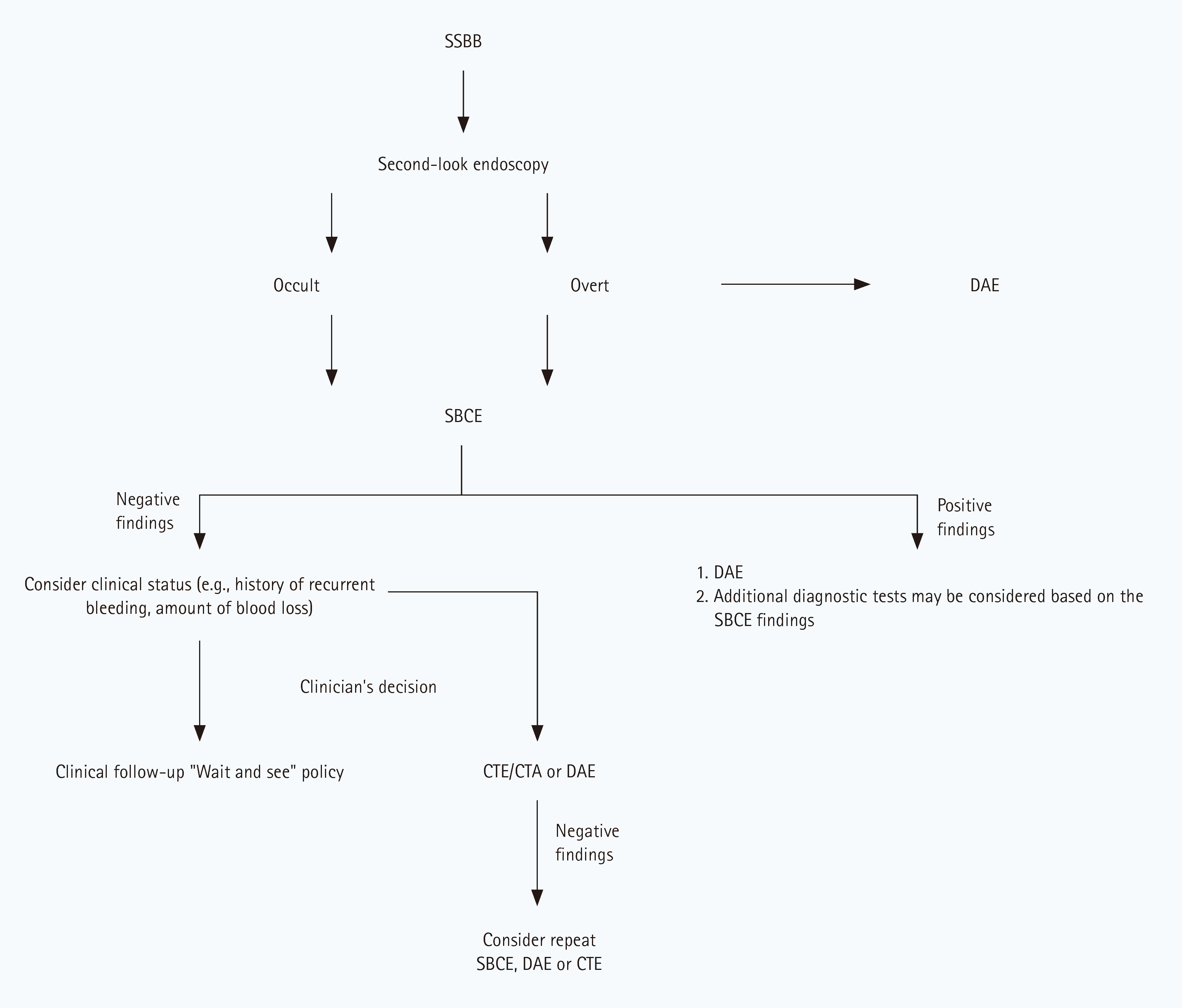
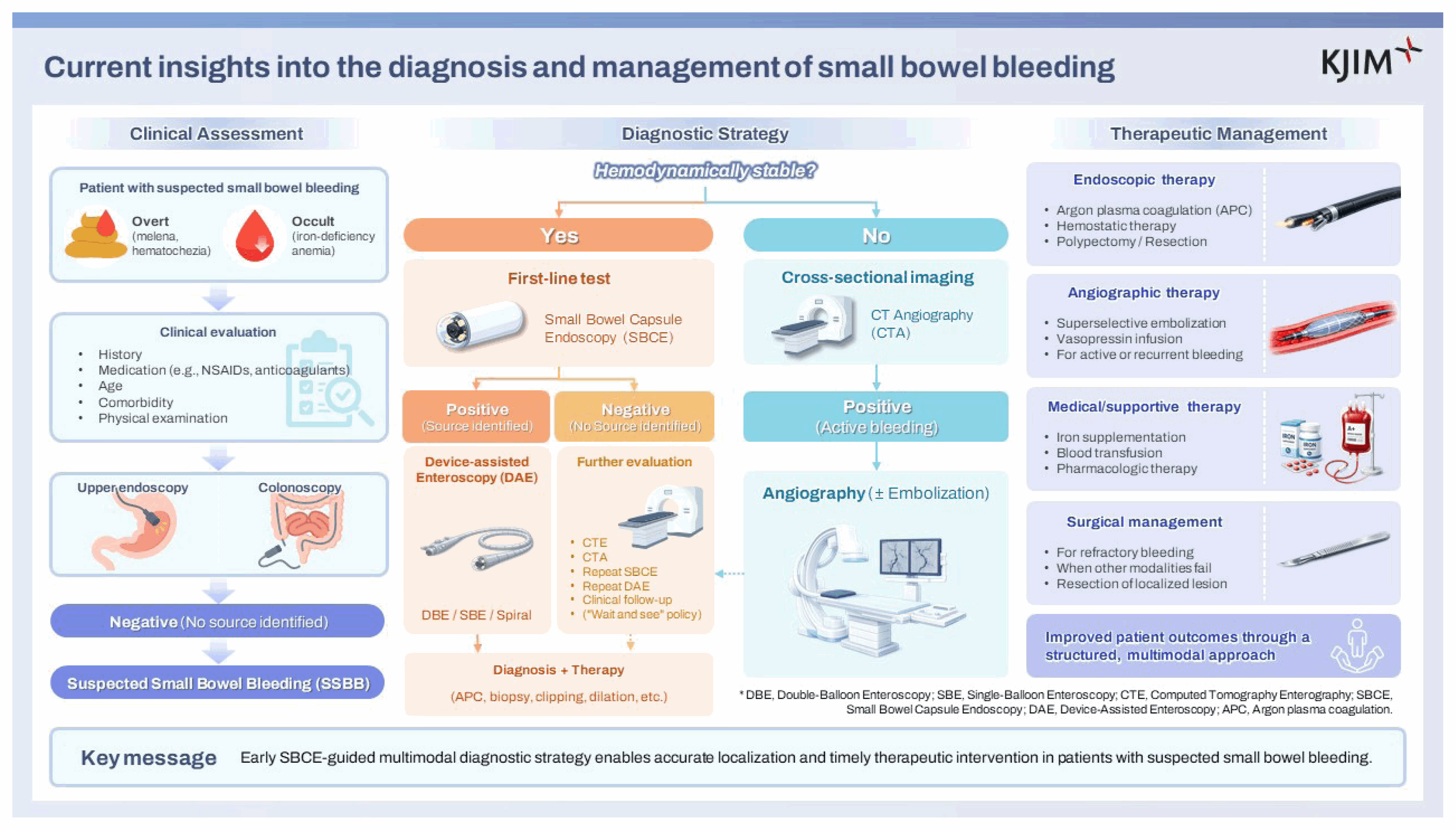
Table 1
Causes of small bowel bleeding
- References
- References
REFERENCES
1. Kuo JR, Pasha SF, Leighton JA. The clinician’s guide to suspected small bowel bleeding. Am J Gastroenterol 2019;114:591–598.
[Article] [PubMed]2. Zuckerman GR, Prakash C, Askin MP, Lewis BS. AGA technical review on the evaluation and management of occult and obscure gastrointestinal bleeding. Gastroenterology 2000;118:201–221.
[Article] [PubMed]3. American Gastroenterological Association medical position statement: evaluation and management of occult and obscure gastrointestinal bleeding. Gastroenterology 2000;118:197–201.
[Article] [PubMed] [PMC]4. Gerson LB, Fidler JL, Cave DR, Leighton JA. ACG clinical guideline: diagnosis and management of small bowel bleeding. Am J Gastroenterol 2015;110:1265–1287quiz 1288.
[Article] [PubMed] [PMC]5. Delvaux M, Fassler I, Gay G. Clinical usefulness of the endoscopic video capsule as the initial intestinal investigation in patients with obscure digestive bleeding: validation of a diagnostic strategy based on the patient outcome after 12 months. Endoscopy 2004;36:1067–1073.
[Article] [PubMed]6. Mata A, Bordas JM, Feu F, et al. Wireless capsule endoscopy in patients with obscure gastrointestinal bleeding: a comparative study with push enteroscopy. Aliment Pharmacol Ther 2004;20:189–194.
[Article] [PubMed]7. ASGE Standards of Practice Committee. Gurudu SR, Bruining DH, et al. The role of endoscopy in the management of suspected small-bowel bleeding. Gastrointest Endosc 2017;85:22–31.
[Article] [PubMed] [PMC]8. Lau WY, Fan ST, Wong SH, et al. Preoperative and intraoperative localisation of gastrointestinal bleeding of obscure origin. Gut 1987;28:869–877.
[Article] [PubMed] [PMC]9. Longstreth GF. Epidemiology and outcome of patients hospitalized with acute lower gastrointestinal hemorrhage: a population-based study. Am J Gastroenterol 1997;92:419–424.
[PubMed]10. Mitsui K, Tanaka S, Yamamoto H, et al. Role of double-balloon endoscopy in the diagnosis of small-bowel tumors: the first Japanese multicenter study. Gastrointest Endosc 2009;70:498–504.
[Article] [PubMed]11. Norton ID, Petersen BT, Sorbi D, Balm RK, Alexander GL, Gostout CJ. Management and long-term prognosis of Dieulafoy lesion. Gastrointest Endosc 1999;50:762–767.
[Article] [PubMed]12. Sone Y, Kumada T, Toyoda H, Hisanaga Y, Kiriyama S, Tanikawa M. Endoscopic management and follow up of Dieulafoy lesion in the upper gastrointestinal tract. Endoscopy 2005;37:449–453.
[Article] [PubMed]13. Ohmiya N. Management of obscure gastrointestinal bleeding: comparison of guidelines between Japan and other countries. Dig Endosc 2020;32:204–218.
[Article] [PubMed]14. Concha R, Amaro R, Barkin JS. Obscure gastrointestinal bleeding: diagnostic and therapeutic approach. J Clin Gastroenterol 2007;41:242–251.
[PubMed]15. Thakral D, Stein DJ, Saltzman JR. Diagnosis of occult and obscure gastrointestinal bleeding. Gastrointest Endosc Clin N Am 2024;34:317–329.
[Article] [PubMed]16. Descamps C, Schmit A, Van Gossum A. “Missed” upper gastrointestinal tract lesions may explain “occult” bleeding”. Endoscopy 1999;31:452–455.
[Article] [PubMed]17. Tang SJ, Christodoulou D, Zanati S, et al. Wireless capsule endoscopy for obscure gastrointestinal bleeding: a single-centre, one-year experience. Can J Gastroenterol 2004;18:559–565.
[PubMed]18. Zaman A, Katon RM. Push enteroscopy for obscure gastrointestinal bleeding yields a high incidence of proximal lesions within reach of a standard endoscope. Gastrointest Endosc 1998;47:372–376.
[Article] [PubMed]19. Fry LC, Bellutti M, Neumann H, Malfertheiner P, Mönkemüller K. Incidence of bleeding lesions within reach of conventional upper and lower endoscopes in patients undergoing double-balloon enteroscopy for obscure gastrointestinal bleeding. Aliment Pharmacol Ther 2009;29:342–349.
[Article] [PubMed]20. Lorenceau-Savale C, Ben-Soussan E, Ramirez S, Antonietti M, Lerebours E, Ducrotté P. Outcome of patients with obscure gastrointestinal bleeding after negative capsule endoscopy: results of a one-year follow-up study. Gastroenterol Clin Biol 2010;34:606–611.
[Article] [PubMed]21. Tee HP, Kaffes AJ. Non-small-bowel lesions encountered during double-balloon enteroscopy performed for obscure gastrointestinal bleeding. World J Gastroenterol 2010;16:1885–1889.
[Article] [PubMed] [PMC]22. van Turenhout ST, Jacobs MA, van Weyenberg SJ, et al. Diagnostic yield of capsule endoscopy in a tertiary hospital in patients with obscure gastrointestinal bleeding. J Gastrointestin Liver Dis 2010;19:141–145.
[PubMed]23. Pennazio M, Rondonotti E, Despott EJ, et al. Small-bowel capsule endoscopy and device-assisted enteroscopy for diagnosis and treatment of small-bowel disorders: European Society of Gastrointestinal Endoscopy (ESGE) guideline-update 2022. Endoscopy 2023;55:58–95.
[Article] [PubMed] [PMC]24. Rondonotti E, Villa F, Mulder CJ, Jacobs MA, de Franchis R. Small bowel capsule endoscopy in 2007: indications, risks and limitations. World J Gastroenterol 2007;13:6140–6149.
[Article] [PubMed] [PMC]25. Ben Soussan E, Antonietti M, Hervé S, et al. Diagnostic yield and therapeutic implications of capsule endoscopy in obscure gastrointestinal bleeding. Gastroenterol Clin Biol 2004;28:1068–1073.
[Article] [PubMed]26. Pennazio M, Santucci R, Rondonotti E, et al. Outcome of patients with obscure gastrointestinal bleeding after capsule endoscopy: report of 100 consecutive cases. Gastroenterology 2004;126:643–653.
[Article] [PubMed]27. Estévez E, González-Conde B, Vázquez-Iglesias JL, et al. Diagnostic yield and clinical outcomes after capsule endoscopy in 100 consecutive patients with obscure gastrointestinal bleeding. Eur J Gastroenterol Hepatol 2006;18:881–888.
[Article] [PubMed]28. Bresci G, Parisi G, Bertoni M, Tumino E, Capria A. The role of video capsule endoscopy for evaluating obscure gastrointestinal bleeding: usefulness of early use. J Gastroenterol 2005;40:256–259.
[Article] [PubMed]29. Singh A, Marshall C, Chaudhuri B, Okoli C, Foley A, Person SD, et al. Timing of video capsule endoscopy relative to overt obscure GI bleeding: implications from a retrospective study. Gastrointest Endosc 2013;77:761–766.
[Article] [PubMed]30. Yamada A, Watabe H, Kobayashi Y, Yamaji Y, Yoshida H, Koike K. Timing of capsule endoscopy influences the diagnosis and outcome in obscure-overt gastrointestinal bleeding. Hepatogastroenterology 2012;59:676–679.
[PubMed]31. Carey EJ, Leighton JA, Heigh RI, et al. A single-center experience of 260 consecutive patients undergoing capsule endoscopy for obscure gastrointestinal bleeding. Am J Gastroenterol 2007;102:89–95.
[Article] [PubMed]32. Lepileur L, Dray X, Antonietti M, et al. Factors associated with diagnosis of obscure gastrointestinal bleeding by video capsule enteroscopy. Clin Gastroenterol Hepatol 2012;10:1376–1380.
[Article] [PubMed]33. Goldstein JL, Eisen GM, Lewis B, et al. Video capsule endoscopy to prospectively assess small bowel injury with celecoxib, naproxen plus omeprazole, and placebo. Clin Gastroenterol Hepatol 2005;3:133–141.
[Article] [PubMed]34. Appleyard M, Fireman Z, Glukhovsky A, et al. A randomized trial comparing wireless capsule endoscopy with push enteroscopy for the detection of small-bowel lesions. Gastroenterology 2000;119:1431–1438.
[Article] [PubMed]35. Lewis BS, Eisen GM, Friedman S. A pooled analysis to evaluate results of capsule endoscopy trials. Endoscopy 2005;37:960–965.
[Article] [PubMed]36. Cheifetz AS, Kornbluth AA, Legnani P, et al. The risk of retention of the capsule endoscope in patients with known or suspected Crohn’s disease. Am J Gastroenterol 2006;101:2218–2222.
[Article] [PubMed]37. Fry LC, De Petris G, Swain JM, Fleischer DE. Impaction and fracture of a video capsule in the small bowel requiring laparotomy for removal of the capsule fragments. Endoscopy 2005;37:674–676.
[Article] [PubMed]38. Pennazio M. Capsule endoscopy: where are we after 6 years of clinical use? Dig Liver Dis 2006;38:867–878.
[Article] [PubMed]39. Lee HH, Kim JS, Goong HJ, et al. Use of device-assisted enteroscopy in small bowel disease: an expert consensus statement by the Korean Association for the Study of Intestinal Diseases. Intest Res 2023;21:3–19.
[Article] [PubMed] [PMC]40. Domagk D, Mensink P, Aktas H, et al. Single- vs. double-balloon enteroscopy in small-bowel diagnostics: a randomized multicenter trial. Endoscopy 2011;43:472–476.
[Article] [PubMed]41. Rahmi G, Samaha E, Vahedi K, et al. Multicenter comparison of double-balloon enteroscopy and spiral enteroscopy. J Gastroenterol Hepatol 2013;28:992–998.
[Article] [PubMed]42. Takano N, Yamada A, Watabe H, et al. Single-balloon versus double-balloon endoscopy for achieving total enteroscopy: a randomized, controlled trial. Gastrointest Endosc 2011;73:734–739.
[Article] [PubMed]43. Gross SA, Stark ME. Initial experience with double-balloon enteroscopy at a U.S. center. Gastrointest Endosc 2008;67:890–897.
[Article] [PubMed]44. Yamamoto H, Kita H, Sunada K, et al. Clinical outcomes of double-balloon endoscopy for the diagnosis and treatment of small-intestinal diseases. Clin Gastroenterol Hepatol 2004;2:1010–1016.
[Article] [PubMed]45. Zhang Q, He Q, Liu J, Ma F, Zhi F, Bai Y. Combined use of capsule endoscopy and double-balloon enteroscopy in the diagnosis of obscure gastrointestinal bleeding: meta-analysis and pooled analysis. Hepatogastroenterology 2013;60:1885–1891.
[PubMed]46. Gay G, Delvaux M, Fassler I. Outcome of capsule endoscopy in determining indication and route for push-and-pull enteroscopy. Endoscopy 2006;38:49–58.
[Article] [PubMed]47. Li X, Chen H, Dai J, Gao Y, Ge Z. Predictive role of capsule endoscopy on the insertion route of double-balloon enteroscopy. Endoscopy 2009;41:762–766.
[Article] [PubMed]48. Mensink PB, Haringsma J, Kucharzik T, et al. Complications of double balloon enteroscopy: a multicenter survey. Endoscopy 2007;39:613–615.
[Article] [PubMed]49. Gerson LB, Tokar J, Chiorean M, et al. Complications associated with double balloon enteroscopy at nine US centers. Clin Gastroenterol Hepatol 2009;7:1177–11821182.e1–3.
[Article] [PubMed]50. Hara AK, Walker FB, Silva AC, Leighton JA. Preliminary estimate of triphasic CT enterography performance in hemodynamically stable patients with suspected gastrointestinal bleeding. AJR Am J Roentgenol 2009;193:1252–1260.
[Article] [PubMed]51. Huprich JE, Fletcher JG, Alexander JA, Fidler JL, Burton SS, McCullough CH. Obscure gastrointestinal bleeding: evaluation with 64-section multiphase CT enterography--initial experience. Radiology 2008;246:562–571.
[Article] [PubMed]52. Dolezal J, Vizd’a J, Bures J. Detection of acute gastrointestinal bleeding by means of technetium-99m in vivo labelled red blood cells. Nucl Med Rev Cent East Eur 2002;5:151–154.
[PubMed]53. Dusold R, Burke K, Carpentier W, Dyck WP. The accuracy of technetium-99m-labeled red cell scintigraphy in localizing gastrointestinal bleeding. Am J Gastroenterol 1994;89:345–348.
[PubMed]54. Abbas SM, Bissett IP, Holden A, Woodfield JC, Parry BR, Duncan D. Clinical variables associated with positive angiographic localization of lower gastrointestinal bleeding. ANZ J Surg 2005;75:953–957.
[Article] [PubMed]55. Rollins ES, Picus D, Hicks ME, Darcy MD, Bower BL, Kleinhoffer MA. Angiography is useful in detecting the source of chronic gastrointestinal bleeding of obscure origin. AJR Am J Roentgenol 1991;156:385–388.
[Article] [PubMed]56. Uchida G, Nakamura M, Yamamura T, et al. Systematic review and meta-analysis of the diagnostic and therapeutic yield of small bowel endoscopy in patients with overt small bowel bleeding. Dig Endosc 2021;33:66–82.
[Article] [PubMed]57. Wang Z, Chen JQ, Liu JL, Qin XG, Huang Y. CT enterography in obscure gastrointestinal bleeding: a systematic review and meta-analysis. J Med Imaging Radiat Oncol 2013;57:263–273.
[PubMed]58. Estevinho MM, Pinho R, Fernandes C, et al. Diagnostic and therapeutic yields of early capsule endoscopy and device-assisted enteroscopy in the setting of overt GI bleeding: a systematic review with meta-analysis. Gastrointest Endosc 2022;95:610–625e9.
[Article] [PubMed]59. Samaha E, Rahmi G, Landi B, et al. Long-term outcome of patients treated with double balloon enteroscopy for small bowel vascular lesions. Am J Gastroenterol 2012;107:240–246.
[Article] [PubMed]60. Jackson CS, Gerson LB. Management of gastrointestinal angiodysplastic lesions (GIADs): a systematic review and meta-analysis. Am J Gastroenterol 2014;109:474–483; quiz 484.
[Article] [PubMed]61. Rutgeerts P, Van Gompel F, Geboes K, Vantrappen G, Broeckaert L, Coremans G. Long term results of treatment of vascular malformations of the gastrointestinal tract by neodymium Yag laser photocoagulation. Gut 1985;26:586–593.
[Article] [PubMed] [PMC]62. Fan GW, Chen TH, Lin WP, et al. Angiodysplasia and bleeding in the small intestine treated by balloon-assisted enteroscopy. J Dig Dis 2013;14:113–116.
[Article] [PubMed]63. Lepère C, Cuillerier E, Van Gossum A, et al. Predictive factors of positive findings in patients explored by push enteroscopy for unexplained GI bleeding. Gastrointest Endosc 2005;61:709–714.
[Article] [PubMed]64. Hongsakul K, Pakdeejit S, Tanutit P. Outcome and predictive factors of successful transarterial embolization for the treatment of acute gastrointestinal hemorrhage. Acta Radiol 2014;55:186–194.
[Article] [PubMed]65. Arakawa D, Ohmiya N, Nakamura M, et al. Outcome after enteroscopy for patients with obscure GI bleeding: diagnostic comparison between double-balloon endoscopy and videocapsule endoscopy. Gastrointest Endosc 2009;69:866–874.
[Article] [PubMed]66. Shinozaki S, Yamamoto H, Yano T, et al. Long-term outcome of patients with obscure gastrointestinal bleeding investigated by double-balloon endoscopy. Clin Gastroenterol Hepatol 2010;8:151–158.
[Article] [PubMed]67. Ge ZZ, Chen HM, Gao YJ, et al. Efficacy of thalidomide for refractory gastrointestinal bleeding from vascular malformation. Gastroenterology 2011;141:1629–1637e1–4.
[Article] [PubMed]68. Nardone G, Compare D, Scarpignato C, Rocco A. Long acting release-octreotide as “rescue” therapy to control angiodysplasia bleeding: a retrospective study of 98 cases. Dig Liver Dis 2014;46:688–694.
[Article] [PubMed]69. Stephens TD, Bunde CJ, Fillmore BJ. Mechanism of action in thalidomide teratogenesis. Biochem Pharmacol 2000;59:1489–1499.
[Article] [PubMed]70. Bonnet S, Douard R, Malamut G, Cellier C, Wind P. Intraoperative enteroscopy in the management of obscure gastrointestinal bleeding. Dig Liver Dis 2013;45:277–284.
[Article] [PubMed]71. Douard R, Wind P, Panis Y, et al. Intraoperative enteroscopy for diagnosis and management of unexplained gastrointestinal bleeding. Am J Surg 2000;180:181–184.
[Article] [PubMed]72. Zaman A, Sheppard B, Katon RM. Total peroral intraoperative enteroscopy for obscure GI bleeding using a dedicated push enteroscope: diagnostic yield and patient outcome. Gastrointest Endosc 1999;50:506–510.
[Article] [PubMed]73. Voron T, Rahmi G, Bonnet S, et al. Intraoperative enteroscopy: is there still a role? Gastrointest Endosc Clin N Am 2017;27:153–170.
[PubMed]