Heart failure with preserved ejection fraction in women: a sex-specific clinical review
-
Soo-Jin Kim1, Mi-Seung Shin2

- Received February 18, 2026; Revised April 26, 2026; Accepted May 28, 2026;
- Abstract
-
Heart failure with preserved ejection fraction (HFpEF) affects approximately half of the current heart failure population and demonstrates marked sex-related differences in pathophysiology, clinical presentation, and therapeutic response. These distinctions reflect the divergent biological pathways that shape disease expression in women and men. In women, disease expression is frequently linked to cardiometabolic stress, vascular dysfunction, and heightened ventricular–arterial coupling, often manifesting as concentric remodeling and a pronounced symptom burden. By contrast, men more commonly exhibit an ischemia-associated phenotype characterized by adverse remodeling and diffuse myocardial fibrosis. These biological differences influence the clinical presentation, biomarker interpretation, imaging findings, and long-term outcomes. Although most pharmacological therapies demonstrate broadly comparable efficacy between sexes, selected neurohormonal interventions and lifestyle-based strategies may yield differential benefits, underscoring the importance of sex- and phenotype-informed management. The recognition of sex-related heterogeneity in HFpEF may refine diagnostic algorithms, improve therapeutic targeting, and inform the design of future precision-based clinical trials.
- Graphical abstract
- Graphical abstract
- INTRODUCTION
- INTRODUCTION
Heart failure with preserved ejection fraction (HFpEF) affects approximately half of the current heart failure population, and its prevalence continues to increase, posing a substantial clinical and public health burden [1,2]. In contrast to heart failure with reduced ejection fraction (HFrEF), for which multiple disease-modifying therapies have demonstrated well-established benefits, HFpEF remains a biologically heterogeneous syndrome with limited evidence-based therapeutic options. Consistent female predominance has been observed across community-based cohorts and clinical trials [3–6].According to the Korean Heart Failure Fact Sheet 2025, the prevalence of heart failure has increased substantially over the past two decades, increasing from 0.97% to 3.41% in women and 0.57% to 3.40% in men between 2002 and 2023. Over the same period, mortality increased from 4.0% to 5.9% in women, but slightly declined from 6.4% to 6.0% in men, underscoring evolving sex-specific differences in heart failure burden and outcomes [7].Current guidelines classify heart failure into three categories according to left ventricular ejection fraction (LVEF): HFrEF (≤ 40%), heart failure with mildly reduced ejection fraction (HFmrEF; 41–49%), and HFpEF (≥ 50%). HFpEF is defined as typical heart failure symptoms and signs accompanied by objective evidence of structural and/or functional cardiac abnormalities, including left ventricular (LV) diastolic dysfunction, left atrial enlargement, concentric remodeling or hypertrophy, or elevated filling pressure. Circulating natriuretic peptide levels further support this diagnosis, with the recommended thresholds of B-type natriuretic peptide (BNP) > 35 pg/mL or N-terminal pro-B-type natriuretic peptide (NT-proBNP) > 125 pg/mL. Contemporary guidelines also recommend adjunctive modalities such as advanced echocardiographic indices, cardiac magnetic resonance (CMR) imaging, and invasive hemodynamic assessment to confirm elevated filling pressures, particularly in patients with equivocal noninvasive findings [8,9].Across national heart failure registries, HFpEF accounts for approximately 45–55% of heart failure hospitalizations, with women comprising 60–70% of patients with HFpEF [6,10]. Similarly, Korean registry data demonstrate that women with HFpEF tend to be older and more frequently have hypertension and metabolic comorbidities, whereas men more commonly present with concomitant coronary artery disease [11–14]. The consistency of these sex-specific patterns across Western and East Asian populations underscores the need to approach HFpEF within a sex-informed framework.While our previous review focused on obesity-related HFpEF in women, the present study expands this perspective by examining sex-divergent biological pathways and their diagnostic and therapeutic implications across the HFpEF spectrum [15,16]. Accordingly, this review synthesizes current evidence from a sex-specific perspective, integrating data across epidemiology, pathophysiology, clinical presentation, diagnosis, therapeutic response, and outcomes to advance precision and equity in HFpEF care (Fig. 1).
- EPIDEMIOLOGY AND RISK FACTORS
- EPIDEMIOLOGY AND RISK FACTORS
Women constitute the majority of patients with HFpEF across community-based cohorts and major clinical trials, including the Olmsted County study (women accounting for approximately 62%), Irbesartan in Heart Failure With Preserved Ejection Fraction (I-PRESERVE, approximately 60%), Candesartan in Heart Failure: Assessment of Reduction in Mortality and Morbidity—Preserved (CHARM-Preserved, approximately 58% in the preserved ejection fraction subgroup), and Treatment of Preserved Cardiac Function Heart Failure With an Aldosterone Antagonist (TOPCAT, 52% overall and nearly 60% in the Americas cohort). Across these studies, women consistently accounted for more than half of patients with HFpEF. A consistent finding is that women present with HFpEF at an older age than men, a pattern observed across diverse geographic regions and study designs [6,11,17–19].The distribution of cardiometabolic risk factors varies across studies, highlighting the clinical heterogeneity of HFpEF populations and trial enrollment. In the I-PRESERVE and Digitalis Investigation Group (DIG) ancillary HFpEF studies, women were more likely to have hypertension, obesity, and diabetes, whereas in the TOPCAT study, these sex differences were attenuated, likely reflecting regional variations in patient characteristics (Table 1) [10,20–23]. Data-driven phenotyping analyses have further identified distinct HFpEF subgroups characterized by different combinations of cardiometabolic, vascular, renal, and cardiac abnormalities [24]. These phenotypes are overlapping rather than mutually exclusive, and their sex distributions vary across cohorts. Metabolic and obesity-related phenotypes are frequently enriched in women, and vascular-aging and hypertensive profiles are also commonly observed in women (Table 2).Sex-related differences extend beyond comorbidity profiles to cardiovascular structure, hemodynamics, and exercise physiology. Women with HFpEF tend to have higher systolic blood pressure (BP), increased arterial stiffness, and more pronounced diastolic dysfunction than men [17,25,26]. Invasive and exercise-based studies have further demonstrated higher exercise filling pressures, lower systemic and pulmonary arterial compliance, and greater limitations in peripheral oxygen utilization in women [25,27]. Women are also more likely to have comorbid conditions, such as iron deficiency, autoimmune diseases, pulmonary vascular dysfunction, and coronary microvascular abnormalities [17,27–29]. In the DIG ancillary HFpEF analysis, women reported a greater symptom burden but had a lower adjusted mortality risk than men (hazard ratio [HR], 0.59), underscoring important sex-related differences in clinical course [21].By contrast, men with HFpEF more commonly exhibit a clinical profile characterized by prior myocardial infarction, obstructive coronary artery disease, and a higher burden of lifestyle-related risk factors such as smoking and alcohol use. This pattern has been consistently reported in CHARM-preserved and Prospective Comparisons of ARNI with ARB Global Outcomes in HF with Preserved Ejection Fraction (PARAGON-HF), in which men have a higher prevalence of ischemic heart disease and greater evidence of ventricular remodeling [4,10,11,30].Taken together, the epidemiological data indicate that HFpEF in women and men is associated with distinct constellations of age, comorbidities, and cardiovascular risk profiles. These sex-specific differences provide a critical foundation for understanding the subsequent variations in HFpEF phenotypes, diagnostic performance, and therapeutic response, which are explored in the following sections.
- PATHOPHYSIOLOGY OF SEX DIFFERENCES IN HFpEF
- PATHOPHYSIOLOGY OF SEX DIFFERENCES IN HFpEF
- Female-predominant pathophysiology: cardiometabolic inflammation, microvascular dysfunction, and diastolic stiffening
- Female-predominant pathophysiology: cardiometabolic inflammation, microvascular dysfunction, and diastolic stiffening
Rather than representing a purely myocardial disorder, HFpEF in many women may arise from a systemic cardiometabolic milieu that secondarily alters myocardial structure and function. Chronic exposure to hypertension, central adiposity, and insulin resistance promotes low-grade systemic inflammation, which affects both coronary microcirculation and peripheral vascular function. This vascular-centered inflammatory cascade leads to microvascular dysfunction, capillary rarefaction, and reduced nitric oxide (NO) bioavailability. The resulting reduction in vasodilatory reserve and augmented ventricular–arterial coupling favors concentric remodeling, impaired myocardial relaxation, and reduced myocardial energy efficiency [29,31].Increased myocardial stiffness in women is accompanied by concentric geometric adaptation and heightened sensitivity to volume shifts, which may explain the disproportionate symptom burden frequently observed despite preserved ejection fraction. Importantly, adipose tissue in this phenotype functions as an active metabolic organ, by contributing circulating inflammatory mediators that further amplify vascular–myocardial interactions rather than acting solely as a passive comorbidity [16,32]. Adipose tissue–derived inflammatory mediators further amplify the microvascular inflammatory cascade, particularly in obese women with increased visceral adiposity. These adipokine-driven signals reduce NO bioavailability and attenuate cyclic guanosine monophosphate–protein kinase G (cGMP–PKG) signaling in cardiomyocytes, thereby increasing titin-mediated passive stiffness and impairing diastolic relaxation [25,31]. Consistent with these clinical phenotypic patterns, preclinical studies have provided additional support for sex-specific mechanisms in HFpEF, demonstrating greater myofilament-based diastolic stiffness in females and more pronounced excitation–contraction coupling abnormalities and electrophysiologic instability in males.Multiomic profiling studies have further clarified this paradigm, demonstrating that women with HFpEF exhibit distinct transcriptomic and proteomic signatures associated with coronary microvascular dysfunction, inflammation, and extracellular matrix regulation. These findings support the microvascular-inflammation-diastolic stiffness axis as a prominent mechanistic pathway in female HFpEF [33,34]. Postmenopausal hormonal changes may further potentiate these effects by diminishing estrogen-mediated vascular protection, thereby contributing to accelerated vascular stiffening, endothelial dysfunction, and microvascular rarefaction [17,26]. Collectively, these interconnected pathways link systemic metabolic and inflammatory stress to myocardial diastolic impairment in women, positioning microvascular dysfunction as a central mechanistic hub, distinct from that associated with epicardial coronary obstruction.In addition to the intrinsic biological mechanisms, modifiable exposure may further amplify microvascular and inflammatory injuries in a sex-specific manner relevant to HFpEF pathogenesis. Alcohol is increasingly recognized as a potentially important contributor in women who may develop myocardial injury at lower cumulative exposure than in men because of sex differences in alcohol pharmacokinetics, including lower body water content, reduced gastric alcohol dehydrogenase activity, and greater effective exposure to ethanol and acetaldehyde [35]. Urbano-Márquez et al. reported that women developed alcoholic cardiomyopathy at a lifetime ethanol dose approximately 60% of that observed in men [36]. In the Atherosclerosis Risk in Communities Study, higher alcohol intake was also associated with subclinical alterations in LV structure and function that were more pronounced in women, supporting its relevance to the concentric remodeling and diastolic dysfunction characteristics of female-predominant HFpEF [37].Beyond average BP levels, BP variability (BPV) may also contribute to the microvascular substrate of HFpEF in women. Elevated long-term BPV predicts cardiovascular events independent of mean BP, suggesting that repetitive hemodynamic fluctuations may promote endothelial dysfunction, oxidative stress, and systemic microvascular injury [38,39]. Notably, an analysis of 23,918 postmenopausal women from the Women’s Health Initiative showed that greater systolic BPV was independently associated with incident HFpEF hospitalization but not with HFrEF even after adjusting for mean BP and interim coronary events [40]. This HFpEF-specific association is biologically plausible in postmenopausal women in whom estrogen withdrawal, accelerated arterial stiffening, and heightened sympathetic reactivity may amplify hemodynamic instability and microvascular stress. These findings suggest that BPV represents a sex-relevant pathway linking postmenopausal vascular aging, microvascular dysfunction, and incident HFpEF.- Male-predominant pathophysiology: ischemic injury and fibrotic remodeling
- Male-predominant pathophysiology: ischemic injury and fibrotic remodeling
By contrast, HFpEF in men appears to be more strongly influenced by macrovascular ischemic injury and myocardial fibrosis. Higher burdens of obstructive coronary artery disease, prior myocardial infarction, and recurrent subclinical ischemia are associated with cardiomyocyte injury and replacement fibrosis. These processes contribute to increased myocardial stiffness and impair both diastolic and systolic reserves, even in the setting of a preserved ejection fraction [31,41].Contemporary CMR studies have provided further mechanistic insights into this fibrosis-driven HFpEF phenotype, demonstrating that men more commonly exhibit diffuse interstitial myocardial fibrosis, as reflected by increased extracellular volume (ECV), rather than focal scarring alone. An elevated ECV has been associated with impaired ventricular compliance, abnormal ventricular–arterial coupling, and adverse clinical outcomes, supporting myocardial fibrosis as a central pathophysiological substrate in male patients with HFpEF. At the tissue level, ischemia-related myocyte loss and fibroblast activation are associated with expansion of the extracellular matrix, with diffuse interstitial fibrosis contributing to increased chamber stiffness and functional limitation [42,43].This remodeling pattern likely reflects a cumulative response to chronic ischemic stress and myocardial injury rather than being primarily driven by microvascular inflammation, thereby distinguishing male-predominant HFpEF biology from the microvascular-inflammatory pathways more commonly observed in women.- Integrative mechanistic framework
- Integrative mechanistic framework
Collectively, these data suggest a sex-divergent mechanistic framework for the development of HFpEF. In women, HFpEF appears to be predominantly influenced by microvascular dysfunction and inflammation-driven impairment of myocardial relaxation. In men, ischemic injury and fibrosis, particularly diffuse interstitial fibrosis, play more prominent roles in determining myocardial stiffness and functional limitations. Importantly, these mechanisms are not mutually exclusive but differ in their relative contributions, providing a biological basis for the observed sex differences in phenotypic expression, diagnostic performance, and therapeutic response discussed in subsequent sections.
- CLINICAL PRESENTATION
- CLINICAL PRESENTATION
Women with HFpEF commonly present with exertional dyspnea, exercise intolerance, pulmonary hypertension, and impaired quality of life. In invasive exercise hemodynamic studies, women demonstrate higher filling pressures during exertion, reduced stroke volume augmentation, and greater functional limitation, collectively contributing to increased symptom burden. Despite this heightened symptomatic profile, multiple HFpEF cohorts, including the DIG ancillary HFpEF trial and pooled meta-analyses, consistently reported lower adjusted mortality in women, whereas heart failure rehospitalization rates are generally comparable between women and men [17,21]. Notably, the clinical expression of HFpEF in women may be further shaped by the sex-specific modifiable exposures discussed above, including heightened myocardial susceptibility to alcohol and greater postmenopausal BPV, which warrant dedicated attention during history taking and ambulatory BP assessment.In contrast, men with HFpEF more frequently present with a more ischemic clinical profile, including exertional chest discomfort and reduced exercise capacity. They have a higher prevalence of coronary artery disease and prior myocardial infarction, which are associated with less favorable long-term outcomes than women [21].Although atrial fibrillation (AF) is common in patients with HFpEF, sex differences in the prevalence of AF are inconsistent. Large randomized trials such as I-PRESERVE, TOPCAT, and PARAGON-HF have reported a lower prevalence of AF in women, whereas community-based cohorts have demonstrated minimal or no significant sex differences. These observations suggest that AF occurrence in patients with HFpEF may be more strongly influenced by age, comorbidity burden, and disease severity than by sex alone [4,10,18–20,23,30].Overall, women with HFpEF tend to present with a symptom-predominant phenotype characterized by exercise intolerance and pulmonary vascular dysfunction despite relatively preserved survival, whereas men more often exhibit ischemia-associated manifestations and experience worse long-term prognosis. These differences reflect the clinical expression of underlying sex-related HFpEF heterogeneity rather than distinct diagnostic entities.
- DIAGNOSIS AND IMAGING FEATURES
- DIAGNOSIS AND IMAGING FEATURES
Diagnostic evaluation of HFpEF reveals sex-specific differences in cardiac structure, functional indices, and biomarkers, reflecting underlying disease heterogeneity. On echocardiography, women more frequently demonstrate smaller indexed LV cavity size, greater concentric remodeling, and higher E/e′ ratios, consistent with elevated filling pressures and diastolic dysfunction. In invasive exercise studies, women exhibited higher pulmonary capillary wedge pressure for a given workload and reduced stroke-volume reserve, which is consistent with their greater exertional limitation and pulmonary vascular burden [29]. Non-invasive exercise echocardiography is a useful adjunct in the evaluation of suspected HFpEF, especially when exertional symptoms are present but resting findings are equivocal. By assessing exercise E/e′ ratio and tricuspid regurgitation velocity, diastolic stress echocardiography can identify exercise-induced increases in LV filling pressure and pulmonary pressures that may not be evident at rest. Current diagnostic algorithms recommend exercise echocardiography as a functional test for patients with an intermediate probability of HFpEF. Nevertheless, because Doppler parameters may be difficult to obtain during exercise and diagnostic sensitivity is imperfect, invasive exercise hemodynamic testing remains the reference standard when uncertainty persists [8,25].In contrast, men more often exhibit larger LV volumes, higher LV mass indices, and ischemia-associated remodeling patterns, consistent with their greater burden of obstructive coronary artery disease and prior myocardial injury [31,44]. CMR studies, although variable across cohorts, generally demonstrate greater myocardial fibrosis in men, whereas women more frequently show subtle abnormalities in diastolic function or left atrial mechanics despite preserved chamber size [31,45].Sex-related differences extend to the circulating biomarker profiles. Women with HFpEF tend to have higher natriuretic peptide concentrations, partly related to smaller ventricular size and higher wall stress, and are more likely to have iron deficiency and lower hemoglobin levels, which may further exacerbate exercise intolerance [46,47]. In contrast, men more often exhibit higher troponin levels and fibrosis-associated biomarkers, which are consistent with greater myocardial injury and extracellular matrix expansion [48].These physiological differences may influence the performance of contemporary diagnostic algorithms for HFpEF, including the Heavy, Hypertensive, atrial Fibrillation, Pulmonary hypertension, Elder, and Filling pressure (H2FPEF) score and the Heart Failure Association–Pre-test assessment, Echocardiography and natriuretic peptide, Functional testing, Final etiology (HFA-PEFF) algorithm. Because the H2FPEF score and the HFA-PEFF algorithm incorporate different clinical, echocardiographic, and biomarker features, sex-related differences in age, body composition, cardiac geometry, filling pressures, and natriuretic peptide levels may influence their diagnostic performance differently [46,49]. In older women, age-related changes in these parameters may further affect diagnostic probability, although the extent to which this reflects true disease biology versus algorithm calibration remains uncertain. Notably, a clinically meaningful subgroup of younger women with HFpEF, particularly those with obesity or metabolic dysfunction, may present with a low-natriuretic-peptide phenotype despite exertional symptoms and objective evidence of elevated filling pressures. This low-NT-proBNP HFpEF phenotype, increasingly recognized in obesity-related HFpEF, may contribute to the underestimation of disease probability when natriuretic peptide–weighted diagnostic algorithms are applied [15,16]. Conversely, ischemic-remodeling phenotypes, which are more commonly observed in men, may be less well captured by current diagnostic frameworks. Together, these observations suggest that the performance of current HFpEF diagnostic algorithms may vary according to sex, age, and phenotype, and further validation in larger sex-balanced cohorts is required [49].Collectively, these observations support the possibility of sex- and phenotype-related differences in diagnostic performance. Existing algorithms may more readily identify presentations characterized by older age and diastolic-stiffness features, whereas obesity-associated low-natriuretic-peptide presentations and ischemic-remodeling phenotypes may be less readily captured. Recognition of these potential limitations is important for improving diagnostic accuracy and for developing sex- and phenotype-sensitive validation strategies for HFpEF evaluation.
- TREATMENT RESPONSE AND OUTCOMES
- TREATMENT RESPONSE AND OUTCOMES
Sex differences in the response to pharmacological treatment in HFpEF appear modest overall, although selected therapies have demonstrated potential sex-related signals. In a secondary analysis of TOPCAT, spironolactone did not show a consistent sex-specific effect on the primary outcome; however, a potential mortality benefit was observed in women, particularly within the Americas cohort [23]. This finding should be considered exploratory and warrants cautious interpretation and external validation.A notable sex-specific benefit was observed with sacubitril/valsartan in the PARAGON-HF trial. Although the primary endpoint was not met in the overall population, women had a significant reduction in heart failure hospitalizations, whereas no significant benefit was observed in men (rate ratio [RR] 0.73 vs. 1.03; p for interaction = 0.017) [30]. Subsequent pooled analyses across the preserved and mildly reduced ejection fraction spectrum suggest that this benefit may extend across a broader range of LVEF in women than in men, further supporting the possibility of sex-related heterogeneity in therapeutic response. This sex-specific signal has been hypothesized to reflect a potentially greater hemodynamic responsiveness to neprilysin inhibition in women, in whom arterial stiffness, impaired diastolic relaxation, and elevated filling pressures are prominent contributors to the pathophysiology of HFpEF, potentially enhancing the hemodynamic and natriuretic effects of sacubitril/valsartan.In contrast, sodium-glucose cotransporter 2 (SGLT2) inhibitors have demonstrated consistent efficacy across sexes. In the Empagliflozin Outcome Trial in Patients with Chronic Heart Failure with Preserved Ejection Fraction (EMPEROR-Preserved) and Dapagliflozin Evaluation to Improve the Lives of Patients with Preserved Ejection Fraction Heart Failure (DELIVER) trials, empagliflozin and dapagliflozin significantly reduced the composite endpoint of cardiovascular death or heart failure hospitalization without evidence of a significant sex-by-treatment interaction [12,13]. These findings support the use of SGLT2 inhibitors as sex-neutral therapies for a broad range of HFpEF phenotypes. Despite this consistent cardiovascular benefit, the safety profiles of SGLT2 inhibitors in HFpEF demonstrate clinically meaningful sex-related differences that warrant further investigation. Glucosuria-mediated genitourinary infections, particularly genital mycotic infections and, to a lesser extent, urinary tract infections, occur more frequently in women than in men; this risk appears to be further amplified in obese women, who represent a large proportion of the HFpEF population [50,51]. Real-world pharmacovigilance and pooled clinical trial analyses have identified these adverse events as important contributors to early treatment discontinuation in female patients [52]. Accordingly, while SGLT2 inhibitors should remain a cornerstone of HFpEF therapy in both sexes, their initiation in women, especially those with obesity, should be accompanied by proactive patient counseling on perineal hygiene, early symptom recognition, and structured monitoring to preserve adherence and maximize the long-term clinical benefits of these agents.Earlier trials of renin–angiotensin system inhibition in patients with HFpEF, including CHARM-Preserved and I-PRESERVE, did not demonstrate consistent sex-based differences in treatment effects. Emerging therapies, such as finerenone and vericiguat, have also not shown consistent sex-specific treatment effects, although formal sex-stratified analyses remain limited (Table 3).In addition to pharmacological treatment response, BPV may have additional prognostic relevance in established HFpEF. In a post hoc analysis of the TOPCAT trial, greater visit-to-visit systolic and diastolic BPV were independently associated with all-cause mortality, stroke, and heart failure hospitalization, even after adjustment for mean BP levels [53]. This finding extends the relevance of BPV from the incident HFpEF risk, particularly in postmenopausal women, to adverse clinical outcomes after the development of HFpEF. However, whether the targeted reduction of BPV improves outcomes remains uncertain. Further sex-stratified analyses and prospective studies are required before BPV can be established as a therapeutic target for sex-informed HFpEF.Beyond pharmacological therapy, lifestyle and weight loss interventions have demonstrated differential functional effects by sex, with potentially greater benefits observed in women with HFpEF. Structured weight reduction and exercise training have been associated with more pronounced improvements in exercise hemodynamics, left atrial strain, E/e′, and pulmonary capillary wedge pressure during exertion in women, underscoring the importance of nonpharmacologic strategies in sex-informed HFpEF management.Overall, current evidence suggests that most pharmacological therapies for HFpEF demonstrate broadly comparable efficacy between women and men. However, the selection of agents, particularly sacubitril/valsartan and possibly mineralocorticoid receptor antagonists, along with lifestyle-based interventions, may confer greater benefits in women, underscoring the need for prospective trials incorporating sex-and phenotype-informed treatment strategies.
- FUTURE PERSPECTIVES
- FUTURE PERSPECTIVES
Future research on HFpEF should incorporate sex-specific designs and analyses to advance precision medicine. Although women constitute a substantial proportion and often the majority of the HFpEF population, trials have not been consistently designed or powered to evaluate sex-dependent therapeutic effects. Ensuring balanced enrollment, prespecified sex-stratified analyses, and adequate statistical power to detect interaction effects are essential for generating robust and generalizable evidence.Beyond pharmacological trials, the development and refinement of phenotype-guided and sex-informed treatment strategies represent a critical research priority. HFpEF encompasses diverse clinical presentations that differ in prevalence between women and men. Therapies may confer differential benefits depending on the dominant disease phenotype rather than sex alone. From a clinical perspective, this underscores the need to move beyond a uniform HFpEF treatment paradigm toward phenotype-directed management, in which diagnostic evaluation and therapeutic selection are informed by sex, age, body composition, ischemic burden, and comorbidity profiles.Advances in diagnostic and analytical technologies, including AI-assisted echocardiography, CMR imaging, strain analysis, and multiomic biomarker profiling, offer opportunities to refine risk stratification and identify biologically distinct HFpEF subgroups. These approaches may help address the limitations of the current diagnostic algorithms that incompletely capture certain HFpEF presentations. Integration of such tools into routine clinical workflows may facilitate the earlier recognition of obesity-related HFpEF phenotypes more commonly observed in women and ischemic–fibrotic phenotypes more frequently observed in men.Finally, multidisciplinary and individualized care models are needed to address the distinct comorbidity clusters commonly observed in patients with HFpEF. In women, these conditions commonly include obesity and metabolic dysfunction, with autoimmune conditions contributing to the inflammatory burden. In men, ischemic heart disease and smoking-related vascular injury are more prevalent. In this context, weight loss interventions, exercise training, and potential anti-inflammatory strategies may represent promising adjunctive approaches for women with HFpEF, whereas comprehensive ischemic assessment and risk factor modification may be particularly relevant for men. Tailoring management strategies for these sex-associated comorbidity patterns, along with guideline-directed heart failure therapy, is essential for improving outcomes in HFpEF.
- CONCLUSION
- CONCLUSION
HFpEF is a biologically heterogeneous syndrome, in which sex is a major determinant of clinical expression, diagnostic performance, and therapeutic responsiveness. In clinical practice, this heterogeneity necessitates a shift toward sex- and phenotype-informed evaluations, with heightened attention paid to vascular stiffness and diastolic burden in women and the systematic assessment of ischemic injury and fibrosis-related risk in men. Moving forward, the integration of sex-aware diagnostic algorithms, phenotype-guided therapeutic strategies, and routine prespecified sex-stratified analyses in clinical trials will be essential to advance precision and equity and to improve outcomes in HFpEF care.
- Notes
- Notes
-
CRedit authorship contributions Soo-Jin Kim: conceptualization, methodology, investigation, writing - original draft, visualization; Mi-Seung Shin: conceptualization, methodology, validation, writing - review & editing, supervision
- Conflicts of Interest
- Conflicts of Interest
-
Conflicts of interest The authors disclose no conflicts.
- Notes
- Notes
-
Funding None
Figure 1
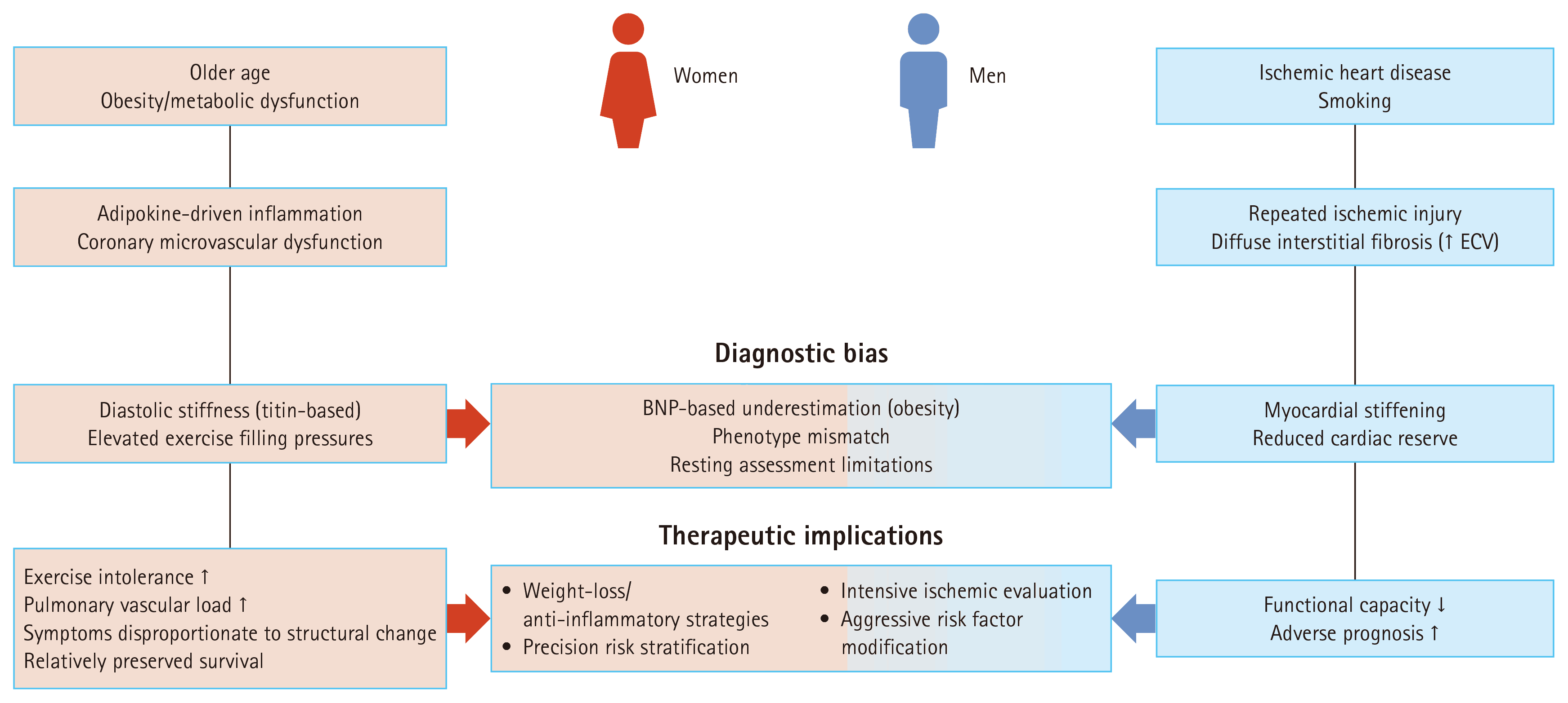
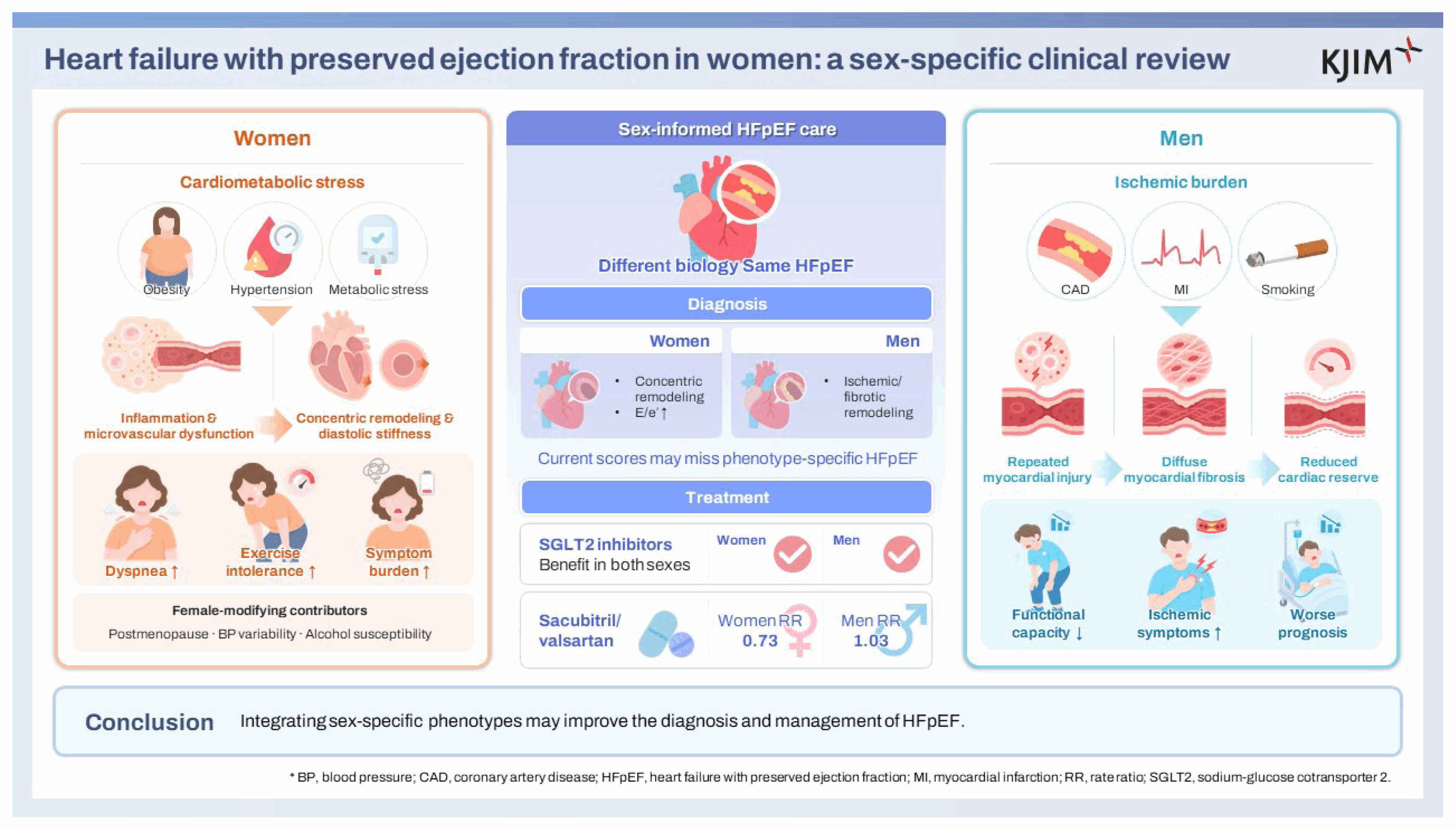
Table 1
| Study dependent difference | Women | Men | Evidence source |
|---|---|---|---|
| Age | ↑ | ↓ | I-PRESERVE [20], TOPCAT [23], DIG ancillary [21] |
| Hypertension | ↑ | ↓ | I-PRESERVE [20], CHARM [10], DIG ancillary [21] |
| Obesity | ↑ | ↓ | Metabolic HFpEF phenotypes [20,32] |
| Diabetes | Variable (↑ in DIG ancillary, → in TOPCAT) | Variable | Study-dependent [21,23] |
| AF | ↓ or → | ↑ in some cohorts | CHARM [10], PARAGON-HF [30] (AF↓ in women) |
| CAD/prior MI | ↓ | ↑↑ | DIG ancillary [21], PARAGON-HF [30] |
| eGFR | ↓ | ↑ | DIG ancillary [21] |
| NT-proBNP | ↑ | ↓ | PARAGON-HF [30], biomarker studies [46–48] |
| Concentric remodeling | ↑ | ↓ | Beale et al. [25] |
| E/e′ | ↑ | ↓ | Beale et al. [25] |
HFpEF, heart failure with preserved ejection fraction; AF, atrial fibrillation; CAD, coronary artery disease; MI, myocardial infarction; eGFR, estimated glomerular filtration rate; NT-proBNP, N-terminal pro-B-type natriuretic peptide; DIG, Digitalis Investigation Group; TOPCAT, Treatment of Preserved Cardiac Function Heart Failure With an Aldosterone Antagonist; I-PRESERVE, Irbesartan in Heart Failure With Preserved Ejection Fraction; CHARM, Candesartan in Heart Failure: Assessment of Reduction in Mortality and Morbidity; PARAGON-HF, Prospective Comparisons of ARNI with ARB Global Outcomes in HF with Preserved Ejection Fraction. Comparison of baseline characteristics between women and men across major HFpEF trials. Arrows indicate relative direction (↑ higher, ↓ lower, → similar).
Table 2
| HFpEF phenotype | Core characteristics | Sex-related pattern |
|---|---|---|
| Vascular-aging/hypertensive phenotype | Older age, hypertension, arterial stiffness, concentric remodeling, diastolic dysfunction | Women-predominant [17,25,26] |
| Metabolic–obese phenotype | Obesity, diabetes, metabolic syndrome, systemic inflammation | Frequently women-predominant [16,20,21,26,32] |
| Ischemic–fibrotic phenotype | CAD/prior MI, ischemic remodeling, myocardial fibrosis | Men-predominant [10,21,26,30,41–43] |
| Pulmonary vascular/RV phenotype | PH, abnormal pulmonary vascular reserve, RV dysfunction/ventricular interaction | Variable; greater functional impairment reported in women [25,27,28] |
| Obesity-associated/low-NP phenotype | Obesity, relatively low BNP/NT-proBNP despite elevated filling pressures | Variable; clinically important in women with obesity [15,16,26,32,46,47] |
| AF/atrial myopathy phenotype | AF burden, LA enlargement/dysfunction, impaired LA strain | Mixed/cohort-dependent [10,20,23,26,30] |
HFpEF, heart failure with preserved ejection fraction; RV, right ventricle; NP, natriuretic peptide; BNP, B-type natriuretic peptide; NTproBNP, N-terminal pro-B-type natriuretic peptide; AF, atrial fibrillation; CAD, coronary artery disease; MI, myocardial infarction; LA, left atrium.
Representative HFpEF phenotypes and sex-related patterns synthesized from observational, phenotyping, and physiological studies. These phenotypes are overlapping clinical constructs rather than mutually exclusive categories, and sex-related patterns indicate relative enrichment. Such patterns may vary according to cohort characteristics and phenotype definitions [24,26].
Table 3
| Trial | Drug/class | Women (HR/RR) | Men (HR/RR) | Interaction |
|---|---|---|---|---|
| TOPCAT [23] | MRA | Signal of ↓ mortality in women (US cohort) | Neutral | NS (overall) |
| PARAGON-HF [30] | ARNI | 0.73 (benefit) | 1.03 (no benefit) | p = 0.017 |
| EMPEROR-Preserved [12] | SGLT2 inhibitor | Benefit | Benefit | NS |
| DELIVER [13] | SGLT2 inhibitor | Benefit | Benefit | NS |
| CHARM-Preserved [10] | ARB | Neutral | Neutral | NS |
| I-PRESERVE [19] | ARB | Neutral | Neutral | NS |
HFpEF, heart failure with preserved ejection fraction; TOPCAT, Treatment of Preserved Cardiac Function Heart Failure With an Aldosterone Antagonist; HR, hazard ratio; RR, rate ratio; PARAGON-HF, Prospective Comparisons of ARNI with ARB Global Outcomes in HF with Preserved Ejection Fraction; EMPEROR-Preserved, Empagliflozin Outcome Trial in Patients with Chronic Heart Failure with Preserved Ejection Fraction; DELIVER, Dapagliflozin Evaluation to Improve the Lives of Patients with Preserved Ejection Fraction Heart Failure; CHARM, Candesartan in Heart Failure: Assessment of Reduction in Mortality and Morbidity; I-PRESERVE, Irbesartan in Heart Failure With Preserved Ejection Fraction; MRA, mineralocorticoid receptor antagonist; ARNI, angiotensin receptor–neprilysin inhibitor; SGLT2, sodium-glucose cotransporter 2; ARB, angiotensin receptor blocker; NS, not significant. Summary of therapeutic effects by sex across major HFpEF trials. PARAGON-HF remains the only trial demonstrating a statistically significant sex–treatment interaction.
- References
- References
REFERENCES
1. Owan TE, Hodge DO, Herges RM, Jacobsen SJ, Roger VL, Redfield MM. Trends in prevalence and outcome of heart failure with preserved ejection fraction. N Engl J Med 2006;355:251–259.
[Article] [PubMed] [PMC]2. Borlaug BA, Paulus WJ. Heart failure with preserved ejection fraction: pathophysiology, diagnosis, and treatment. Eur Heart J 2011;32:670–679.
[Article] [PubMed] [PMC]3. Lam CSP, Gamble GD, Ling LH, et al. Mortality associated with heart failure with preserved vs. reduced ejection fraction in a prospective international multi-ethnic cohort study. Eur Heart J 2018;39:1770–1780.
[Article] [PubMed] [PMC]4. Solomon SD, McMurray JJV, Anand IS, et al. Angiotensin-neprilysin inhibition in heart failure with preserved ejection fraction. N Engl J Med 2019;381:1609–1620.
[PubMed]5. van Riet EE, Hoes AW, Wagenaar KP, Limburg A, Landman MA, Rutten FH. Epidemiology of heart failure: the prevalence of heart failure and ventricular dysfunction in older adults over time. A systematic review. Eur J Heart Fail 2016;18:242–252.
[Article] [PubMed]6. Gerber Y, Weston SA, Redfield MM, et al. A contemporary appraisal of the heart failure epidemic in Olmsted County, Minnesota, 2000 to 2010. JAMA Intern Med 2015;175:996–1004.
[Article] [PubMed] [PMC]7. Korean Society of Heart Failure Fact Sheet & Data Management Committee. Korean Heart Failure Fact Sheet 2025 [Internet] Seoul: Korean Society of Heart Failure, c2025. [cited 2026 Aug 15]. Available from: https://www.kshf.or.kr/heart/heart_04_04.php .8. Cho JY, Cho DH, Youn JC, et al. Korean Society of Heart Failure Guidelines for the management of heart failure: definition and diagnosis. Int J Heart Fail 2023;5:51–65.
[Article] [PubMed] [PMC]9. McDonagh TA, Metra M, Adamo M, et al. 2021 ESC Guidelines for the diagnosis and treatment of acute and chronic heart failure. Eur Heart J 2021;42:3599–3726.
[Article] [PubMed]10. O’Meara E, Clayton T, McEntegart MB, et al. Sex differences in clinical characteristics and prognosis in a broad spectrum of patients with heart failure: results of the candesartan in heart failure: assessment of reduction in mortality and morbidity (CHARM) program. Circulation 2007;115:3111–3120.
[Article] [PubMed]11. Yusuf S, Pfeffer MA, Swedberg K, et al. Effects of candesartan in patients with chronic heart failure and preserved left-ventricular ejection fraction: the CHARM-Preserved Trial. Lancet 2003;362:777–781.
[Article] [PubMed]12. Anker SD, Butler J, Filippatos G, et al. Empagliflozin in Heart Failure with a Preserved Ejection Fraction. N Engl J Med 2021;385:1451–1461.
[PubMed]13. Solomon SD, McMurray JJV, Claggett B, et al. Dapagliflozin in heart failure with mildly reduced or preserved ejection fraction. N Engl J Med 2022;387:1089–1098.
[PubMed]14. Kim HM, Kim HL, Kim MA, Lee HY, Park JJ, Choi DJ. Sex differences in clinical characteristics and long-term outcome in patients with heart failure: data from the KorAHF registry. Korean J Intern Med 2024;39:95–109.
[Article] [PubMed] [PMC]15. Jung MH, Shin MS. Obesity-related heart failure with preserved ejection fraction: diagnostic and therapeutic challenges. Korean J Intern Med 2023;38:157–166.
[Article] [PubMed] [PMC]16. Shin MS. Obesity and heart failure with preserved ejection fraction in women: the interplay of sex-specific pathophysiology and clinical implications. Cardiometab Syndr J 2026;6:3–11.
[Article]17. Sabbatini AR, Kararigas G. Menopause-related estrogen decrease and the pathogenesis of HFpEF: JACC review topic of the week. J Am Coll Cardiol 2020;75:1074–1082.
[PubMed]18. Pitt B, Pfeffer MA, Assmann SF, et al. Spironolactone for heart failure with preserved ejection fraction. N Engl J Med 2014;370:1383–1392.
[Article] [PubMed] [PMC]19. Massie BM, Carson PE, McMurray JJ, et al. Irbesartan in patients with heart failure and preserved ejection fraction. N Engl J Med 2008;359:2456–2467.
[Article] [PubMed]20. Lam CS, Carson PE, Anand IS, et al. Sex differences in clinical characteristics and outcomes in elderly patients with heart failure and preserved ejection fraction: the irbesartan in heart failure with preserved ejection fraction (I-PRESERVE) trial. Circ Heart Fail 2012;5:571–578.
[PubMed] [PMC]21. Deswal A, Bozkurt B. Comparison of morbidity in women versus men with heart failure and preserved ejection fraction. Am J Cardiol 2006;97:1228–1231.
[Article] [PubMed]22. Ahmed A, Rich MW, Fleg JL, et al. Effects of digoxin on morbidity and mortality in diastolic heart failure: the ancillary digitalis investigation group trial. Circulation 2006;114:397–403.
[Article] [PubMed] [PMC]23. Merrill M, Sweitzer NK, Lindenfeld J, Kao DP. Sex differences in outcomes and responses to spironolactone in heart failure with preserved ejection fraction: a secondary analysis of TOPCAT trial. JACC Heart Fail 2019;7:228–238.
[PubMed] [PMC]24. Kao DP, Lewsey JD, Anand IS, et al. Characterization of subgroups of heart failure patients with preserved ejection fraction with possible implications for prognosis and treatment response. Eur J Heart Fail 2015;17:925–935.
[Article] [PubMed] [PMC]25. Beale AL, Nanayakkara S, Segan L, et al. Sex differences in heart failure with preserved ejection fraction pathophysiology: a detailed invasive hemodynamic and echocardiographic analysis. JACC Heart Fail 2019;7:239–249.
[PubMed]26. Shah SJ, Katz DH, Deo RC. Phenotypic spectrum of heart failure with preserved ejection fraction. Heart Fail Clin 2014;10:407–418.
[Article] [PubMed] [PMC]27. Verwerft J, Foulkes S, Bekhuis Y, et al. The oxygen cascade according to HFpEF likelihood: a focus on sex differences. JACC Adv 2024;3:101039.
[PubMed] [PMC]28. Parasuraman SK, Loudon BL, Lowery C, et al. Diastolic ventricular interaction in heart failure with preserved ejection fraction. J Am Heart Assoc 2019;8:e010114.
[PubMed] [PMC]29. Shah SJ, Lam CS, Svedlund S, et al. Prevalence and correlates of coronary microvascular dysfunction in heart failure with preserved ejection fraction: PROMIS-HFpEF. Eur Heart J 2018;39:3439–3450.
[Article] [PubMed] [PMC]30. McMurray JJ, Jackson AM, Lam CS, et al. Effects of sacubitril-valsartan versus valsartan in women compared with men with heart failure and preserved ejection fraction: insights from PARAGON-HF. Circulation 2020;141:338–351.
[PubMed]31. Paulus WJ, Tschöpe C. A novel paradigm for heart failure with preserved ejection fraction: comorbidities drive myocardial dysfunction and remodeling through coronary microvascular endothelial inflammation. J Am Coll Cardiol 2013;62:263–271.
[PubMed]32. Kitzman DW, Shah SJ. The HFpEF obesity phenotype: the elephant in the room. J Am Coll Cardiol 2016;68:200–213.
[PubMed]33. Versnjak J, Kuehne T, Fahjen P, et al. Deep phenotyping of heart failure with preserved ejection fraction through multi-omics integration. Eur J Heart Fail 2025;27:3243–3259.
[Article] [PubMed] [PMC]34. Binek A, Janssens JV, Sundararaman N, et al. Multicenter HFpEF study identifies sex disparity linked with two discrete cardiac proteomic signatures. bioRxiv 2025:2025.12.18.695212.
[Article] [PubMed]35. Piano MR, Thur LA, Hwang CL, Phillips SA. Effects of alcohol on the cardiovascular system in women. Alcohol Res 2020;40:12.
[Article] [PubMed] [PMC]36. Urbano-Márquez A, Estruch R, Fernández-Solá J, Nicolás JM, Paré JC, Rubin E. The greater risk of alcoholic cardiomyopathy and myopathy in women compared with men. JAMA 1995;274:149–154.
[Article] [PubMed]37. Gonçalves A, Jhund PS, Claggett B, et al. Relationship between alcohol consumption and cardiac structure and function in the elderly: the Atherosclerosis Risk In Communities Study. Circ Cardiovasc Imaging 2015;8:e002846.
[PubMed] [PMC]38. Stevens SL, Wood S, Koshiaris C, et al. Blood pressure variability and cardiovascular disease: systematic review and meta-analysis. BMJ 2016;354:i4098.
[Article] [PubMed] [PMC]39. Ma Y, Song A, Viswanathan A, et al. Blood pressure variability and cerebral small vessel disease: a systematic review and meta-analysis of population-based cohorts. Stroke 2020;51:82–89.
[Article] [PubMed] [PMC]40. Haring B, Hunt RP, Manson JE, et al. Blood pressure variability and heart failure hospitalization: results from the women’s health initiative. Am J Prev Med 2022;63:410–418.
[Article] [PubMed]41. Zile MR, Gottdiener JS, Hetzel SJ, et al. Prevalence and significance of alterations in cardiac structure and function in patients with heart failure and a preserved ejection fraction. Circulation 2011;124:2491–2501.
[Article] [PubMed]42. Rommel KP, von Roeder M, Latuscynski K, et al. Extracellular volume fraction for characterization of patients with heart failure and preserved ejection fraction. J Am Coll Cardiol 2016;67:1815–1825.
[Article] [PubMed]43. Quarta G, Gori M, Iorio A, et al. Cardiac magnetic resonance in heart failure with preserved ejection fraction: myocyte, interstitium, microvascular, and metabolic abnormalities. Eur J Heart Fail 2020;22:1065–1075.
[Article] [PubMed]44. Chung AK, Das SR, Leonard D, et al. Women have higher left ventricular ejection fractions than men independent of differences in left ventricular volume: the Dallas Heart Study. Circulation 2006;113:1597–1604.
[Article] [PubMed]45. Föll D, Jung B, Schilli E, et al. Magnetic resonance tissue phase mapping of myocardial motion: new insight in age and gender. Circ Cardiovasc Imaging 2010;3:54–64.
[PubMed]46. Redfield MM, Rodeheffer RJ, Jacobsen SJ, Mahoney DW, Bailey KR, Burnett JC Jr. Plasma brain natriuretic peptide concentration: impact of age and gender. J Am Coll Cardiol 2002;40:976–982.
[Article] [PubMed]47. Hsich EM, Grau-Sepulveda MV, Hernandez AF, et al. Relationship between sex, ejection fraction, and B-type natriuretic peptide levels in patients hospitalized with heart failure and associations with inhospital outcomes: findings from the Get With The Guideline-Heart Failure Registry. Am Heart J 2013;166:1063–1071.
[Article] [PubMed]48. Gohar A, Chong JPC, Liew OW, et al. The prognostic value of highly sensitive cardiac troponin assays for adverse events in men and women with stable heart failure and a preserved vs. reduced ejection fraction. Eur J Heart Fail 2017;19:1638–1647.
[Article] [PubMed]49. Li X, Achten A, Nassiri S, et al. Sex-specific performance of clinical diagnostic algorithms for HFpEF across two independent cohorts. Neth Heart J 2025;33:412–420.
[Article] [PubMed] [PMC]50. McGill JB, Subramanian S. Safety of sodium-glucose co-transporter 2 inhibitors. Am J Cardiol 2019;124:Suppl 1. S45–52.
[Article] [PubMed]51. Liu J, Li L, Li S, et al. Effects of SGLT2 inhibitors on UTIs and genital infections in type 2 diabetes mellitus: a systematic review and meta-analysis. Sci Rep 2017;7:2824.
[Article] [PubMed] [PMC]